This document discusses the secondary and tertiary structures of RNA. It begins by defining RNA and its types, including mRNA, rRNA, tRNA, snRNAs, miRNAs, siRNAs and others. It then explains the primary, secondary and tertiary structures of RNA. The secondary structure involves base pairing to form stems and loops, including bulge loops, internal loops, multi loops and hairpin loops. Tertiary structure involves interactions between these secondary structure elements. Specific interactions discussed include coaxial stacking and the role of magnesium ions. The structures are important for RNA's catalytic, regulatory and structural roles in cells.
RNA (Ribonucleic Acid)
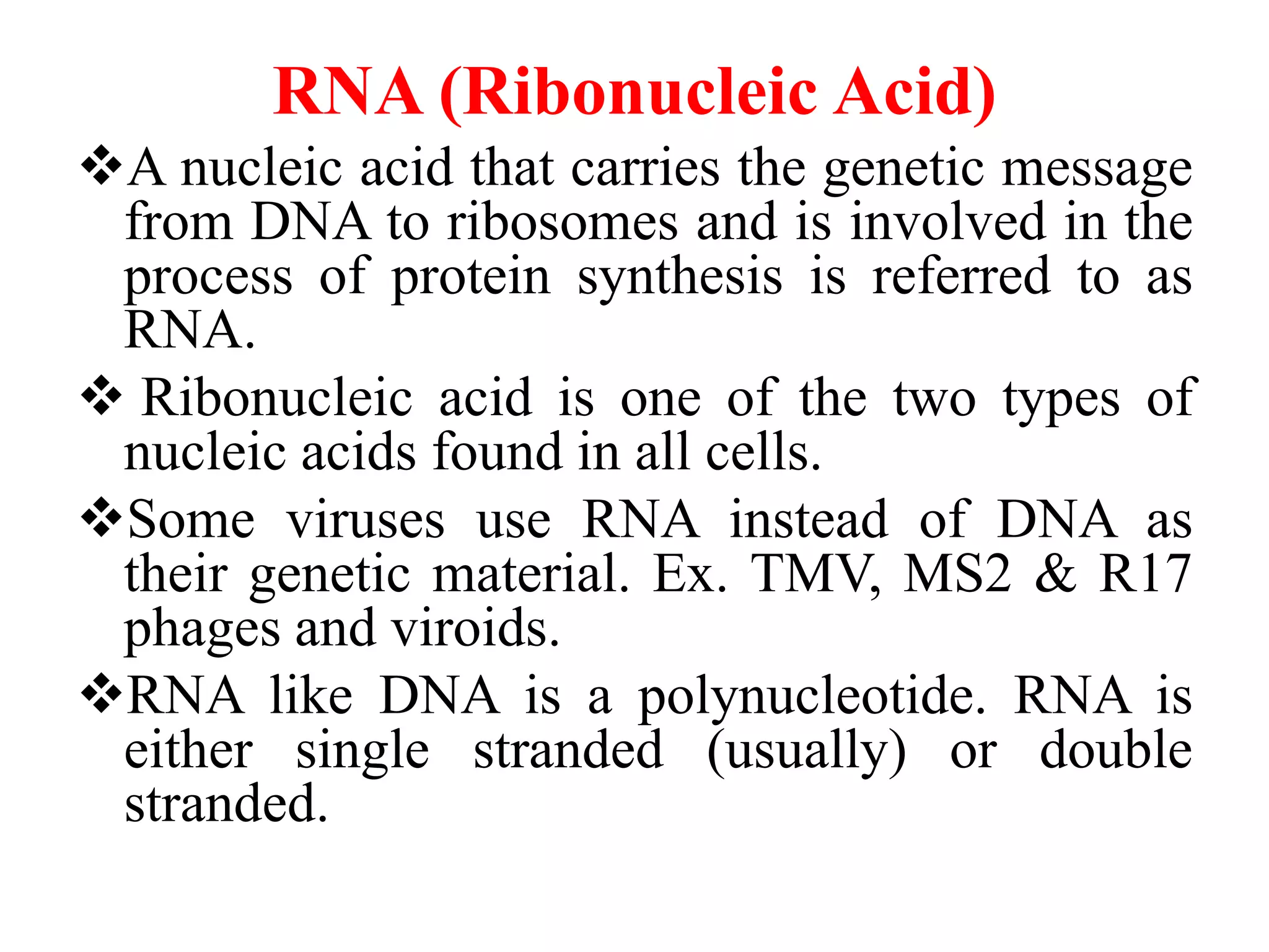
Anucleic acid that carries the genetic message
from DNA to ribosomes and is involved in the
process of protein synthesis is referred to as
RNA.
Ribonucleic acid is one of the two types of
nucleic acids found in all cells.
Some viruses use RNA instead of DNA as
their genetic material. Ex. TMV, MS2 & R17
phages and viroids.
RNA like DNA is a polynucleotide. RNA is
either single stranded (usually) or double
stranded.
3.
Basic structure ofRNA
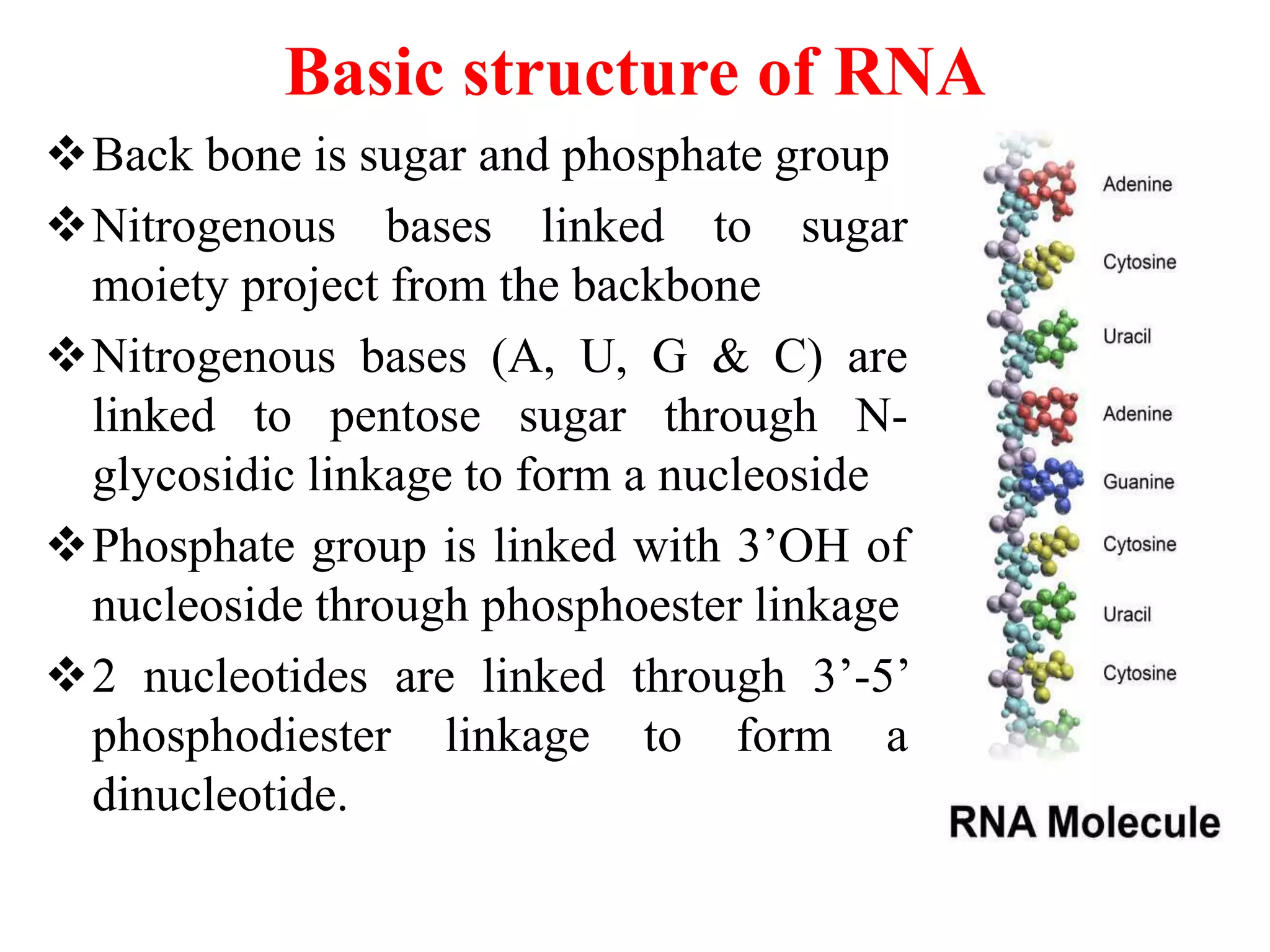
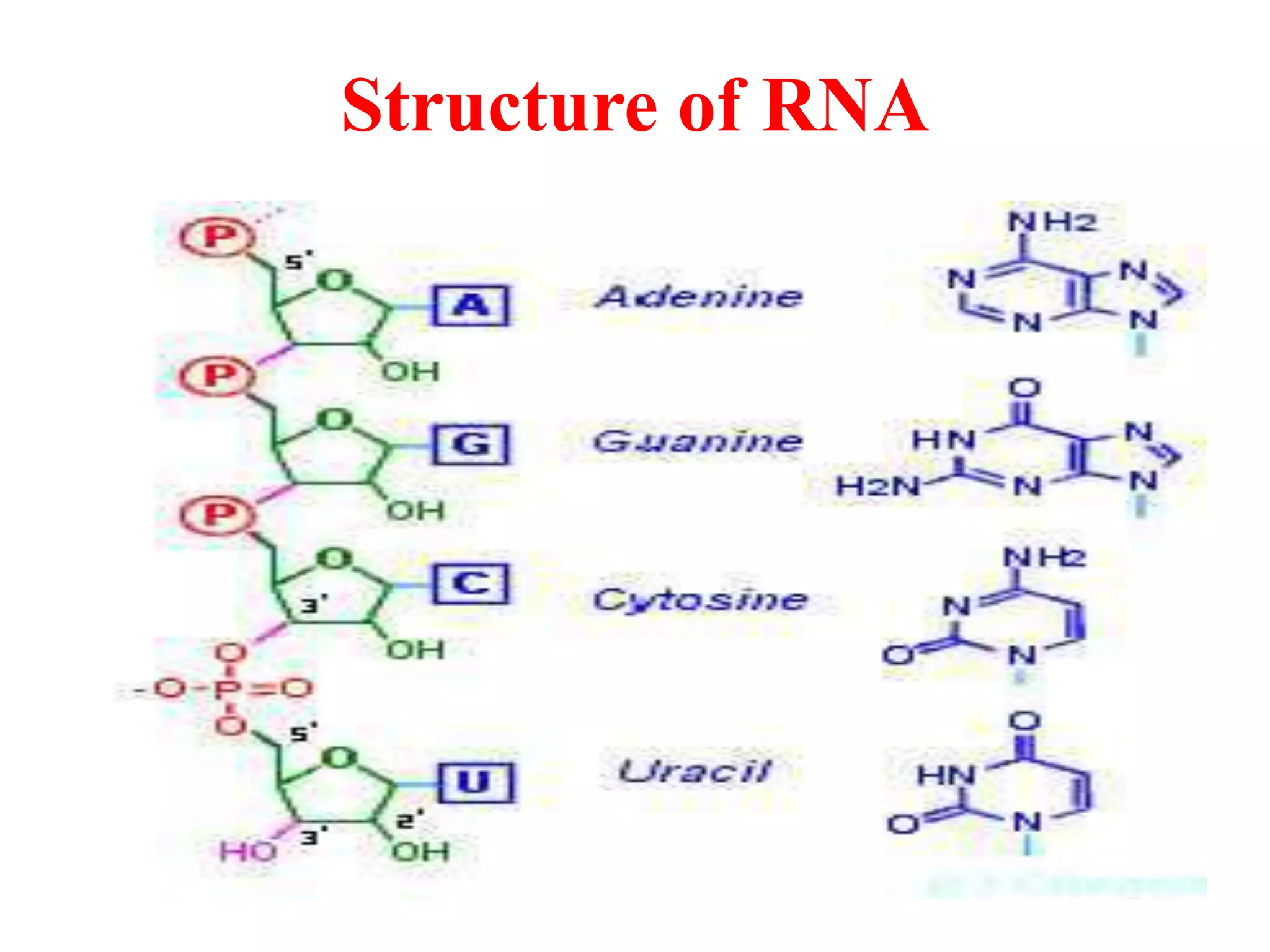
Back bone is sugar and phosphate group
Nitrogenous bases linked to sugar
moiety project from the backbone
Nitrogenous bases (A, U, G & C) are
linked to pentose sugar through N-
glycosidic linkage to form a nucleoside
Phosphate group is linked with 3’OH of
nucleoside through phosphoester linkage
2 nucleotides are linked through 3’-5’
phosphodiester linkage to form a
dinucleotide.
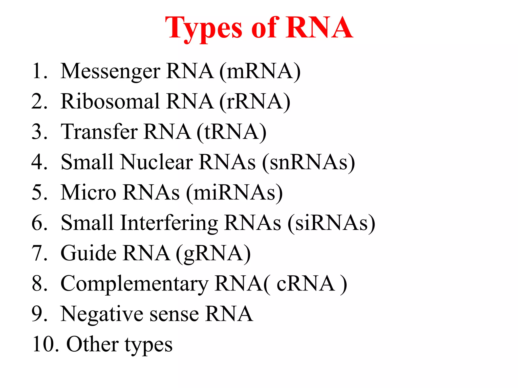
Types of RNA
1.Messenger RNA (mRNA)
2. Ribosomal RNA (rRNA)
3. Transfer RNA (tRNA)
4. Small Nuclear RNAs (snRNAs)
5. Micro RNAs (miRNAs)
6. Small Interfering RNAs (siRNAs)
7. Guide RNA (gRNA)
8. Complementary RNA( cRNA )
9. Negative sense RNA
10. Other types
6.
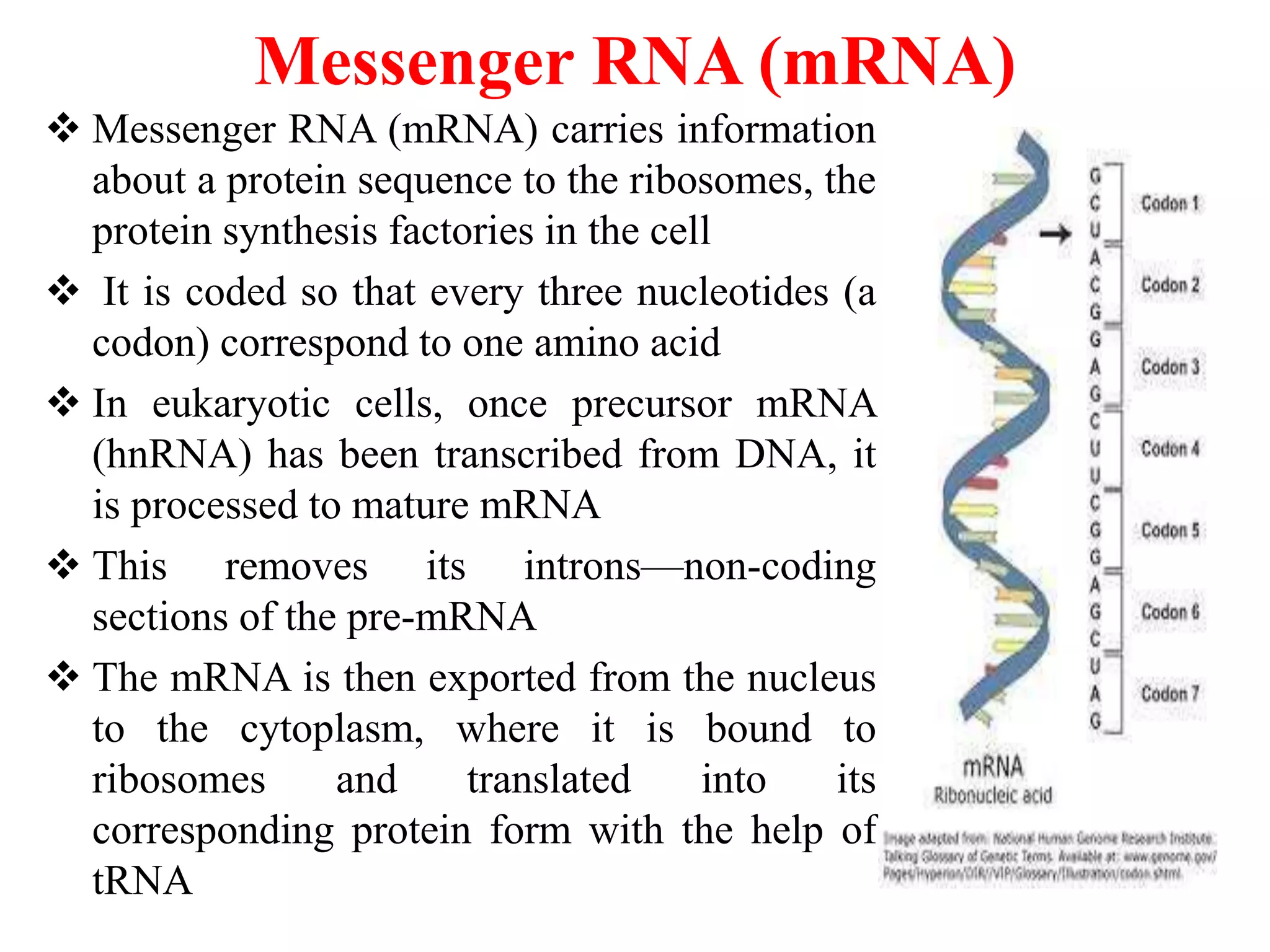
Messenger RNA (mRNA)
Messenger RNA (mRNA) carries information
about a protein sequence to the ribosomes, the
protein synthesis factories in the cell
It is coded so that every three nucleotides (a
codon) correspond to one amino acid
In eukaryotic cells, once precursor mRNA
(hnRNA) has been transcribed from DNA, it
is processed to mature mRNA
This removes its introns—non-coding
sections of the pre-mRNA
The mRNA is then exported from the nucleus
to the cytoplasm, where it is bound to
ribosomes and translated into its
corresponding protein form with the help of
tRNA
7.
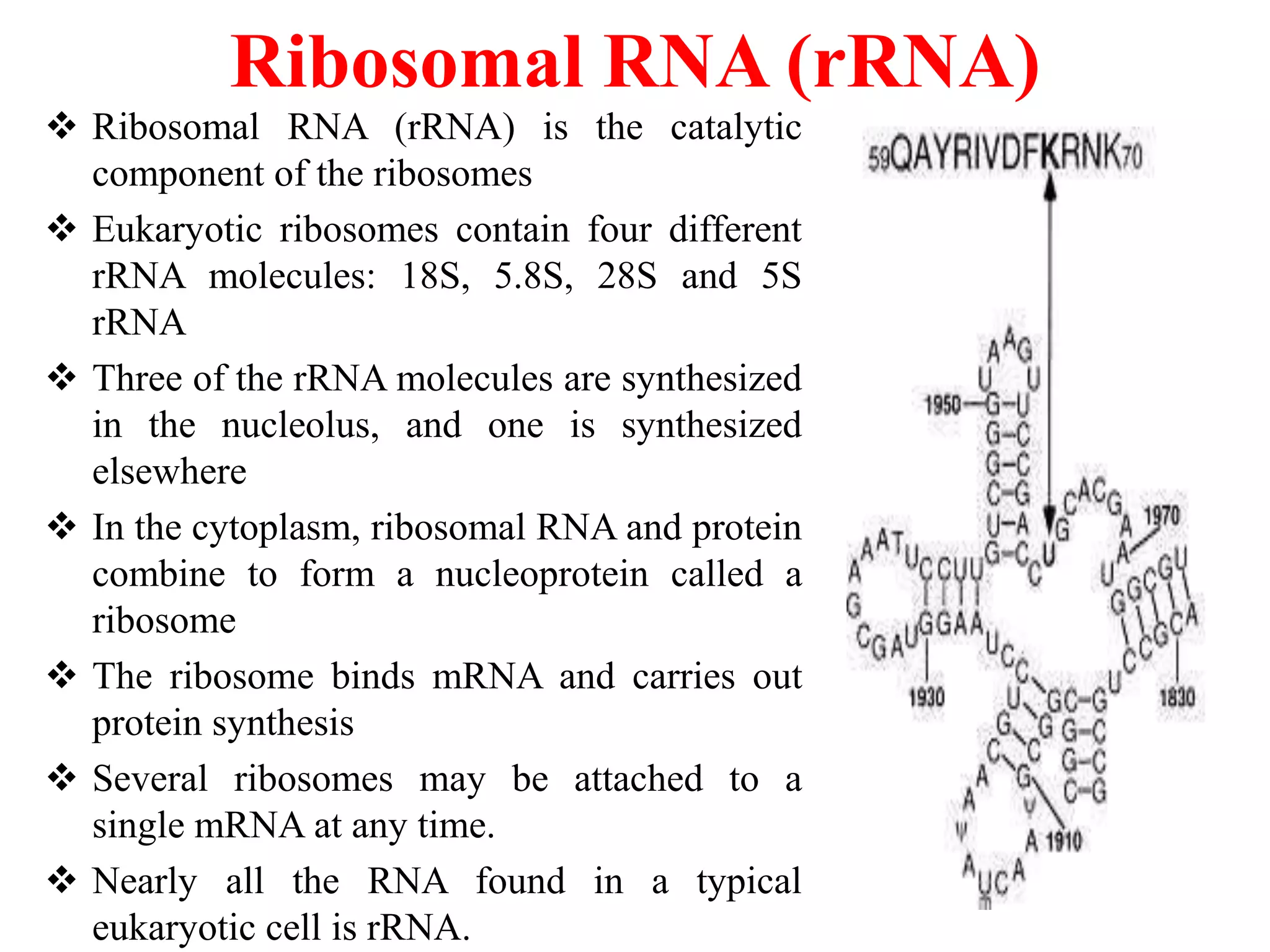
Ribosomal RNA (rRNA)
Ribosomal RNA (rRNA) is the catalytic
component of the ribosomes
Eukaryotic ribosomes contain four different
rRNA molecules: 18S, 5.8S, 28S and 5S
rRNA
Three of the rRNA molecules are synthesized
in the nucleolus, and one is synthesized
elsewhere
In the cytoplasm, ribosomal RNA and protein
combine to form a nucleoprotein called a
ribosome
The ribosome binds mRNA and carries out
protein synthesis
Several ribosomes may be attached to a
single mRNA at any time.
Nearly all the RNA found in a typical
eukaryotic cell is rRNA.
8.
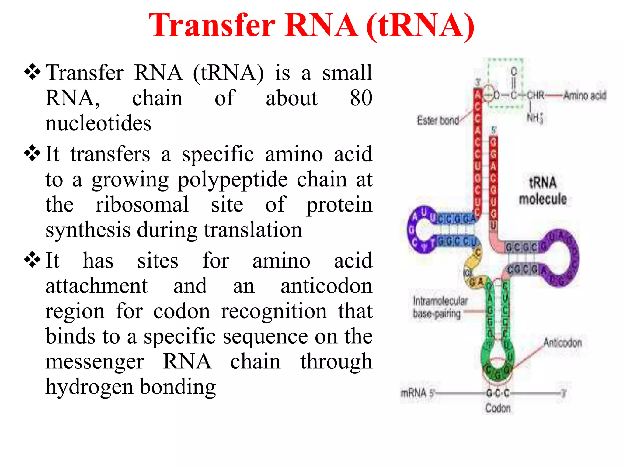
Transfer RNA (tRNA)
TransferRNA (tRNA) is a small
RNA, chain of about 80
nucleotides
It transfers a specific amino acid
to a growing polypeptide chain at
the ribosomal site of protein
synthesis during translation
It has sites for amino acid
attachment and an anticodon
region for codon recognition that
binds to a specific sequence on the
messenger RNA chain through
hydrogen bonding
9.
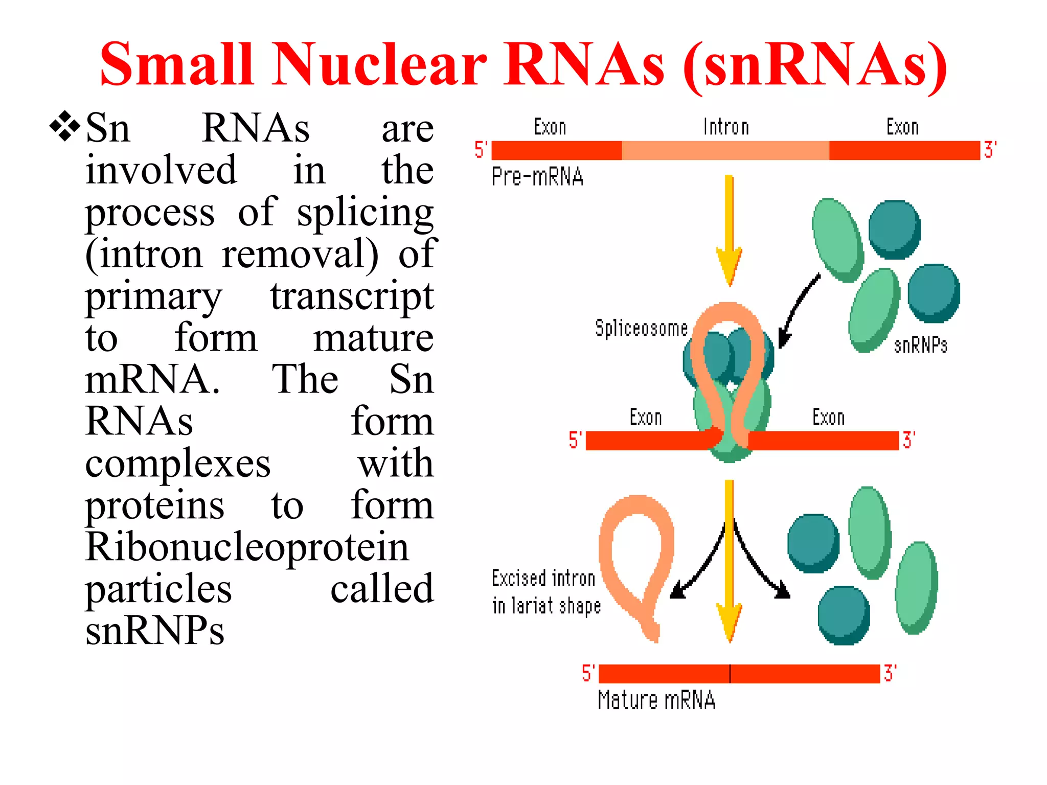
Small Nuclear RNAs(snRNAs)
Sn RNAs are
involved in the
process of splicing
(intron removal) of
primary transcript
to form mature
mRNA. The Sn
RNAs form
complexes with
proteins to form
Ribonucleoprotein
particles called
snRNPs
10.
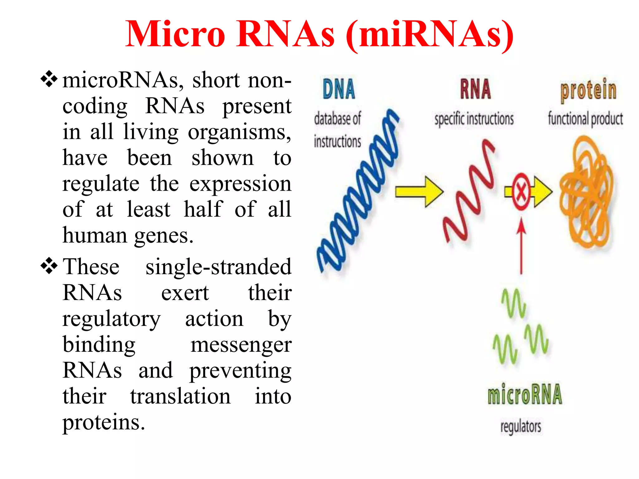
Micro RNAs (miRNAs)
microRNAs,short non-
coding RNAs present
in all living organisms,
have been shown to
regulate the expression
of at least half of all
human genes.
These single-stranded
RNAs exert their
regulatory action by
binding messenger
RNAs and preventing
their translation into
proteins.
11.
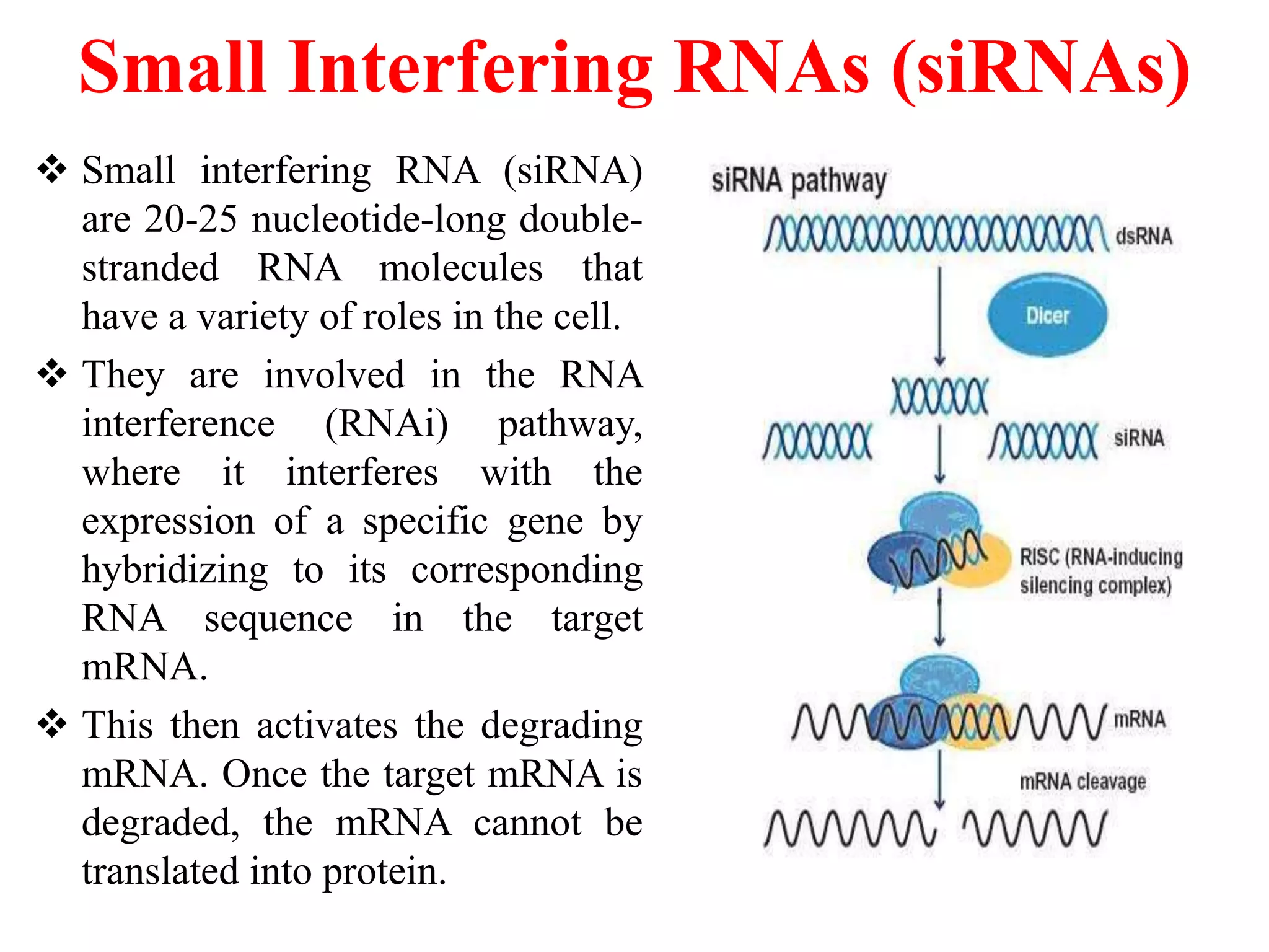
Small Interfering RNAs(siRNAs)
Small interfering RNA (siRNA)
are 20-25 nucleotide-long double-
stranded RNA molecules that
have a variety of roles in the cell.
They are involved in the RNA
interference (RNAi) pathway,
where it interferes with the
expression of a specific gene by
hybridizing to its corresponding
RNA sequence in the target
mRNA.
This then activates the degrading
mRNA. Once the target mRNA is
degraded, the mRNA cannot be
translated into protein.
12.
Guide RNA (gRNA)
RNA genes that function in RNA editing, found in mitochondria
by inserting or deleting stretches of uridylates (Us).
The gRNA forms part of editosome and contain sequences to
hybridize to matching sequences in the mRNA to guide the
mRNA modifications.
Complementary RNA( cRNA )
Viral RNA that is transcribed from
negative sense RNA and serves as a template for protein
synthesis.
Negative sense RNA
Viral RNA with a base sequence complementary to that
of mRNA during replication it serves as a template to the
transcription of viral
complementary RNA
13.
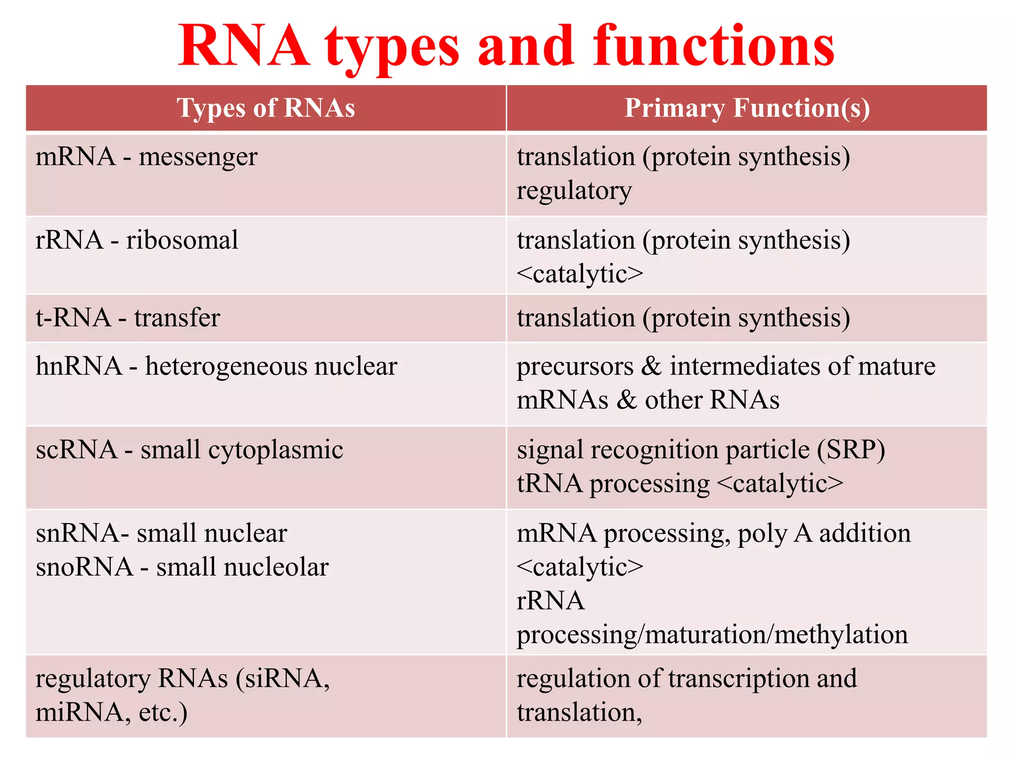
RNA types andfunctions
Types of RNAs Primary Function(s)
mRNA - messenger translation (protein synthesis)
regulatory
rRNA - ribosomal translation (protein synthesis)
<catalytic>
t-RNA - transfer translation (protein synthesis)
hnRNA - heterogeneous nuclear precursors & intermediates of mature
mRNAs & other RNAs
scRNA - small cytoplasmic signal recognition particle (SRP)
tRNA processing <catalytic>
snRNA- small nuclear
snoRNA - small nucleolar
mRNA processing, poly A addition
<catalytic>
rRNA
processing/maturation/methylation
regulatory RNAs (siRNA,
miRNA, etc.)
regulation of transcription and
translation,
14.
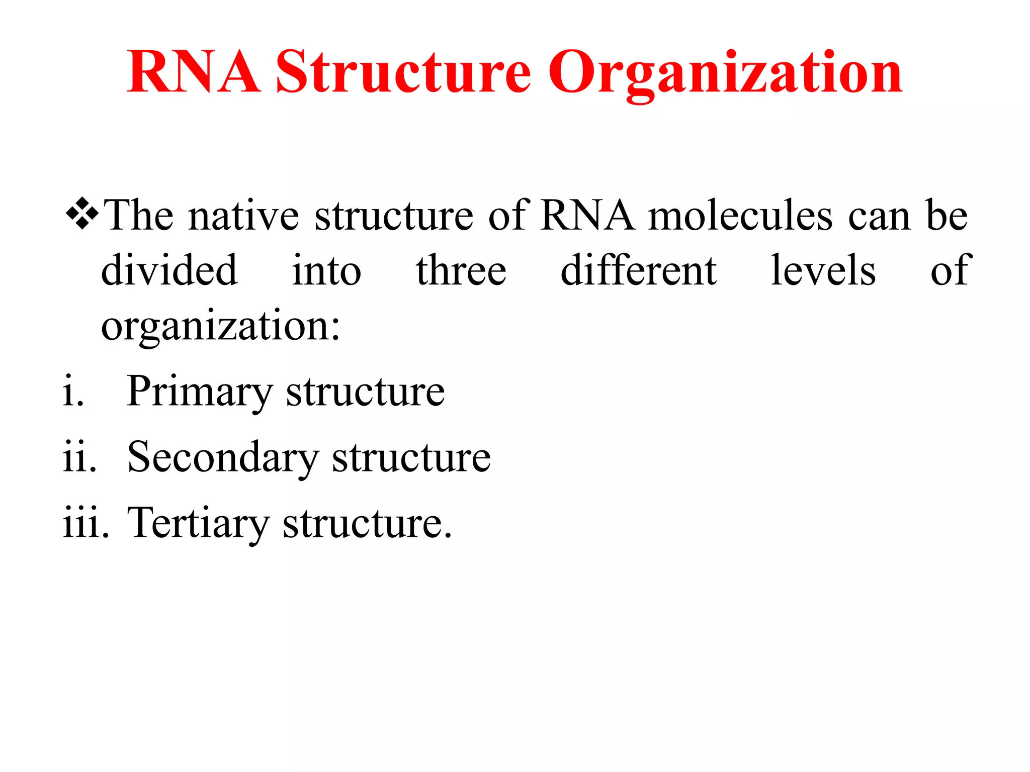
RNA Structure Organization
Thenative structure of RNA molecules can be
divided into three different levels of
organization:
i. Primary structure
ii. Secondary structure
iii. Tertiary structure.
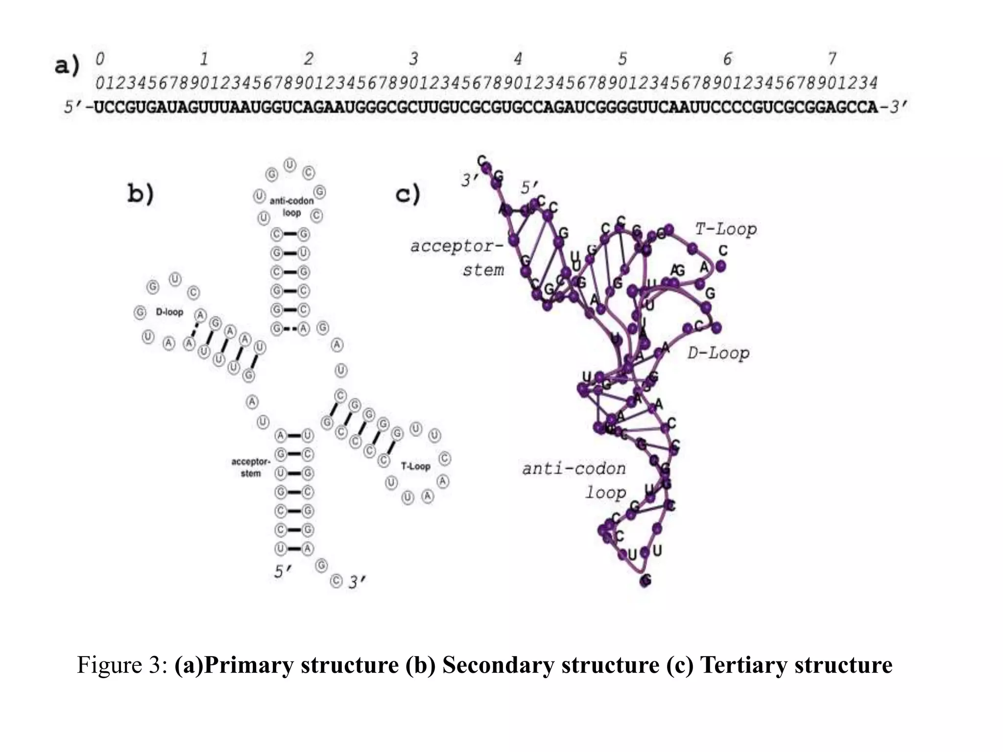
Primary structure
It denotesthe ribo-nucleotide sequence
(commonly referred to as base sequence) of
the molecule.
Usually, the base-sequence of an RNA
molecule only consists of a combination of the
bases A, G, C, U.
Furthermore, modified bases such as
pseudouracil (ψ) are represented by their most-
similar standard base.
17.
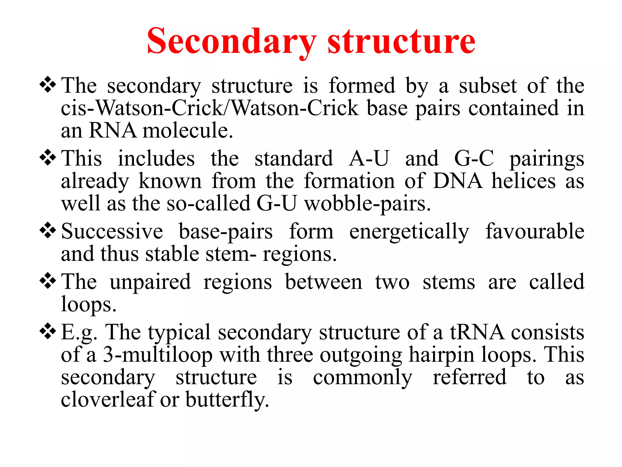
Secondary structure
The secondarystructure is formed by a subset of the
cis-Watson-Crick/Watson-Crick base pairs contained in
an RNA molecule.
This includes the standard A-U and G-C pairings
already known from the formation of DNA helices as
well as the so-called G-U wobble-pairs.
Successive base-pairs form energetically favourable
and thus stable stem- regions.
The unpaired regions between two stems are called
loops.
E.g. The typical secondary structure of a tRNA consists
of a 3-multiloop with three outgoing hairpin loops. This
secondary structure is commonly referred to as
cloverleaf or butterfly.
18.
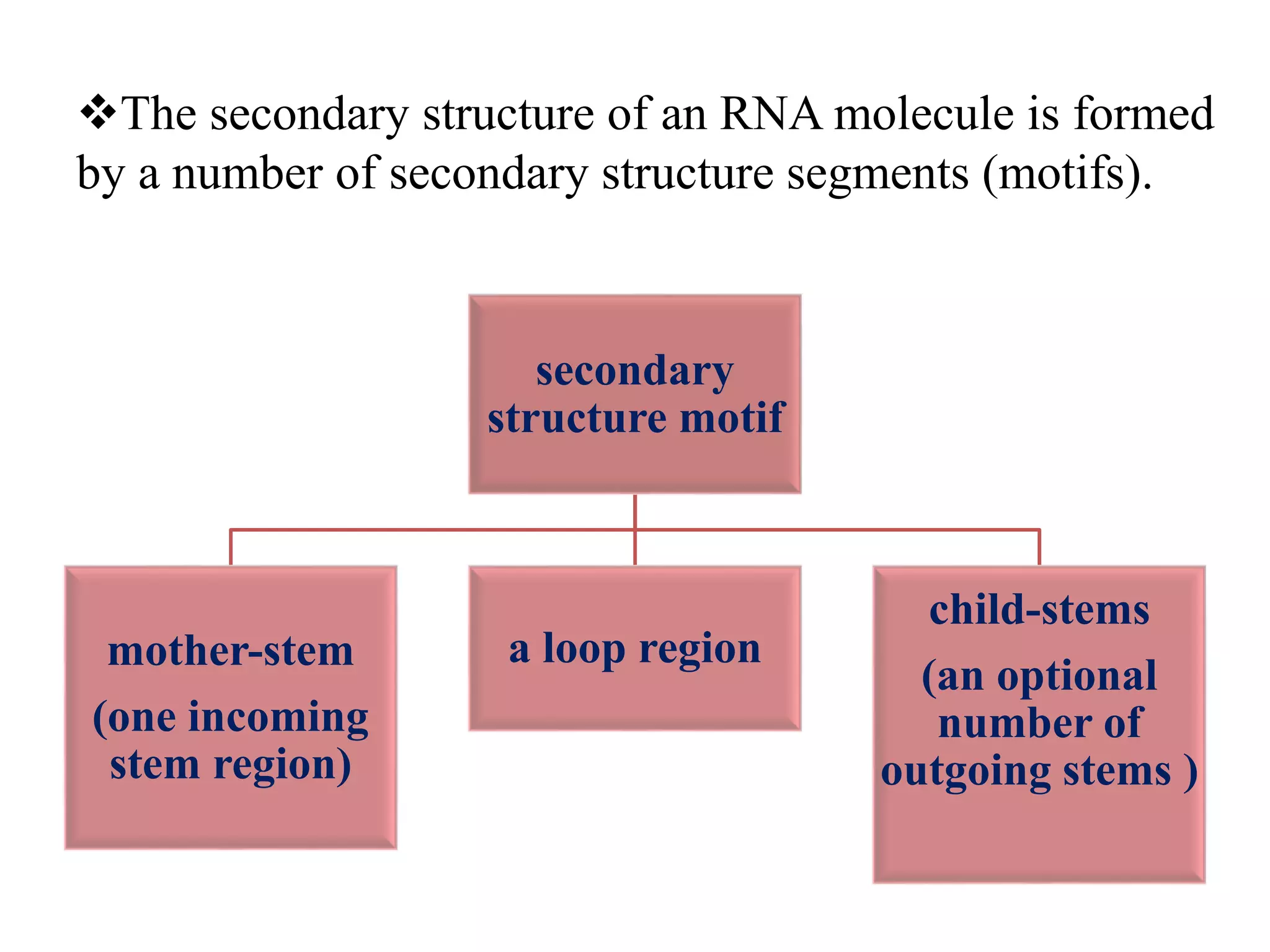
The secondary structureof an RNA molecule is formed
by a number of secondary structure segments (motifs).
secondary
structure motif
mother-stem
(one incoming
stem region)
a loop region
child-stems
(an optional
number of
outgoing stems )
19.
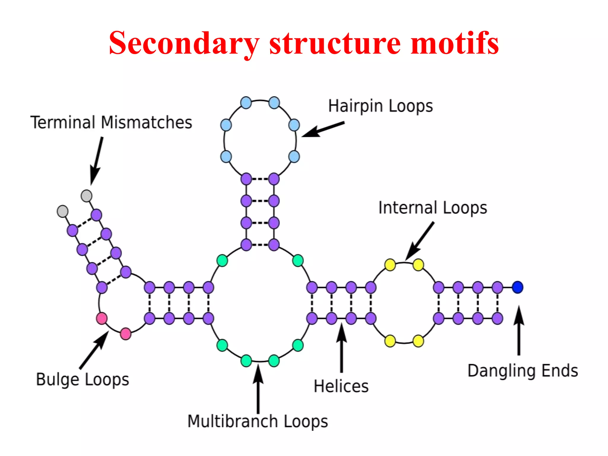
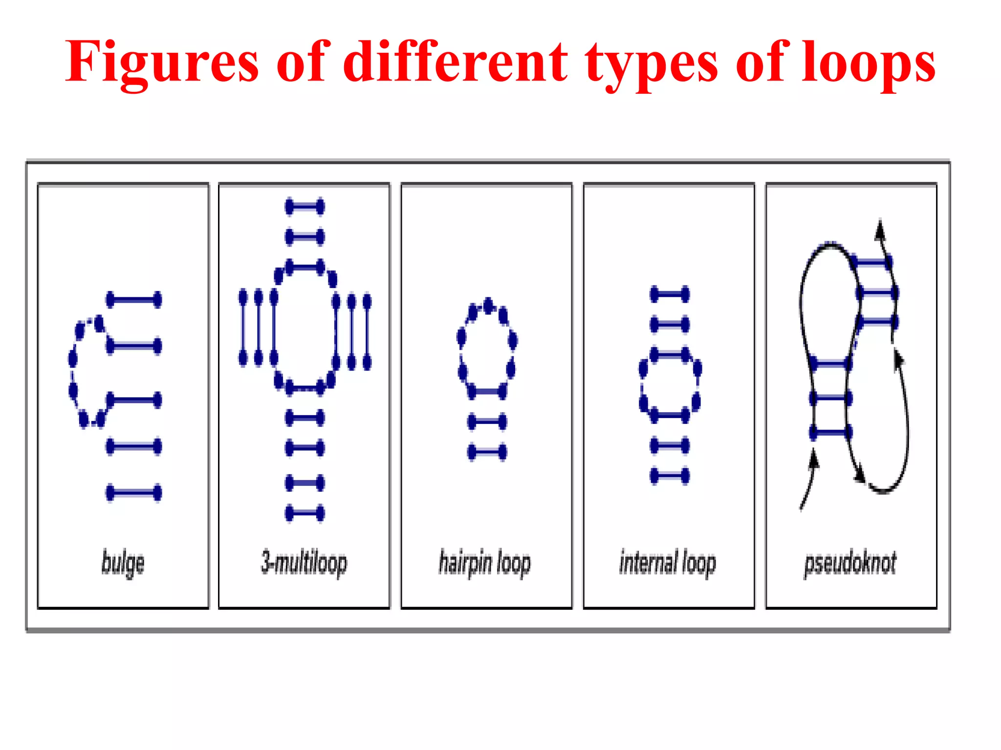
Secondary structure motifscan be classified
into following loop classes:
Loop classes
Regular loops (4
classes)
[do not interfere
with
the 3-D structure of
the molecule]
Interior loop
Ex. Bulge, Internal
loop and Multi loop
External loop
Ex. Hairpin loop
5th class
[creates a change of
the 3-D structure]
Ex. Pseudoknot
A bulge loop
Bulgeloops have unpaired bases on only one
strand in a double-stranded region, whereas the
other strand only has paired bases [5].
The size of the bulge loop is at least the size of
one unpaired base, but in principle there is no
upper limit [5].
They have the ability to bend a stem and
thereby influence the three-dimensional
structure.
22.
An internal loop
Internalloops have unpaired bases on both
strands in a double-stranded region.
The thermodynamical stability of the loops
depends on the types and the number of the
unpaired bases [5].
If the number of the unpaired bases in both
strands are of equal size, the internal loop is
called symmetric[5].
Nevertheless the loop can be very inflexible
due to stacking and/or hydrogen bonds.
23.
A multi loop
Loopswhich connect more than two helices are
called multi loops.
In between the helices unpaired bases can be
found.
Together with the closing base pair, the unpaired
bases are decisive for the stacking of the helices
and thereby they form the three-dimensional
structure [5].
Very often it can be observed that four helices are
connected within a multi loop, for instance in
tRNA, but also more or less helices can be
connected [5].
24.
A hairpin loop
Ahairpin loop describes the structure of a
sequence that folds back on itself, usually a
stem or a double helix and thereby forming an
unpaired loop. Such a loop is called a hairpin
loop and is formed relatively quick [5].
The time needed to grow the loop is at its
minimum in the range of only a few
microseconds and is growing with the length
of the unpaired loop [5].
25.
Continue…
The thermodynamical stabilityof the loop
depends on the sequence of the loop, on the type
of the closing base-pair and on the size of the loop
[5].
A hairpin loop needs at least four unpaired bases
and often loops of five unpaired bases are the
most stable ones [5].
Very stable tetraloop hairpins can be found in
rRNA and even bigger hairpin loops can for
instance be found in tRNA: The anticodon loops
consist of seven bases [5].
26.
A pseudoknot
A pseudoknotis a tertiary
structural element of RNA.
It is formed by base-pairing
between an already existing
secondary structure loop and a
free ending [5].
Nucleotides within a hairpin
loop form base pairs with
nucleotides outside the stem
[7]. Hence base pairs occur that
overlap each other in their
sequence position.
Fig. formation of a
pseudoknot with
coaxial stacking of the
two helices
Tertiary structure
Base-pairs thatdo not belong to the secondary
structure together with pseudo-base-pairs form the
tertiary structure of the molecule. This includes
other atomic interactions such as vanderwaals
forces, electrostatic and hydrophobic interactions
and hydrogen-bonds between e.g. base and ribose
residues.
Tertiary contacts are interactions between distinct
secondary structure elements.
They induce local and/or global structure folds
and as such are dominantly responsible for the
overall three-dimensional structure of an RNA
molecule [4].
29.
Continue….
Tertiary interactions canoccur between two
helical motifs (stem-stem), between two unpaired
(loop-loop), and between an unpaired region and
a stem region (loop-stem) [4].
In the three-dimensional structure of a tRNA
molecule, the stems of the D-loop and the T-loop,
as well as the acceptor-stem and the stem of the
anticodon-loop stack upon another (coaxial
stacking stem-stem interaction).
The typical L-shape of a tRNA molecule is
yielded by the stacked stem regions as well as the
kissing hairpin loop-loop interaction between the
D-loop and T-loop hairpins.
30.
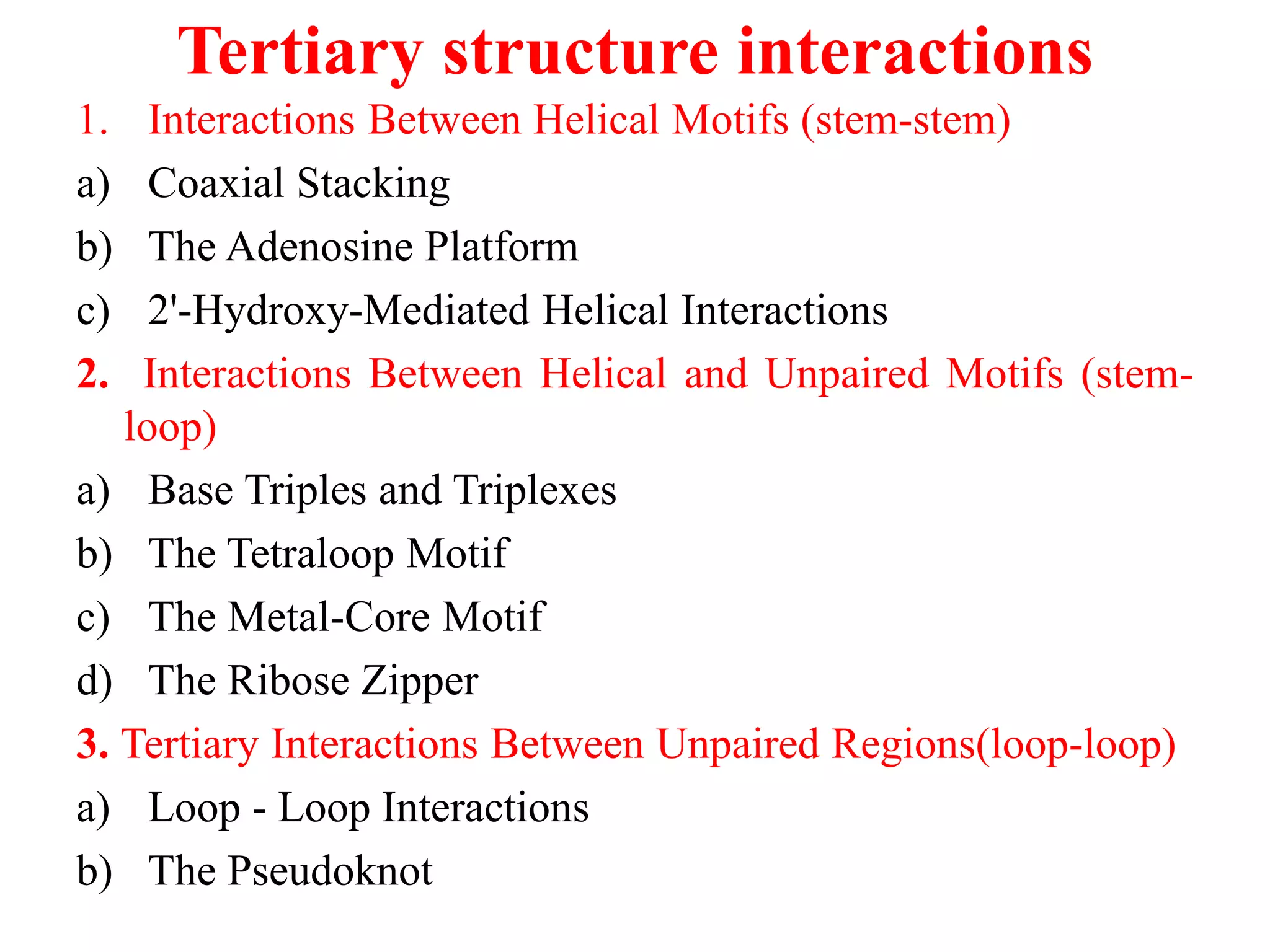
Tertiary structure interactions
1.Interactions Between Helical Motifs (stem-stem)
a) Coaxial Stacking
b) The Adenosine Platform
c) 2'-Hydroxy-Mediated Helical Interactions
2. Interactions Between Helical and Unpaired Motifs (stem-
loop)
a) Base Triples and Triplexes
b) The Tetraloop Motif
c) The Metal-Core Motif
d) The Ribose Zipper
3. Tertiary Interactions Between Unpaired Regions(loop-loop)
a) Loop - Loop Interactions
b) The Pseudoknot
31.
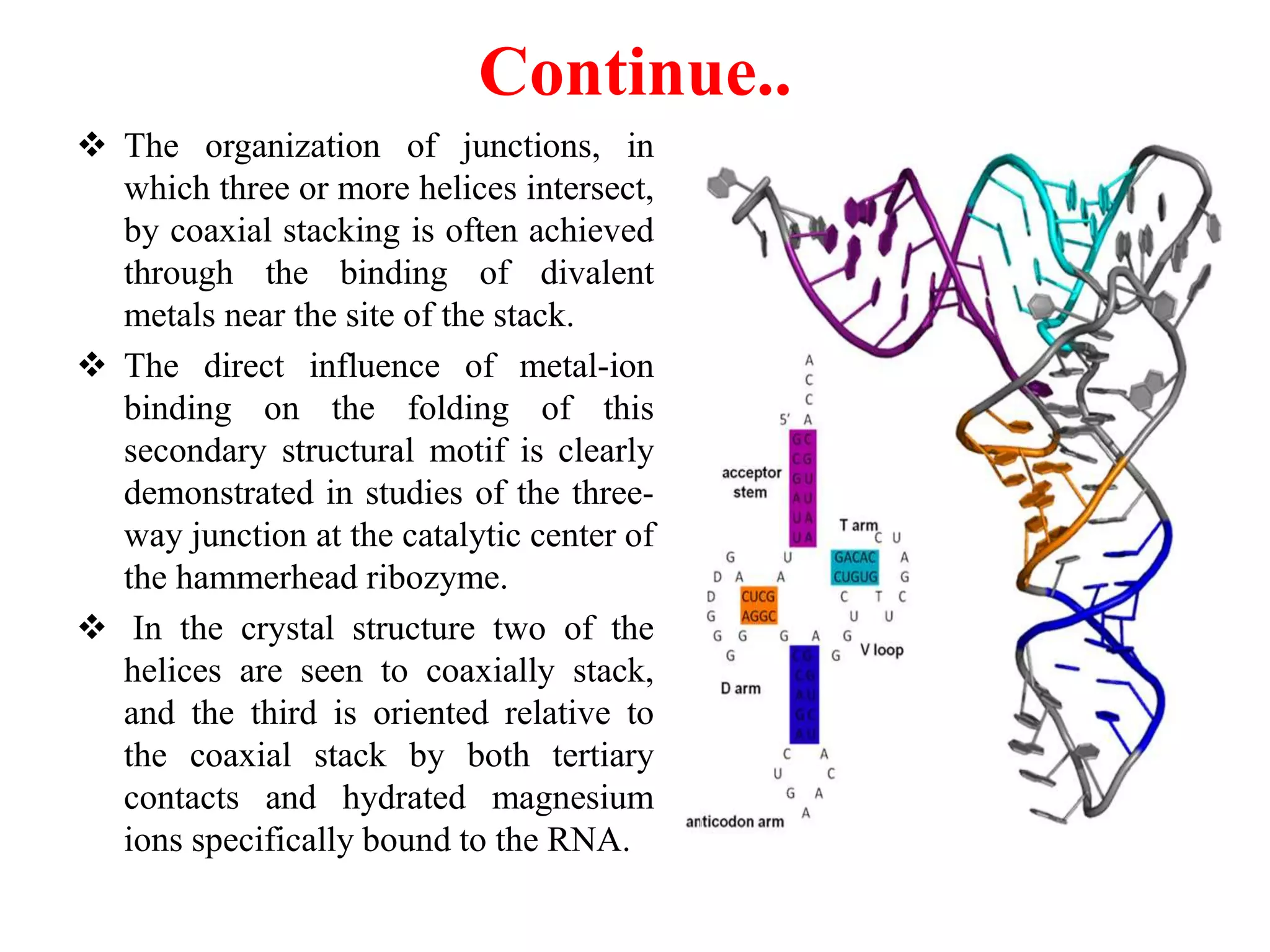
Coaxial Stacking
Themost fundamental method by which RNA achieves
higher order organization, is a consequence of the highly
favorable energetic contributions of stacking interactions
between the pie-electron system of the nucleotide bases to
the overall stability of nucleic structure.
The contribution of coaxial stacking to the global fold of an
RNA was first observed in the crystal structure of
tRNAPhe.[6, 8, 9] In the 3-D structure the stems of the D-
and anticodon arms stack upon one another as do the stems
of the T-arm and aminoacyl acceptor arm [9].
These two coaxial stacks are oriented perpendicularly with
respect to one another by tertiary interactions between the D
and T-loops to yield the overall L-shape of the molecule.
The predominance of coaxial stacking in the organization of
RNA structure is also evident in the structures of the P4-P6
domain and the hepatitis delta ribozyme.
32.
Continue..
The organizationof junctions, in
which three or more helices intersect,
by coaxial stacking is often achieved
through the binding of divalent
metals near the site of the stack.
The direct influence of metal-ion
binding on the folding of this
secondary structural motif is clearly
demonstrated in studies of the three-
way junction at the catalytic center of
the hammerhead ribozyme.
In the crystal structure two of the
helices are seen to coaxially stack,
and the third is oriented relative to
the coaxial stack by both tertiary
contacts and hydrated magnesium
ions specifically bound to the RNA.
33.
Role of secondaryand tertiary structures of RNA
The different structures are important for catalytic,
regulatory or structural roles within the cells.
RNA secondary structure prediction has applications to
the design of nucleic acid probes [10]. It is also used by
molecular biologists to help predict conserved
structural elements in non-coding regions of gene
transcripts [10].
There is also an application in predicting structures that
are conserved during evolution [10].
Tertiary structure prediction is important for
understanding structure–function relationships for
RNAs whose structures are unknown and for
characterizing RNA states recalcitrant to direct
analysis.
34.
Conclusion
Ribonucleic acids arenegatively charged polymers
assembled from four different types of monomers. Each
monomer is made of an invariant phosphorylated sugar to
which is attached one of the four standard nucleic acid
bases; the pyrimidines uracil and cytosine, and the
purines guanine and adenine. The first level of
organization is thus the sequence of bases attached to the
sugar–phosphate backbone.
In salty water, the RNA molecules fold back on
themselves via Watson–Crick base pairing between the
bases (A with U, G with C or U) leading to double-
stranded helices interrupted by single-stranded regions in
internal loops or hairpin loops. The enumeration of the
base-paired regions or helices constitutes a description of
the second level of organization, the secondary structure.
35.
Continue…
Under appropriate conditions,structured RNA
molecules undergo a transition to a three-
dimensional (3D) fold in which the helices and
the unpaired regions are precisely organized in
space. This folding process usually depends on
the presence of divalent ions, such as
magnesium ions, and on the temperature. The
tertiary structure is the level of organization
relevant for biological function of structured
RNA molecules.
36.
References
[1] Christine E.Hajdin1, Feng Ding2, Nikolay V. Dokholyan2 and Kevin M. Weeks1
2010. On the significance of an RNA tertiary structure prediction. RNA, 16: 1340-
1349.
[2] Christian Schudoma (3680 750) 2014. A Fragment Based Approach to RNA
Threading.
[3] Philip C. Bevilacqua,1,2,3 Laura E. Ritchey,1,3 Zhao Su,4 and Sarah M. Assmann4
2016. Genome-Wide Analysis of RNA Secondary Structure. Annu. Rev. Genet..
50:235–66.
[4] Batey, R. T. and Rambo, R. B. and Doudna, J. A. 1999. Tertiary Motifs in RNA
Structure and Folding. Angew. Chem. Int. Ed., 38:2326–2343.
[5] Steger G. 2003. "Bioinformatik- Methoden zur Vorhersage von RNA- und
Proteinstrukturen", Birkh auser Verlag.
[6] S. H. Kim, F. L. Suddath, G. J. Quigley, A. McPherson, J. L. Sussman, A. Wang, N.
C. Seeman, A. Rich 1974. Science, 185, 435 ± 440.
[7]Rivas E., Eddy S.R. 1999."A Dynamic Programming Algorithm for RNA
StructurePrediction Including Pseudoknots", Academic Press.
[8] J. D. Robertus, J. E. Ladner, J. T. Finch, D. Rhodes, R. D. Brown, B. F. C. Clark, A.
Klug 1974. Nature, 250, 546 ± 551.
[9] A. Jack, J. E. Lander, A. Klug 1976. J. Mol. Biol., 108, 619 ± 649.
[10] ESI Special Topic, "Fast Breaking Comments by Michael Zuker",http :
m==www:esi topics:com=fbp=2004=august04 MichaelZuker:html,16.08.2008

![Secondary structure motifs can be classified
into following loop classes:
Loop classes
Regular loops (4
classes)
[do not interfere
with
the 3-D structure of
the molecule]
Interior loop
Ex. Bulge, Internal
loop and Multi loop
External loop
Ex. Hairpin loop
5th class
[creates a change of
the 3-D structure]
Ex. Pseudoknot](https://image.slidesharecdn.com/secondaryandtertiarystructureofrna-200204095131/75/Secondary-and-tertiary-structure-of-RNA-19-2048.jpg)
![A bulge loop
Bulge loops have unpaired bases on only one
strand in a double-stranded region, whereas the
other strand only has paired bases [5].
The size of the bulge loop is at least the size of
one unpaired base, but in principle there is no
upper limit [5].
They have the ability to bend a stem and
thereby influence the three-dimensional
structure.](https://image.slidesharecdn.com/secondaryandtertiarystructureofrna-200204095131/75/Secondary-and-tertiary-structure-of-RNA-21-2048.jpg)
![An internal loop
Internal loops have unpaired bases on both
strands in a double-stranded region.
The thermodynamical stability of the loops
depends on the types and the number of the
unpaired bases [5].
If the number of the unpaired bases in both
strands are of equal size, the internal loop is
called symmetric[5].
Nevertheless the loop can be very inflexible
due to stacking and/or hydrogen bonds.](https://image.slidesharecdn.com/secondaryandtertiarystructureofrna-200204095131/75/Secondary-and-tertiary-structure-of-RNA-22-2048.jpg)
![A multi loop
Loops which connect more than two helices are
called multi loops.
In between the helices unpaired bases can be
found.
Together with the closing base pair, the unpaired
bases are decisive for the stacking of the helices
and thereby they form the three-dimensional
structure [5].
Very often it can be observed that four helices are
connected within a multi loop, for instance in
tRNA, but also more or less helices can be
connected [5].](https://image.slidesharecdn.com/secondaryandtertiarystructureofrna-200204095131/75/Secondary-and-tertiary-structure-of-RNA-23-2048.jpg)
![A hairpin loop
A hairpin loop describes the structure of a
sequence that folds back on itself, usually a
stem or a double helix and thereby forming an
unpaired loop. Such a loop is called a hairpin
loop and is formed relatively quick [5].
The time needed to grow the loop is at its
minimum in the range of only a few
microseconds and is growing with the length
of the unpaired loop [5].](https://image.slidesharecdn.com/secondaryandtertiarystructureofrna-200204095131/75/Secondary-and-tertiary-structure-of-RNA-24-2048.jpg)
![Continue…
The thermodynamical stability of the loop
depends on the sequence of the loop, on the type
of the closing base-pair and on the size of the loop
[5].
A hairpin loop needs at least four unpaired bases
and often loops of five unpaired bases are the
most stable ones [5].
Very stable tetraloop hairpins can be found in
rRNA and even bigger hairpin loops can for
instance be found in tRNA: The anticodon loops
consist of seven bases [5].](https://image.slidesharecdn.com/secondaryandtertiarystructureofrna-200204095131/75/Secondary-and-tertiary-structure-of-RNA-25-2048.jpg)
![A pseudoknot
A pseudoknot is a tertiary
structural element of RNA.
It is formed by base-pairing
between an already existing
secondary structure loop and a
free ending [5].
Nucleotides within a hairpin
loop form base pairs with
nucleotides outside the stem
[7]. Hence base pairs occur that
overlap each other in their
sequence position.
Fig. formation of a
pseudoknot with
coaxial stacking of the
two helices](https://image.slidesharecdn.com/secondaryandtertiarystructureofrna-200204095131/75/Secondary-and-tertiary-structure-of-RNA-26-2048.jpg)
![Tertiary structure
Base-pairs that do not belong to the secondary
structure together with pseudo-base-pairs form the
tertiary structure of the molecule. This includes
other atomic interactions such as vanderwaals
forces, electrostatic and hydrophobic interactions
and hydrogen-bonds between e.g. base and ribose
residues.
Tertiary contacts are interactions between distinct
secondary structure elements.
They induce local and/or global structure folds
and as such are dominantly responsible for the
overall three-dimensional structure of an RNA
molecule [4].](https://image.slidesharecdn.com/secondaryandtertiarystructureofrna-200204095131/75/Secondary-and-tertiary-structure-of-RNA-28-2048.jpg)
![Continue….
Tertiary interactions can occur between two
helical motifs (stem-stem), between two unpaired
(loop-loop), and between an unpaired region and
a stem region (loop-stem) [4].
In the three-dimensional structure of a tRNA
molecule, the stems of the D-loop and the T-loop,
as well as the acceptor-stem and the stem of the
anticodon-loop stack upon another (coaxial
stacking stem-stem interaction).
The typical L-shape of a tRNA molecule is
yielded by the stacked stem regions as well as the
kissing hairpin loop-loop interaction between the
D-loop and T-loop hairpins.](https://image.slidesharecdn.com/secondaryandtertiarystructureofrna-200204095131/75/Secondary-and-tertiary-structure-of-RNA-29-2048.jpg)
![Coaxial Stacking
The most fundamental method by which RNA achieves
higher order organization, is a consequence of the highly
favorable energetic contributions of stacking interactions
between the pie-electron system of the nucleotide bases to
the overall stability of nucleic structure.
The contribution of coaxial stacking to the global fold of an
RNA was first observed in the crystal structure of
tRNAPhe.[6, 8, 9] In the 3-D structure the stems of the D-
and anticodon arms stack upon one another as do the stems
of the T-arm and aminoacyl acceptor arm [9].
These two coaxial stacks are oriented perpendicularly with
respect to one another by tertiary interactions between the D
and T-loops to yield the overall L-shape of the molecule.
The predominance of coaxial stacking in the organization of
RNA structure is also evident in the structures of the P4-P6
domain and the hepatitis delta ribozyme.](https://image.slidesharecdn.com/secondaryandtertiarystructureofrna-200204095131/75/Secondary-and-tertiary-structure-of-RNA-31-2048.jpg)
![Role of secondary and tertiary structures of RNA
The different structures are important for catalytic,
regulatory or structural roles within the cells.
RNA secondary structure prediction has applications to
the design of nucleic acid probes [10]. It is also used by
molecular biologists to help predict conserved
structural elements in non-coding regions of gene
transcripts [10].
There is also an application in predicting structures that
are conserved during evolution [10].
Tertiary structure prediction is important for
understanding structure–function relationships for
RNAs whose structures are unknown and for
characterizing RNA states recalcitrant to direct
analysis.](https://image.slidesharecdn.com/secondaryandtertiarystructureofrna-200204095131/75/Secondary-and-tertiary-structure-of-RNA-33-2048.jpg)
![References
[1] Christine E. Hajdin1, Feng Ding2, Nikolay V. Dokholyan2 and Kevin M. Weeks1
2010. On the significance of an RNA tertiary structure prediction. RNA, 16: 1340-
1349.
[2] Christian Schudoma (3680 750) 2014. A Fragment Based Approach to RNA
Threading.
[3] Philip C. Bevilacqua,1,2,3 Laura E. Ritchey,1,3 Zhao Su,4 and Sarah M. Assmann4
2016. Genome-Wide Analysis of RNA Secondary Structure. Annu. Rev. Genet..
50:235–66.
[4] Batey, R. T. and Rambo, R. B. and Doudna, J. A. 1999. Tertiary Motifs in RNA
Structure and Folding. Angew. Chem. Int. Ed., 38:2326–2343.
[5] Steger G. 2003. "Bioinformatik- Methoden zur Vorhersage von RNA- und
Proteinstrukturen", Birkh auser Verlag.
[6] S. H. Kim, F. L. Suddath, G. J. Quigley, A. McPherson, J. L. Sussman, A. Wang, N.
C. Seeman, A. Rich 1974. Science, 185, 435 ± 440.
[7]Rivas E., Eddy S.R. 1999."A Dynamic Programming Algorithm for RNA
StructurePrediction Including Pseudoknots", Academic Press.
[8] J. D. Robertus, J. E. Ladner, J. T. Finch, D. Rhodes, R. D. Brown, B. F. C. Clark, A.
Klug 1974. Nature, 250, 546 ± 551.
[9] A. Jack, J. E. Lander, A. Klug 1976. J. Mol. Biol., 108, 619 ± 649.
[10] ESI Special Topic, "Fast Breaking Comments by Michael Zuker",http :
m==www:esi topics:com=fbp=2004=august04 MichaelZuker:html,16.08.2008](https://image.slidesharecdn.com/secondaryandtertiarystructureofrna-200204095131/75/Secondary-and-tertiary-structure-of-RNA-36-2048.jpg)