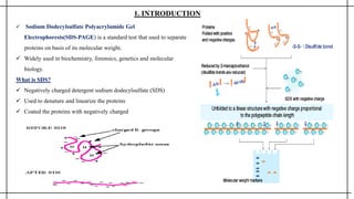
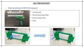
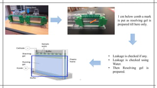
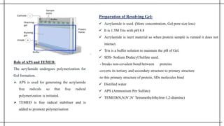
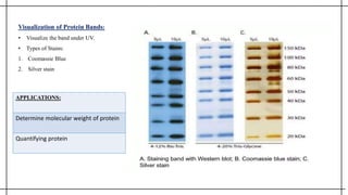
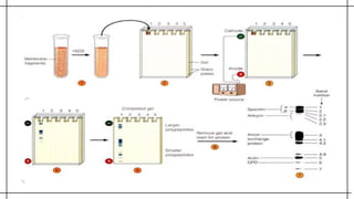
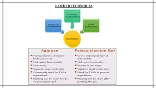
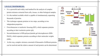
SDS-PAGE is a technique used to separate proteins according to their electrophoretic mobility. It involves treating proteins with SDS detergent and running them on a polyacrylamide gel with an electric current, allowing smaller proteins to migrate faster. The document provides details on the history, principles, required materials, procedure, applications, and advantages/disadvantages of SDS-PAGE. It is commonly used to determine protein molecular weights, compare protein compositions, and analyze purity.
SDS-PAGE is a technique used to separate proteins based on their size. It involves coating proteins with negatively charged SDS detergent to linearize them before running them through a discontinuous polyacrylamide gel with an electric current. As proteins migrate through the gel pores at different rates depending on their size, bands will form that can then be visualized to analyze the proteins. SDS-PAGE is widely used in biochemistry, molecular biology, and forensics to study protein structure and purity.
Introduction, Principle, Instrumentation and Applications of SDS-PAGEMohammed Mubeen
The following presentation contains helpful information regarding SDS-PAGE, including the history, introduction, principle, instrumentation, advantages and applications of SDS-PAGE.
Sodium dodecyl sulphate - Polyacrylamide Gel Electrophoresis (SDS - PAGE) is a technique used for the separation of deoxyribonucleic acid (DNA) ,Ribonucleic acid (RNA) And protein molecules according to their size and electrical charge.
SDS PAGE, or sodium dodecyl sulfate polyacrylamide gel electrophoresis, is a technique used to separate proteins by molecular weight. Proteins are denatured and given a negative charge by SDS, allowing them to be separated by size as they migrate through a polyacrylamide gel under an electric field. Key steps include sample preparation with SDS and reducing agents, casting the polyacrylamide gel, running electrophoresis to separate proteins by size, and staining to visualize the separated protein bands.
This document discusses various types of electrophoresis techniques. It begins by explaining that electrophoresis involves charged molecules like proteins and nucleic acids migrating in response to an electrical field. It then discusses different gel types like agarose and polyacrylamide gels that are used as supporting matrices. It also covers techniques like SDS-PAGE, isoelectric focusing, two-dimensional gel electrophoresis, and capillary electrophoresis. The document provides details on how each technique separates molecules based on properties like size, charge, and isoelectric point.
SDS-PAGE is a standard technique used to separate proteins based on their molecular weight. It involves coating proteins with SDS detergent to linearize them, then running them through a polyacrylamide gel with an electric current which causes smaller proteins to migrate faster, allowing separation based on size. The document provides details on the principle, gel preparation, running the electrophoresis, visualizing results with stains like Coomassie blue or silver stain, and applications like determining protein purity or identification.
Polyacrylamide gel electrophoresis (PAGE) is a technique used to separate proteins based on their size and charge. PAGE uses a polyacrylamide gel with a tight matrix and small pore sizes, allowing for the separation of smaller proteins. Sodium dodecyl sulfate-PAGE (SDS-PAGE) is a common type that uses the detergent SDS to denature proteins and impart a uniform negative charge, allowing separation based solely on molecular weight. The gel consists of a stacking gel that concentrates proteins, and a resolving gel where separation occurs. Proteins are visualized after electrophoresis by staining.
This document provides information on electrophoresis techniques. It discusses how electrophoresis separates charged molecules like proteins and nucleic acids using an electric current. The key techniques covered are:
1. SDS-PAGE, which uses sodium dodecyl sulfate to denature proteins and give them a uniform negative charge for separation by size in a polyacrylamide gel.
2. Native PAGE, which separates intact proteins by their charge-to-size ratio.
3. Isoelectric focusing, which separates proteins based on their isoelectric point in a pH gradient gel.
It also discusses two-dimensional electrophoresis, which combines isoelectric focusing and SDS-PAGE to better resolve complex protein mixtures. The document
The document provides information on gel electrophoresis techniques. It discusses different types of gels like agarose, polyacrylamide, and starch that can be used for electrophoresis. Agarose gel is commonly used to separate macromolecules like nucleic acids and proteins due to its large pore size. Polyacrylamide gel is used to separate smaller molecules and provides better resolution than agarose. SDS-PAGE allows separation based on molecular weight by making all proteins negatively charged. The document outlines the procedures for agarose and polyacrylamide gel electrophoresis and their applications in analyzing proteins and nucleic acids.
Electrophoresis is a method used to separate macromolecules like DNA, RNA, and proteins based on their size and charge. There are several electrophoretic techniques including gel electrophoresis using agarose, polyacrylamide, or gradient gels, as well as capillary electrophoresis, isoelectric focusing, and microchip electrophoresis. Two-dimensional gel electrophoresis separates proteins in two steps based on isoelectric point and molecular weight to analyze complex protein mixtures.
This document summarizes the process of 2-D gel electrophoresis used to separate proteins. It begins with an introduction explaining that 2-D gel electrophoresis couples isoelectric focusing in the first dimension based on isoelectric point and SDS-PAGE in the second dimension based on molecular mass. The document then covers the principles, processes, and techniques involved in isoelectric focusing, SDS-PAGE, blotting, and concludes by discussing the applications and references for 2-D gel electrophoresis.
Electrophoresis and its types and its importance in Genetic engineeringSwaatiSharma2
This document discusses electrophoresis, which is a technique used to separate biomolecules like DNA, RNA, and proteins based on their size and charge. It moves through two common media - agarose gel and polyacrylamide gel. Agarose gel is used for larger molecules while polyacrylamide gel provides better resolution for smaller molecules. The document explains the principles, factors affecting separation, different types like agarose gel electrophoresis and polyacrylamide gel electrophoresis. It also discusses the requirements, steps involved and applications of agarose and polyacrylamide gel electrophoresis.
Gel electrophoresis is a technique used to separate macromolecules like DNA, RNA, and proteins based on their size and charge. It involves placing samples onto an agarose or polyacrylamide gel and applying an electric current, which causes the molecules to migrate through the gel at different rates depending on their size and charge. There are two main types of gel electrophoresis - agarose gel electrophoresis and polyacrylamide gel electrophoresis (PAGE). Agarose gels are used to separate larger molecules like DNA fragments, while PAGE separates smaller molecules like proteins based on their molecular weight. Gel electrophoresis is widely used in research and clinical laboratories.
Electrophoresis is a technique used to separate charged particles such as proteins or nucleic acids. It involves applying an electric field to migrate these particles through a buffer or gel based on their size and charge. There are several types of electrophoresis including paper, gel, capillary, and moving boundary which utilize different supporting media and techniques to achieve high resolution separations. Electrophoresis is widely used in biochemistry and molecular biology for analytical purposes.
Electrophoresis is a laboratory technique used to separate DNA, RNA, or protein molecules based on their size and electrical charge.
Different types of electrophoresis.
Gel electrophoresis; Agarose Gel electrophoresis; polyacrylamide gel electrophoresis; pulsed-field gel electrophoresis
Electrophoresis is a technique used to separate biomolecules like DNA, RNA, or proteins based on their size and charge. It works by applying an electric current to move the molecules through a gel or medium. Smaller molecules move faster through the pores in the gel than larger molecules, allowing separation. There are different types of electrophoresis that use different gel materials like agarose or polyacrylamide gels and techniques like pulsed field gel electrophoresis to separate different sized molecules. Electrophoresis is widely used in areas like molecular biology, genetics, and clinical testing.
Electrophoresis-PAPER ELECTROPHORESIS,GEL ELCTROPHORESIS, PAGE-SDS AND NON-SDSAmrutha Hari
The document discusses different electrophoresis techniques including paper electrophoresis, gel electrophoresis, polyacrylamide gel electrophoresis (PAGE), sodium dodecyl sulfate–polyacrylamide gel electrophoresis (SDS-PAGE), and non-SDS PAGE. It describes the basic principles, apparatus, procedures, applications and advantages/disadvantages of each technique. Paper electrophoresis is a simple method that uses filter paper as the support medium while gel electrophoresis uses gels like agarose or polyacrylamide as the support medium. SDS-PAGE and PAGE are variations that separate molecules based on their molecular weight or size.
Electrophoresis is a technique used to separate charged molecules such as proteins and nucleic acids. It works by applying an electric field to move these molecules through a medium such as a gel or paper. There are different types of electrophoresis based on the medium used and whether the molecules are separated by size alone or by their charge. Zone electrophoresis separates molecules into discrete zones based on their charge and size, while moving boundary electrophoresis separates molecules continuously. SDS-PAGE is a common electrophoresis method that uses SDS detergent to denature proteins and give them a uniform charge-to-size ratio for separation based only on their molecular weight.
Electrophoresis is a technique used to separate charged molecules like proteins and nucleic acids. It works by applying an electric field to migrate these molecules through a buffer or gel based on their size and charge. This document discusses different electrophoresis techniques including paper, gel, capillary electrophoresis and isoelectric focusing. It provides details on how each technique works, advantages and applications.
Electrophoresis is a technique used to separate charged molecules like proteins and nucleic acids based on their size and charge. It works by applying an electric current to move these molecules through a medium like a gel or paper. The document discusses different types of electrophoresis like gel electrophoresis, paper electrophoresis, and isolectric focusing. It also explains how various factors like the molecule's charge, size, and shape affect its movement during electrophoresis.
Electrophoresis is a technique used to separate charged biomolecules like proteins and nucleic acids. It works by applying an electric field to migrate these molecules through a gel or liquid medium based on their size and charge. There are different types of electrophoresis depending on the supporting medium used like agarose gel, polyacrylamide gel or free solution. Gel electrophoresis is commonly used and involves using a gel like agarose or polyacrylamide as a sieving matrix to separate molecules based on size. The separated molecules can then be visualized using stains and have various applications in research and clinical diagnostics.
This presentation includes the principle involved, chemistry, procedure, and application of various advance molecular biology like SDS PAGE, Western Blotting, and ELISA.
SDS PAGE is widely used to analyze the proteins in complex extracts.
The polyacrylamide gels are used to separate proteins.
Polyacrylamide is inert, and hence, shows no interaction with the protein being separated and forms a matrix.
Size of the pores in the gel can be controlled by adjusting the concentration of acrylamide.
Acrylamide undergoes polymerization in order to form a gel. Hence, APS (ammonium per sulphate) & TEMED (N,N,N’,N’-tetramethylethylenediamine) are added to initiate the process of polymerization.
It's application includes separation of protein mixture on separating gel and their identification using different techniques like western blotting.
Western blotting, also known as immunoblotting or protein blotting, is a core technique in cell and molecular biology. In most basic terms, it is used to detect the presence of a specific protein in a complex mixture extracted from cells.
Western blots are effective in detecting low nanogram to low picogram amounts of target protein, depending on the antibodies used and the detection substrate chosen. If the target is suspected to be of very low abundance, or if there is no detectible signal on the blot, then it may be necessary to concentrate, immunoprecipitate, or fractionate the starting material.
This technique is used to study cell signalling pathways, cell cycle pathways, drug action pathways, protein-protein interaction.
ELISA (enzyme-linked immunosorbent assay) is a plate-based assay technique designed for detecting and quantifying peptides, proteins, antibodies, and hormones.
In an ELISA, an antigen must be immobilized to a solid surface and then complexed with an antibody that is linked to an enzyme.
Detection is accomplished by assessing the conjugated enzyme activity via incubation with a substrate to produce a measurable product.
The most crucial element of the detection strategy is a highly specific antibody-antigen interaction.
ELISA begins with a coating step, in which the first layer, consisting of a target antigen or antibody, is adsorbed onto a 96-well polystyrene plate.
This is followed by a blocking step in which all unbound sites are coated with a blocking agent.
Following a series of washes, the plate is incubated with enzyme-conjugated antibody.
Another series of washes removes all unbound antibody.
A substrate is then added, producing a calorimetric signal. Finally, the plate is read.
It's types include Direct ELISA, Indirect ELISA, Sandwich ELISA and competitive ELISA. This technique is used to determine serum antibody concentrations, potential food allergens (milk, peanuts, almonds), detection of antigens and antibodies, disease outbreaks.
Gel Electrophoresis, ITS FACTOR AFFECTING, ITS TYPES,NORMAL METHODOLOGY, PAGERitamMukherjee11
ELECTROPHORESIS may be defined as the migration of the charged particle through a solution under the influence of an external electric field.
GEL ELECTROPHORESIS is a technique used to separate biomacromolecules (such as DNA, RNA, proteins, etc.) and their fragments based on their size and charge, by applying an electric field to a gel with small pores.
INTRODUCTION - Separation is brought about through molecular sieving techniques, based on the molecular size of the substances. Gel material acts as a “ molecular sieve”
It is important that support media is electrically neutral.
There are different types of gel that can be used, they are Agarose, Polyacrylamide, Starch, and Sephadex.
A porous gel acts as a sieve by retarding or, in some cases by completely obstructing the movement of macromolecules which allowed smaller molecules to migrate freely.
PRINCIPLE - According to charge: When charged molecules are placed in an electric field, they migrate toward either the positive (anode) or negative (cathode) pole according to their charge.
According to size: The smaller molecules move more swiftly than the larger-sized ones, as they can travel through the pores more easily than the later.
According to Molecular weight: The smaller molecular weight will move faster than the larger molecular weight compound.
Characterization of nucleic acids and protein by electrophoresisapeksha40
This document discusses various electrophoresis techniques used to separate and characterize nucleic acids and proteins. It describes the basic principles of electrophoresis, including how charged molecules migrate in an electric field based on their size, shape, and charge. It then focuses on different gel electrophoresis methods like polyacrylamide gel electrophoresis and agarose gel electrophoresis that are commonly used to separate DNA, RNA, and protein samples based on these properties. It also briefly mentions some other specialized electrophoresis techniques like pulsed field gel electrophoresis, isoelectric focusing, and two-dimensional electrophoresis.
Vertical Gel Electrophoresis (SDS-PAGE)Srikanth H N
Vertical gel electrophoresis has several advantages over horizontal gel electrophoresis. It allows for the use of a discontinuous buffer system to separate proteins, which is not possible with horizontal gels. The technique involves pouring an acrylamide gel between glass plates to a thickness of less than 2 mm. Samples are loaded and subjected to an electric current, with cations moving toward the cathode and anions toward the anode. Proteins are separated based on their size and charge using techniques like SDS-PAGE, which involves denaturing proteins to impart a uniform charge.
Similar to mpr-SDS PAGE and other advanced techniques.pptx (20)
Bacterial enzymes and industrial enzymes are important for many industries. Bacterial enzymes like amylase, protease, and cellulase are produced through fermentation of bacteria like Bacillus subtilis. The production process involves selecting a microorganism, isolating it in pure culture, improving the strain, formulating growth media, fermentation, and recovering the enzymes. Industrial enzymes have various applications in industries like textiles, detergents, food, and pulp/paper. Examples are amylases for desizing fabrics and dish detergents, proteases for removing stains, and cellulases for biopolishing textiles.
Candidate young stellar objects in the S-cluster: Kinematic analysis of a sub...Sérgio Sacani
Context. The observation of several L-band emission sources in the S cluster has led to a rich discussion of their nature. However, a definitive answer to the classification of the dusty objects requires an explanation for the detection of compact Doppler-shifted Brγ emission. The ionized hydrogen in combination with the observation of mid-infrared L-band continuum emission suggests that most of these sources are embedded in a dusty envelope. These embedded sources are part of the S-cluster, and their relationship to the S-stars is still under debate. To date, the question of the origin of these two populations has been vague, although all explanations favor migration processes for the individual cluster members. Aims. This work revisits the S-cluster and its dusty members orbiting the supermassive black hole SgrA* on bound Keplerian orbits from a kinematic perspective. The aim is to explore the Keplerian parameters for patterns that might imply a nonrandom distribution of the sample. Additionally, various analytical aspects are considered to address the nature of the dusty sources. Methods. Based on the photometric analysis, we estimated the individual H−K and K−L colors for the source sample and compared the results to known cluster members. The classification revealed a noticeable contrast between the S-stars and the dusty sources. To fit the flux-density distribution, we utilized the radiative transfer code HYPERION and implemented a young stellar object Class I model. We obtained the position angle from the Keplerian fit results; additionally, we analyzed the distribution of the inclinations and the longitudes of the ascending node. Results. The colors of the dusty sources suggest a stellar nature consistent with the spectral energy distribution in the near and midinfrared domains. Furthermore, the evaporation timescales of dusty and gaseous clumps in the vicinity of SgrA* are much shorter ( 2yr) than the epochs covered by the observations (≈15yr). In addition to the strong evidence for the stellar classification of the D-sources, we also find a clear disk-like pattern following the arrangements of S-stars proposed in the literature. Furthermore, we find a global intrinsic inclination for all dusty sources of 60 ± 20◦, implying a common formation process. Conclusions. The pattern of the dusty sources manifested in the distribution of the position angles, inclinations, and longitudes of the ascending node strongly suggests two different scenarios: the main-sequence stars and the dusty stellar S-cluster sources share a common formation history or migrated with a similar formation channel in the vicinity of SgrA*. Alternatively, the gravitational influence of SgrA* in combination with a massive perturber, such as a putative intermediate mass black hole in the IRS 13 cluster, forces the dusty objects and S-stars to follow a particular orbital arrangement. Key words. stars: black holes– stars: formation– Galaxy: center– galaxies: star formation
Discovery of An Apparent Red, High-Velocity Type Ia Supernova at 𝐳 = 2.9 wi...Sérgio Sacani
We present the JWST discovery of SN 2023adsy, a transient object located in a host galaxy JADES-GS
+
53.13485
−
27.82088
with a host spectroscopic redshift of
2.903
±
0.007
. The transient was identified in deep James Webb Space Telescope (JWST)/NIRCam imaging from the JWST Advanced Deep Extragalactic Survey (JADES) program. Photometric and spectroscopic followup with NIRCam and NIRSpec, respectively, confirm the redshift and yield UV-NIR light-curve, NIR color, and spectroscopic information all consistent with a Type Ia classification. Despite its classification as a likely SN Ia, SN 2023adsy is both fairly red (
�
(
�
−
�
)
∼
0.9
) despite a host galaxy with low-extinction and has a high Ca II velocity (
19
,
000
±
2
,
000
km/s) compared to the general population of SNe Ia. While these characteristics are consistent with some Ca-rich SNe Ia, particularly SN 2016hnk, SN 2023adsy is intrinsically brighter than the low-
�
Ca-rich population. Although such an object is too red for any low-
�
cosmological sample, we apply a fiducial standardization approach to SN 2023adsy and find that the SN 2023adsy luminosity distance measurement is in excellent agreement (
≲
1
�
) with
Λ
CDM. Therefore unlike low-
�
Ca-rich SNe Ia, SN 2023adsy is standardizable and gives no indication that SN Ia standardized luminosities change significantly with redshift. A larger sample of distant SNe Ia is required to determine if SN Ia population characteristics at high-
�
truly diverge from their low-
�
counterparts, and to confirm that standardized luminosities nevertheless remain constant with redshift.
Microbial interaction
Microorganisms interacts with each other and can be physically associated with another organisms in a variety of ways.
One organism can be located on the surface of another organism as an ectobiont or located within another organism as endobiont.
Microbial interaction may be positive such as mutualism, proto-cooperation, commensalism or may be negative such as parasitism, predation or competition
Types of microbial interaction
Positive interaction: mutualism, proto-cooperation, commensalism
Negative interaction: Ammensalism (antagonism), parasitism, predation, competition
I. Mutualism:
It is defined as the relationship in which each organism in interaction gets benefits from association. It is an obligatory relationship in which mutualist and host are metabolically dependent on each other.
Mutualistic relationship is very specific where one member of association cannot be replaced by another species.
Mutualism require close physical contact between interacting organisms.
Relationship of mutualism allows organisms to exist in habitat that could not occupied by either species alone.
Mutualistic relationship between organisms allows them to act as a single organism.
Examples of mutualism:
i. Lichens:
Lichens are excellent example of mutualism.
They are the association of specific fungi and certain genus of algae. In lichen, fungal partner is called mycobiont and algal partner is called
II. Syntrophism:
It is an association in which the growth of one organism either depends on or improved by the substrate provided by another organism.
In syntrophism both organism in association gets benefits.
Compound A
Utilized by population 1
Compound B
Utilized by population 2
Compound C
utilized by both Population 1+2
Products
In this theoretical example of syntrophism, population 1 is able to utilize and metabolize compound A, forming compound B but cannot metabolize beyond compound B without co-operation of population 2. Population 2is unable to utilize compound A but it can metabolize compound B forming compound C. Then both population 1 and 2 are able to carry out metabolic reaction which leads to formation of end product that neither population could produce alone.
Examples of syntrophism:
i. Methanogenic ecosystem in sludge digester
Methane produced by methanogenic bacteria depends upon interspecies hydrogen transfer by other fermentative bacteria.
Anaerobic fermentative bacteria generate CO2 and H2 utilizing carbohydrates which is then utilized by methanogenic bacteria (Methanobacter) to produce methane.
ii. Lactobacillus arobinosus and Enterococcus faecalis:
In the minimal media, Lactobacillus arobinosus and Enterococcus faecalis are able to grow together but not alone.
The synergistic relationship between E. faecalis and L. arobinosus occurs in which E. faecalis require folic acid
Anti-Universe And Emergent Gravity and the Dark UniverseSérgio Sacani
Recent theoretical progress indicates that spacetime and gravity emerge together from the entanglement structure of an underlying microscopic theory. These ideas are best understood in Anti-de Sitter space, where they rely on the area law for entanglement entropy. The extension to de Sitter space requires taking into account the entropy and temperature associated with the cosmological horizon. Using insights from string theory, black hole physics and quantum information theory we argue that the positive dark energy leads to a thermal volume law contribution to the entropy that overtakes the area law precisely at the cosmological horizon. Due to the competition between area and volume law entanglement the microscopic de Sitter states do not thermalise at sub-Hubble scales: they exhibit memory effects in the form of an entropy displacement caused by matter. The emergent laws of gravity contain an additional ‘dark’ gravitational force describing the ‘elastic’ response due to the entropy displacement. We derive an estimate of the strength of this extra force in terms of the baryonic mass, Newton’s constant and the Hubble acceleration scale a0 = cH0, and provide evidence for the fact that this additional ‘dark gravity force’ explains the observed phenomena in galaxies and clusters currently attributed to dark matter.
2. SDS PAGE AND OTHER ADVANCED TECHNIQUES
Assignment of
METHODS IN PHARMACEUTICAL RESEARCH (MPR)
PHS CC 1201
Session 2023-2024
Department of Pharmaceutical Sciences
Dr. Harisingh Gour Vishwavidyalaya,Sagar, (M.P.)
(A Central University)
Supervisors:
PROF.ASMITAGAJBHIYE
DR. UDITA AGRAWAL
MR.SHIVAM KORI
Submitted by:
ARYA OJHA
Y23254025
3. ACKNOWLEGEMENT
Throughout my assignment, I truly appreciate the help and encouragement I received from my teachers
PROF. ASMITA GAJBHIYE, DR. UDITA AGRAWAL and MR. SHIVAM KORI.
4. TABLE OF CONTENT :
1. INTRODUCTION
2. ABOUT SDS-PAGE
3. GEL PREPARATION
4. PROCEDURE
5. OTHER TECHNIQUES
5. 1. INTRODUCTION
Sodium Dodecylsulfate Polyacrylamide Gel
Electrophoresis(SDS-PAGE) is a standard test that used to separate
proteins on basis of its molecular weight.
Widely used in biochemistry, forensics, genetics and molecular
biology.
What is SDS?
Negatively charged detergent sodium dodecylsulfate (SDS)
Used to denature and linearize the proteins
Coated the proteins with negatively charged
6. 1. Firstly, the instrument of SDS-PAGE is prepared.
GEL PREPARATION
Casting stand
Tall and Short Glass Plate
Green Casting Frame
Comb
7. 1 cm below comb a mark
is put as resolving gel is
prepared till here only.
• Leakage is checked if any.
• Leakage is checked using
Water.
• Then Resolving gel is
prepared.
8. Preparation of Resolving Gel:
Acrylamide is used. (More concentration, Gel pore size less)
It is 1.5M Tris with pH 8.8
Acrylamide is inert material so when protein sample is runned it does not
interact.
Tris is a buffer solution to maintain the pH of Gel.
SDS- Sodium Dodecyl Sulfate used.
- breaks non-covalent bond between proteins
-coverts its tertiary and secondary structure to primary structure
-to this primary structure of protein, SDs molecules bind
Distilled water
APS (Ammonium Per Sulfate)
TEMED(N,N,N’,N’ Tetramethylrthylrnr-1,2-diamine)
Role of APS and TEMED:
The acrylamide undergoes polymerization for
Gel formation.
APS is used for generating the acrylamide
free radicals so that free radical
polymerization is initiated.
TEMED is free radical stabiliser and is
added to promote polymerisation
9. • The solution was left
to get solidified.
• It was filled upto the
black mark shown
earlier.
• Now, Stacking gel is
prepared.
• The ingredient is same as
resolving gel, but the
concentration of
Acrylamide is changed.
Preparation of Stacking Gel:
Acrylamide is used. (Less concentration, Gel pore size
more)
It is 1.5M Tris with pH 6.8
SDS
Distilled water
APS (Ammonium Per Sulfate)
TEMED(N,N,N’,N’ Tetramethylrthylrnr-1,2-diamine)
10. The Stacking gel is poured from side of the comb.
It is ensured that bubble formation is not there.
The Gel is left to solidify.
Then the comb is removed because of which Well-Formation takes place.
In this well only, Sample is prepared.
Running Buffer is prepared using Tris-Glycine with pH 8.3.
Preparation Sample:
Tris-HCl maintains pH as well as provides
Cl ions.
ᵦ - Mercaptoethanol breaks SH bond of
proteins.
Bromophenol Blue for Sample visualisation.
Glycerol for providing viscosity to sample
solution as sample can come out of well.
11. • Well formation in
which sample is loaded.
• Lid is closed by
taking care of anode
and cathode.
• Electric current is
passed through it.
• Running buffer is added.
12. PRINCIPLE OF SDS-PAGE:
Separates proteins in an electric field.
Migrate through a liquid or semisolid medium when subjected to an electric field
from anode to cathode terminal.
Molecules flow at different rates depend on the molecular size of proteins.
Sds-coated large proteins migrate slowly through the gel matrix and small proteins
migrate quickly through the matrix.
The Tris-HCl provides Cl- ions.
In Stacking Gel movement:
Cl-
SDS-Protein
Glycine as pH of stacking is 6.8
In Running Gel movement:
Cl-
Glycine as pH of running is 8.8 so Glycine starts becoming negatively charged
SDS-Page
More acrylamide, less gel pore size so smaller proteins moves first than larger
proteins and then separate out.
13. Visualization of Protein Bands:
• Visualize the band under UV.
• Types of Stains:
1. Coomassie Blue
2. Silver stain
APPLICATIONS:
Determine molecular weight of protein
Quantifying protein
16. AGAROSE GEL
ELECTROPHOR
ESIS:
Commonly
used support
medium
Less expensive than
cellulose acetate
Equally good
separation
Agar is a complex acidic
polysaccharide containing
monomers of sulfated
galactose
Agarose is a sulfate
free fraction of Agar
Gel is prepared in
buffer and spread
over a microscopic
slide
A small sample of
serum or biological
fluid is applied by
cutting into the gel
with a sharp edge
The electrophoretic
rum takes about 90
minutes
17. STARCH GEL ELECTROPHORESIS:
• A suspension of granular starch should be boiled in a buffer to give a clear colloidal suspension.
• The suspension on cooling sets as a semisolid gel due to intertwining of the branched chains of amylopectin.
• To avoid swelling and shrinking petroleum jelly is used.
• ADVANTAGES:
-High resolving power and sharp zones are obtained.
-The components resolved can be recovered in reasonable yield, especially proteins.
-Can be used for analytical as well as preparative electrophoresis.
• DISADVANTAGES:
-Electro osmotic effect.
-Variation in pore size from batch to batch.
18. 2-D ELECTROPHORESIS:
• It is a powerful and widely used method for the analysis of complex
protein mixtures extracted from cells, tissues, or other biological samples.
• It is the method available which is capable of simultaneously separating
thousands of proteins.
• This technique separate proteins in two steps, according to two
independent properties:
• First-dimension is isoelectric focusing (IEF), which separates proteins
according to their isoelectric points (pI);
• Second-dimension is SDS-polyacrylamide gel electrophoresis (SDS-
PAGE), which separates proteins according to their molecular weights
(MW).
• In this way, complex mixtures consisted of thousands of different proteins
can be resolved and the relative amount of each protein can be determined.
19. 1. Shankar S, " Textbook of Pharmaceutical Analysis", Rx Publications , 5th edition, 2018, Pg no: 28-1:28-8
2. Gondo Y, Fukumura R, Murata T, Makino S. Next-generation gene targeting in the mouse for functional genomics.
BMB Rep. 2009:42:315-23.
3. Wang X. A PCR-based platform for microRNA expression profiling studies. RNA. 2009:15:716-23.
4. Dholariya S, Sonagra A, “Electrophoresis”, Statpearls publishing, 2023, Pg.no 100-167
5. Lee PY, Costumbrado J, Hsu CY, Kim YH. Agarose gel electrophoresis for the separation of DNA fragments. J Vis
Exp. 2012 Apr 20;(62):3923
REFERENCES: