Download as PDF, PPTX
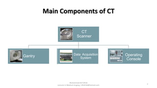
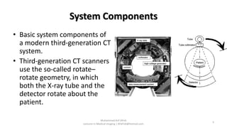
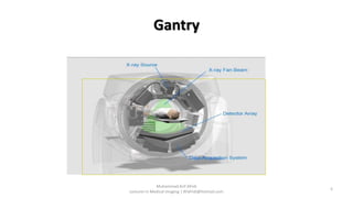
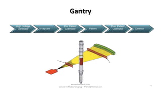
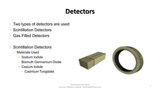
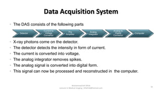
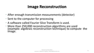
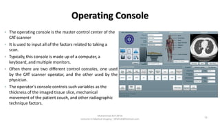
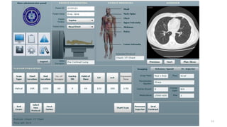


The document discusses the main components of a CT scanner system. It describes the key components as including the x-ray source, high-powered generator, detector, data transmission systems, and computer system for image reconstruction. It provides details on the gantry, detectors, data acquisition system, slip-ring technology that allows continuous rotation, and operating console as the main control center.