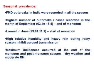
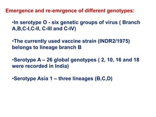
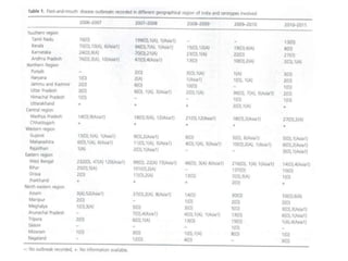
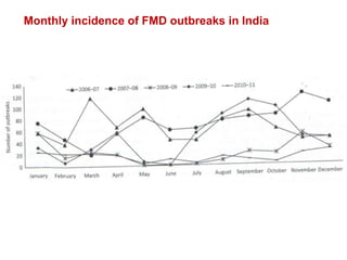
This document summarizes information about foot-and-mouth disease (FMD). It describes FMD virus, the clinical signs of disease, diagnosis, and pathogenesis. Key points include:
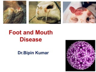
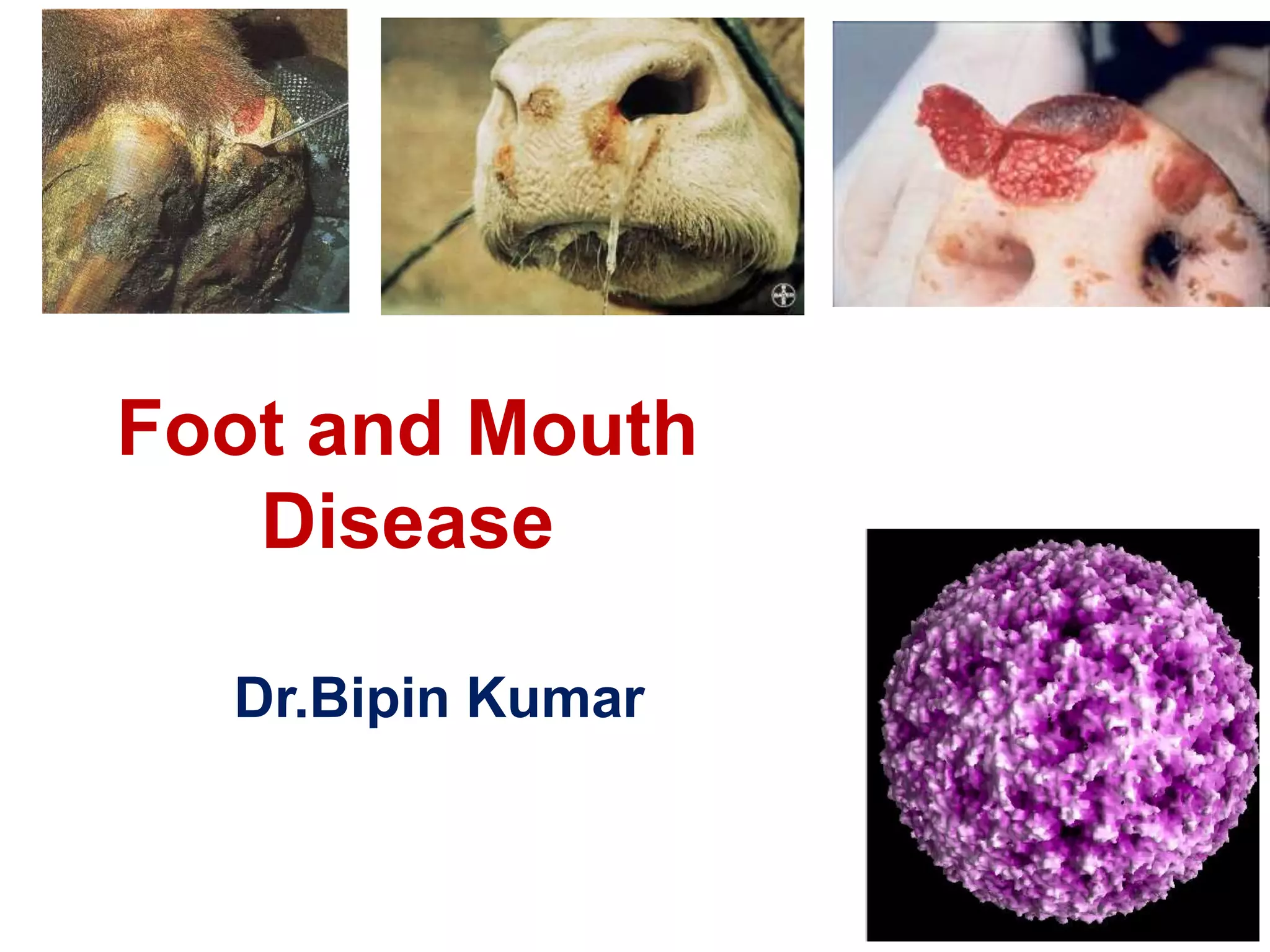
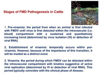
- FMD virus causes an acute, highly contagious disease in cloven-hoofed animals characterized by fever and vesicles around the mouth and feet.
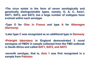
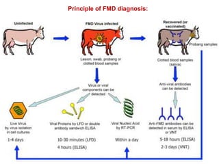
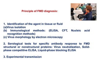
- The virus exists in 7 types and many subtypes that are diagnosed using techniques like virus isolation, ELISA, and PCR.
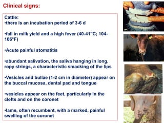
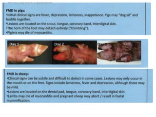
- Clinical signs vary by species but include lameness, vesicles, and decreased milk production. The disease has significant economic impacts.
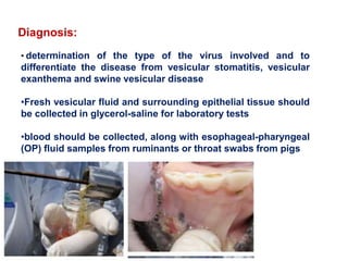
- Diagnosis involves detecting viral antigen or antibodies. Techniques include virus isolation, ELISA, and PCR which are
2. •Foot-and-mouth disease virus (FMDV; family
Picornaviridae; genus Aphthovirus) causes an acute
disease of cloven-hoofed animals characterized by
fever, lameness and vesicular lesions of the feet,
tongue, snout and teats.
• other names: Aphthous fever, Aftosa, Epizootic
aphthae
3. •The history of FMD may be traced to era of
Hieronymus Fracastorius, a monk who described a
disease outbreak in 1546 A.D. that occurred in cattle
near Verona, Italy
•Almost 400 years later, in 1897, Friedrich Loeffler and
Paul Frosch demonstrated that a filterable agent is
responsible for FMD
•foot-and-mouth disease virus (FMDV) is classified
within the genus Aphthovirus in the family
Picornaviridae
4. •The virus exists in the form of seven serologically and
genetically distinguishable types, namely, O, A, C, Asia1,
SAT1, SAT2, and SAT3, but a large number of subtypes have
evolved within each serotype
•Type O for Oise in France and type A for Allemagne
(Germany)
•Later type C was recognized as an additional type in Germany
•Pirbright laboratory in England demonstrated 3 novel
serotypes of FMDV in sample collected from the FMD outbreak
in South Africa and called SAT1, SAT2, and SAT3
•seventh serotype, that is, Asia 1 was first recognized in a
sample from Pakistan
5. •FMDV is a single stranded (ss) positive sense RNA virus with the
whole virus particles having sedimentation coefficient of 146S
•The viral genome is translated as a single polyprotein, which is
posttranslationally cleaved by viral proteases into four structural
proteins (VP1, VP2, VP3, and VP4) and several nonstructural
proteins (L, 2A, 2B, 2C, 3A, 3B, 3C, and 3D)
•Among the 4 structural polypeptides, VP1 is the most
immunogenic protein of FMDV having its G-H loop protruded
from the surface , and is maximally exposed on the capsid
surface forming large part (54%) of virus surface
6.
7. •The highly contagious nature of the virus and severity
of economic impacts associated with the disease
determine FMD’s status as the most important disease
limiting trade of animals and animal products
throughout the world
•FMD affects extensive areas worldwide and is
included in the list of diseases notifiable to the World
Organization for Animal Health
(http://www.oie.int/eng/en index.htm)
• The disease affects domestic cloven- hoofed animals,
including cattle, swine, sheep, and goats, as well as
more than 70 species of wild animals, including deer,
and is characterized by fever, lameness, and vesicular
lesions on the tongue, feet, snout, and teats
8. Resistance to physical and chemical action:
Temperature:
•Preserved by refrigeration and freezing
•temperature of 70°C for at least 30 minutes inactivates the virus
•survive for more than 60 days in bull semen frozen to -79°C (-l lO°F)
pH: Quickly inactivated by pH <6.0 or >9.0
Disinfectants:
•Inactivated by sodium hydroxide (2%), sodium carbonate (4%), citric
acid (0.2%), acetic acid (2%), sodium hypochlorite (3%), potassium
peroxymonosulfate/sodium chloride (1%), and chlorine dioxide
•Resistant to iodophores, quaternary ammonium compounds, and
phenol, especially in the presence of organic matter
9. Survival:
•Survives in lymph nodes and bone marrow at neutral pH, but
destroyed in muscle at pH <6.0 i.e. after rigor mortis
•Survives in frozen bone marrow or lymph nodes
• Residual virus survives in milk and milk products during
regular pasteurisation, but is inactivated by ultra high-
temperature pasteurisation
• Can persist in contaminated fodder and the environment for
up to 1 month
10. Geographic Distribution :
•Foot and mouth disease is endemic in parts of Asia,
Africa, the Middle East and South America
•In parts of Africa, virus persistence in wild African
buffalo makes eradication unfeasible
•North America, New Zealand, Australia, Greenland,
Iceland and most of Europe are free of this disease
11.
12. •Out of the possible seven, only four serotypes, e.g., ‘O’, ‘A’, ‘C’
and Asia 1 were ever recorded in India.
•Serotype ‘C’ too has not been recorded in the country since
1995. Vaccination against FMD is grossly inadequate in the
country.
•Similarly in the North-Eastern region of India, the frequency of
FMD is highest with type “O”, which is always followed by other
types including the type “Asia-1”
•There are records of about 5,000 outbreaks to occur in India
annually affecting nearly three lakh animals with an estimated
staggering loss of Rs. 4,300 cores to the economy annually
FMD in India
13. •In India zoning approach with properly campaigned 6 monthly
vaccinations of cattle and buffalo followed by rigorous
surveillance and sero-monitoring done to create disease free
zones (DFZ)
• A trivalent (O,A,Asia 1) vaccine is currently used in vaccination
programme
• FMD surveillance in India carried out by 8 regional FMD centres
and 15 FMD network units and a central FMD laboratory at
Muktehwar
•Serotyping of the clinical materials is done by using sandwich
ELISA
FMD in India
14. • ELISA negative samples are subjected to multiplex PCR for further
diagnosis
•Molecular epidemiological analysis based on P1/1D gene sequence
and studies of antigenic relationship of the field outbreakstrains with
currently used vaccine strains
FMD in India
15. Regional prevalence:
•Maximum proportions (43%) occurred in eastern region
•Southern region – 31.5%
•North eastern region – 11.6%
•Central region – 5%
•Western region – 4.4%
•Northern region – 4%
(S.Subramaniam et al., 2013)
17. Seasonal prevalence:
•FMD outbreaks in India were recorded in all the season
•Highest number of outbreaks / cases recorded in the
month of September (63.4± 18.4) – end of monsoon
•Lowest in June (23.6± 11.1) – start of monsoon
•High relative humidity and heavy rain during rainy
season inhibit aerosol transmission
•Maximum incidences occurred at the end of the
monsoon and post-monsoon season – dry weather and
moderate RH
18. Emergence and re-emrgence of different genotypes:
•In serotype O - six genetic groups of virus ( Branch
A,B,C-I,C-II, C-III and C-IV)
•The currently used vaccine strain (INDR2/1975)
belongs to lineage branch B
•Serotype A – 26 global genotypes ( 2, 10, 16 and 18
were recorded in India)
•Serotype Asia 1 – three lineages (B,C,D)
21. Transmission:
•by inhalation
•by ingestion
•Carriers (cattle, sheep and goats may become carriers, but pigs do
not)
•Wild fauna may serve as FMD reservoir
•Humans are often a vehicle for transmission
•movement of infected animals, or indirectly by the transportation of
virus on inanimate objects
22. Factors involves in aerosol transmission:
•sufficient virus particles
•strength of the virus source (infected pigs excrete 1000-3000
times)
• the wind speed
• Humidity (critical RH range of 55-60%)
• diurnal variation
• sea over land (60 km over land 250 km over sea)
23.
24. Animated FMD virus plumes. Red squares indicate for the positions of
infected farms, brown points for the more than 1000 susceptible farms
in the 25 km x 25 km model domain. Yellow particles are located in the
lowest 10 m above ground. Green, cyan, blue, magenta and black
points mark viruses at higher vertical layers (upper panel). Risk areas
for cattle (green), sheep (blue) and swine (red) based on critical FMD
virus concentrations (lower panel).
25. Stages of FMD Pathogenesis in Cattle
1. Pre-viraemia: the period from when an animal is first infected
with FMDV until virus is first detected within the intravascular (i.e.
blood) compartment with a sustained and quantitatively
increasing trend (determined by virus isolation (VI) or detection of
viral RNA)
2. Establishment of viraemia: temporally occurs within pre-
viraemia. However, because of the importance of this transition, it
will be treated as a distinct event
3. Viraemia: the period during which FMDV can be detected within
the intravascular compartment with kinetics suggestive of active
viral replication (determined by VI or detection of viral RNA). This
period typically coincides with the clinical phase of disease.
26. 4. Post-viraemia: the period following viraemia starting with the first
negative assay on blood (determined by VI or detection of viral RNA)
which includes:
(i) Resolution of clinical signs
(ii) Short-term persistence of infectious virus, antigen and/or RNA in
specific tissues
(iii) Persistent infection (carrier state): the period after 28 dpi in
which infectious FMDV may be detected on at least one of multiple
oesophageal–pharyngeal (OP) samples (as defined by OIE)
(iv) Chronic long-term sequelae including hirsutism, heat-intolerance
(panting) and thyroid dysfunction, which have been reported in
recovered cattle
27.
28. Pathogenesis in Swine:
•Pigs are the second most commonly studied subjects for FMD
pathogenesis research
• Recent and historic reviews have supported the notions that
compared to cattle, pigs are more refractory to aerogenous infection,
more susceptible to infection via the gastrointestinal route and
generate greater quantities of aerosolized virus
29. Clinical signs:
Cattle:
•there is an incubation period of 3-6 d
•fall in milk yield and a high fever (40-41"C; 104-
106°F)
•Acute painful stomatitis
•abundant salivation, the saliva hanging in long,
ropy strings, a characteristic smacking of the lips
•Vesicles and bullae (1-2 cm in diameter) appear on
the buccal mucosa, dental pad and tongue
•vesicles appear on the feet, particularly in the
clefts and on the coronet
•lame, often recumbent, with a marked, painful
swelling of the coronet
30.
31. Sequelae to FMD:
• complex of clinical signs variably referred to as ‘heat-intolerance
syndrome (HIS)’, ‘hairy panters’, ‘asoleadas’ (in Argentina) and
‘peludas’ (in Brazil; M. Hugh-Jones, personal communication)
• intolerance to increased environmental temperatures
•Pronounced panting during hot weather is the striking feature
accompanied by increased body temperature and pulse rate (Minett,
1948; Maqsood et al., 1958)
•abnormal hair growth described as either hirsutism (Ghanem and
Abdel-Hamid, 2010) or
• hypertrichosis as a result of failure of cyclic seasonal shedding of
hair (Minett, 1948; Maqsood et al., 1958)
•anaemia, hyperphosphataemia, lactic acidosis, hypoproteinaemia,
and hypocalcaemia (Mullick, 1949)
32. •serum Na, Cl, Mg, Zn, Fe, albumin, cholesterol, and cortisol
were significantly reduced
•Serum levels of ALT, AST, and monoaldehyde were significantly
increased in affected cows
•serum glucose was also significantly increased although
hyperglycaemia was considered mild and below the cut-off for
the diagnosis of diabetes mellitus in affected cattle
•The morphology was generally consistent with thyroid
hyperplasia and suggestive of hyperthyroidism
•Disruption of these endocrine organs could, in part, account
for the biochemical changes reported by Ghanem and Abdel-
Hamid in heat intolerant cattle and the hyperglycaemia and
hypoinsulinaemia detected during clinical FMD (Yeotikar et al.,
2003; Ghanem and Abdel- Hamid, 2010; Nahed, 2010)
33. •Proper assessment of thyroid function requires evaluation of
serum T3/T4 levels in the context of serum TSH and TRH
•As such data have never been reported for HIS cattle, the
relationship between HIS and thyroid function remains
undetermined
•Early investigators’ efforts to elucidate the functional
derangements of HIS have included administration of thyroxin
which aggravated the symptoms (Mullick, 1949) and
administration of thiouracil which alleviated the symptoms
(Maqsood et al., 1958).
34. Myotropism of FMDV
• a malignant form of the disease in adults in which acute
myocardial failure occur
•typical course initially but a sudden relapse occurs on days 5-6
with dyspnea, a weak and irregular heart action, and death during
convulsions
•myocardial lesions in cattle which died shortly after infection,
including inflammatory infiltrates and cardiomyocyte degeneration
and necrosis (Huguenin, 1924)
•In addition to young cattle, FMDV- associated myocardial necrosis
and myocarditis has been documented in pigs (Potel, 1967), sheep
(Salyi, 1939), goats (Bhalla and Sharma, 1965), adult cattle, gazelle
(Shimshony, et al., 1986)
35. Diagnosis:
• determination of the type of the virus involved and to
differentiate the disease from vesicular stomatitis, vesicular
exanthema and swine vesicular disease
•Fresh vesicular fluid and surrounding epithelial tissue should
be collected in glycerol-saline for laboratory tests
•blood should be collected, along with esophageal-pharyngeal
(OP) fluid samples from ruminants or throat swabs from pigs
37. 1. Identification of the agent in tissue or fluid
(a)Virus isolation
(b) Immunological methods: (ELISA, CFT, Nucleic acid
recognition methods)
(c) Virus morphology by electron microscopy
2. Serological tests for specific antibody response to FMD
structural or nonstructural proteins: Virus neutralization, Solid-
phase competitive ELISA, Liquid-phase blocking ELISA
3. Experimental transmission
Principle of FMD diagnosis:
38. Virus Isolation:
•by inoculation into cell cultures or unweaned mice
•The FMD virus is cultivable on tissue culture and in hen eggs
•Primary cell culture of bovine, ovine and porcine origin has
exhibited susceptibility to FMDV from infected tissues
•Some stable cell lines, like IBRS-2, MVPK-1 clone 7 , LFBK cell
line and BHK-21 are also susceptible to FMDV and so are most
desirable for diagnostic system
•the cell culture system is laborious, time consuming, and
relatively low sensitive
• It also requires careful handling of specimens and a biosafety
laboratory
39. Complement Fixation Test (CFT):
•used extensively for distinguishing different strains of FMDV
• CFT was a fast method it needed high virus load and results were
sometimes affected by pro-and anti-complementary activities of the
test sample
•uses the activation of the classical complement pathway by
antibody to antigen complexes
•affected by pro- or anti-complementary factors
40. Enzyme-Linked Immunosorbent Assay (ELISA):
•ELISA and its various modifications were applied for detection,
typing, and strain differentiation of FMDV isolates with better
sensitivity than CFT
•At the FAO/WRL for FMD, the preferred procedure for the detection
of FMDV antigen and identification of viral serotypes is ELISA
•a sandwich ELISA using convalescent bovine immunoglobulin
(Igs) as capture
•LPBE is being used for the detection of antibody titers against the
FMD vaccinated animals
41. Reverse Transcription-Polymerase Chain Reaction
(RTPCR):
•RT-PCR to amplify RNA targets - a reliable tool for FMD diagnosis
•A specific RT-PCR was developed and validated for the detection
of the polymerase gene (3D) of FMD with an analytical sensitivity
equal to 1000 times higher than that of a single passage virus
isolation
42. Multiplex Polymerase Chain
Reaction (mPCR):
•variant of the test in which more than
one target sequence is amplified
using more than one pair of primers
•mPCR was used for the detection and
differentiation of multiple
pathogens/different strains of the same
pathogen
•the multiplex system is designed to
survey multiple regions of the genome
simultaneously, thereby increasing the
probability of detection The test result by multiplex-RT-PCR. Line
1 and line 2 were two negative samples;
line 3 was positive sample; M was DNA
marker ladder
43. Real-Time Polymerase Chain Reaction:
•Real-time PCR assays recommended by the World Organization
for animal health (OIE) for detection of FMDV incorporate
universal primers and fluorescent-labeled probes that recognized
conserved region within the 5 UTR or conserved gene regions
within the RNA-dependent RNA polymerase gene (3Dpol)
• The use of a specific probe facilitates an increase in specificity
compared to conventional agarose-gel-based PCR assays
•the problem of carry-over is significantly reduced because of the
real-time measuring principle, which is based on closed tube
system
44. Recombinant Antigen-Based Diagnosis:
•The 3AB protein of FMDV was expressed in E. coli or in P.
pastoris and has been used for the diagnosis of FMD infection in
cattle
•Similarly, 3ABC proteins expressed in heterologous systems
were used in ELISA (3ABC ELISA) for serodiagnosis of FMD
45. DIVA-Based Companion Diagnostic Approach:
The ability to identify and selectively delete genes from a
pathogen has allowed the development of “marker vaccines”
that, combined with suitable diagnostic assays, allow
differentiating infected from vaccinated animals (DIVA) by
differentiation of antibody responses induced by the vaccine
(no antibodies generated to deleted genes) from those induced
during infection with the wild-type virus
46. Microarray-Based Diagnosis of FMDV:
•DNAmicroarrays are becoming increasingly useful for
the analysis of gene expression and single nucleotide
polymorphisms (SNPs)
47. •A rapid test which gives results in 5-10
minutes of time
•A laboratory based test- ELISA which
gives results in 3 hours
•Both the tests can be used to detect FMD-
NSP antibodies in all species of animals
susceptible to FMD.
Recent technology by IVRI:
48. Necropsy finding:
•vesicles and erosions in the mouth and on
the feet and udder
•vesicles may extend to the pharynx,
esophagus, fore stomachs, and intestines as
well as trachea and bronchi
•Ventricular walls appear streaked with
patches of yellow tissue interspersed with
apparently , normal myocardium, giving the
typical 'tiger heart' appearance
•heart is enlarged and flabby
Focal myocarditis, 'tiger
striping' on the heart of
a 4-week old piglet
which died due to FMD.
49. •homeopathic preparation of Tarentula cubensis
(Theranekron®) was conducted during an outbreak of FMD in
cattle in Iran
•Ampicillinand Cloxacillin combination @ 10 mg/kg body
weightbid intramuscularly for 3 days
•Enrofloxacin @ 5 mg/kgsid intramuscularly for 3 days
•Oxytetracycline @ 10 mg/kgsid intramuscularly for 3 days
•Gentamicin @ 2.5 mg/kg bidintramuscularly for 3 days and
• Potassium permanganate(1ppm) on the lesions
•local application of antiseptic solutions viz., 2% NaOH, 2%
NaHCO 3 , 4%Na 2CO3 and 5% Povidon iodine respectively
•Meloxicam and paracetamol combination (Melonexplus, Intas)
@ 1ml/25 kg
•Administration of flunixin meglumine
Treatment:
50. Control:
(a) control by eradication and
(b) control by vaccination, or
(c) a combination of the two
•all cloven-footed animals in the exposed groups should be
immediately slaughtered and burned or buried on site
•proper disinfection of human clothing, motor vehicles and farm
machinery
• Barns and small yards must be cleaned and disinfected with 1-2 %
sodium hydroxide or formalin or 4% sodium carbonate solution
•the farm should be left unstocked for 6 months and restocking
permitted only when 'sentinel' test animals are introduced and remain
uninfected
•Immediate quarantine must be imposed on all farms within a radius of
16-24 km (10-15 miles) of the outbreak
51. Vaccination:
•Killed trivalent (containing 0, A, and C strains) vaccines are in
general use
•Locally isolated virus is becoming a more common practice
formalin, binary ethylene immine (BEl)
•oil-adjuvanted induce higher levels of antibody than aluminum
hydroxide gel-saponin adjuvanted vaccines
•Attenuated vaccines have been produced by passage through
white mice, embryonated hen eggs, rabbits and tissue culture
•genetically engineered FMD vaccine polypeptide vaccine
(protein VPl)
52. Alternatives to general vaccination are modified
programs including 'ring‘ vaccination to contain
outbreaks/frontier‘ vaccination to produce a buffer area
between infected and free countries and vaccination of
selected herds on a voluntary basis when an outbreak is
threatened
53. •high contagiousness,
•wide geographical distribution,
•broad host range,
•ability to establish carrier status,
•Antigenic diversity leading to poor cross-immunity, and
•relatively short duration of immunity
The main constraints in controlling this disease and why it is considered as
the most dreaded viral disease
CONCLUSION: