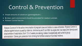
COLIBACILLOSIS
PRESENTED BY: NAQQASH RIAZ
ROLL NO. 49
naqqash.saroya@gmail.com
ETIOLOGY
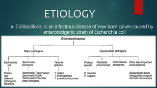
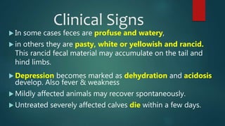
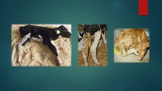
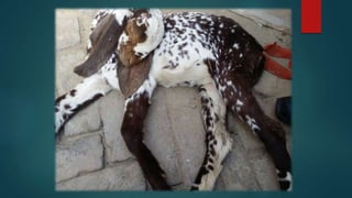
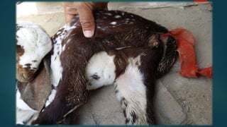
 Colibacillosis is an infectious disease of new born calves caused by
enterotoxigenic strain of Escherichia coli
EPIDEMIOLOGY
 The prevalence of colibacillosis has increased in recent
years.
 There are several possible reasons of this, including: size
of herd, shortage of qualified labor, increased population
density.
 Colibacillosis occurs mostly in new born calves, piglets,
foals, lambs & goat kids.
 The prevalence of enterotoxigenic E. coli in diarrheic
calves varies widely geographically, between herds and
depending on the age of the animals.
 The prevalence can be as high as 50-60% in diarrheic
calves under 3 days of age
 and only 5-10% in diarrheic calves 8 days of age.
 In some countries the prevalence is only 5-8% in diarrheic
calves under 3 days of age.
MORBIDITY &
MORTALITY RATE
 Dairy Calves: Poorly managed calves have 75% morbidity
but usually its 30%.
Case fatality ranges from 10-50% depending on level of
clinical management.
 Beef Calves: Morbidity rate is vary from 10-50% & case
fatality from 5-10%
 Mortality in both calves ranges from 3-60% depending on
herd management
Pathogenesis
 Entcrotoxigenic strains of ETEC, possessing fimbrial adhesins
such as K88 and K99, are of particular importance in neonatal
diarrhea.
 These strains colonize the distal small intestine by attaching
to receptors which are present on the enterocytes of
neonates.
 They produce enterotoxins (LT and STa) which stimulate
hypersecretory diarrhoea and interfere with fluid absorption
without major morphologically-detectable damage to
enterocytes.
Clinical Signs
 In some cases feces are profuse and watery,
 in others they are pasty, white or yellowish and rancid.
This rancid fecal material may accumulate on the tail and
hind limbs.
 Depression becomes marked as dehydration and acidosis
develop. Also fever & weakness
 Mildly affected animals may recover spontaneously.
 Untreated severely affected calves die within a few days.
Diagnosis
 On blood agar the colonics are greyish, round and shiny
with a characteristic smell.
 On MacConkey agar colonies are bright pink.
 IMViC tests can be used for confirmation
 The colonies of some E. coii strains have a metallic
sheen on EMB agar.
 Polymerase chain reaction (PCR)
 Enterotoxins in the small intestine can be detected,
using methods employing monoclonal antibodies
Differential Diagnosis
ENTEROPATHOGENS AGE (days)
Enterotoxigenic E. coli <3
Rotavirus 5-15
Coronavirus 5-21
Other viruses ( Breda virus, parvovirus, bovine virus,
diarrhea virus )
14-30 (and older up to several weeks)
Cryptosporidium spp. 5-35
Salmonella spp. 5-42
Clostridium perfringens type B & C 5-15
Eimeria spp. >30
Giardia spp. 10-30
Factors which may predispose young farm
animals to infection with pathogenic Escherichia
coli strains
Insufficient or no colostral
immunity
Build-up of pathogenic E. coli
strains
Overcrowding and poor
hygiene, facilitating increased
transmission of organisms
Normal flora of neonates not
fully established
Naive immune system in neonates
Receptors for ETEC adhesins are present only during first week of life in
calves
Pigs retain receptors for some adhesins past weaning age (post-weaning
diarrhoea)
Digestive tract of young pigs equipped only for easily digested foods.
Accumulation of undigested and unabsorbed nutrients encourages
replication of E. coli
Stress factors such as cold ambient temperatures and frequent mixing of
animals
Treatment
 Local or oral ( Amoxicillin, Sulfadimidine-but resistant
now ) &
Parenteral ( Enrofloxacin, Potentiate sulfonamide + TMP,
Gentamicin, ceftiofur ) Antibiotic therapy
 ( Flunixin meglumin + dexamethasone + tirilazad
mesylate ) Combination used to minimize the
shock
 Fluid therapy oral or infusion
Control & Prevention
 Ample amounts of colostrum (gammaglobulin )
 A clean, warm environment should be provided for newborn animals.
 Rotavec Corona Vaccine
Coliform mastitis
 Infection of the mammary glands of cows and sows by
members of the Enterobacteriacae, including E. coli,
occurs opportunistically.
 In dairy cows, the source of infection is fecal
contamination of the skin of the mammary gland and
relaxation of the teat sphincter following milking
increases vulnerability to infection.
 Cows with low somatic cell counts are particularly
susceptible to infection.
 No specific serotypes of E, coli have been linked with this
form of mastitis.
 The acute form of the disease is characterized by
endotoxemia and can be life-threatening.
 Per-acute disease may be fatal in 24 to 48 hours. Affected
animals are severely depressed with drooping ears and
sunken eyes. Mammary secretions are watery and contain
white flecks.
Control
 THIS IS PAKSITAN'S FIRST
MASTITIS VACCINE, NOW
COMMERCIALLY AVAILABLE.
RESEACH WAS CONDUCTED
IN DEPARTMENT OF CLINICAL
MEDICINE AND SURGERY,
FACULTY OF VETERINARY
SCIENCE, UNIVERSITY OF
AGRICULTURE, FAISALABAD
BY DR. IMAAD RASHEED,
DR.ALI RAZA, UNDER THE
SUPERVISION OF PROF.
DR.GHULAM MUHAMAMD) .
 Pre milking teat dipping
 Post milking teat dipping
 Dry cow therapy
 Prompt treatment of clinical cases
 Culling of chronic mastitic animals from the
herd
Colibacillosis in Poultry
 Etiology: Avian pathogenic E coli (APEC) strains are commonly of the O1, O2, and O78
serogroups
 Clinical Findings:
 Respiratory signs, coughing, sneezing.
 Snick.
 Dejection.
 Reduced appetite.
 Poor growth.
 Omphalitis.
Post-mortem lesions:
 Airsacculitis.
 Pericarditis.
 Perihepatitis.
 Swollen liver and spleen.
 Peritonitis.
 Salpingitis.
 Omphalitis.
 Synovitis.
 Arthritis.
 Enteritis.
 Granulomata in liver and spleen.
 Cellulitis over the abdomen or in the leg.
 Lesions vary from acute to chronic in the various forms of the disease.
Diagnosis, Treatment & Conrol
 Diagnosis: Isolation, sero-typing, pathology.
Aerobic culture yields colonies of 2-5mm on both
blood and McConkey agar after 18 hours - most
strains are rapidly lactose-fermenting producing
brick-red colonies on McConkey agar.
 Treatment: Amoxycillin, tetracyclines, neomycin
(intestinal activity only), gentamycin or ceftiofur
(where hatchery borne), potentiated
flouroquinolones.
 Control: Good hygiene in handling of hatching
eggs, hatchery hygiene, good sanitation of house,
feed and water. Well-nourished embryo and
optimal incubation to maximise day-old viability.
Severe peri-hepatitis, liver is covered with
substantial layer of fibrin & puss.
Clear airsac of an uninfected bird Foamy exudate seen early in
infection
Caseous exudate later in
infection
Thank You for listening

Colibacillosis

  • 1.
    COLIBACILLOSIS PRESENTED BY: NAQQASHRIAZ ROLL NO. 49 naqqash.saroya@gmail.com
  • 2.
    ETIOLOGY  Colibacillosis isan infectious disease of new born calves caused by enterotoxigenic strain of Escherichia coli
  • 4.
    EPIDEMIOLOGY  The prevalenceof colibacillosis has increased in recent years.  There are several possible reasons of this, including: size of herd, shortage of qualified labor, increased population density.  Colibacillosis occurs mostly in new born calves, piglets, foals, lambs & goat kids.
  • 5.
     The prevalenceof enterotoxigenic E. coli in diarrheic calves varies widely geographically, between herds and depending on the age of the animals.  The prevalence can be as high as 50-60% in diarrheic calves under 3 days of age  and only 5-10% in diarrheic calves 8 days of age.  In some countries the prevalence is only 5-8% in diarrheic calves under 3 days of age.
  • 6.
    MORBIDITY & MORTALITY RATE Dairy Calves: Poorly managed calves have 75% morbidity but usually its 30%. Case fatality ranges from 10-50% depending on level of clinical management.  Beef Calves: Morbidity rate is vary from 10-50% & case fatality from 5-10%  Mortality in both calves ranges from 3-60% depending on herd management
  • 7.
    Pathogenesis  Entcrotoxigenic strainsof ETEC, possessing fimbrial adhesins such as K88 and K99, are of particular importance in neonatal diarrhea.  These strains colonize the distal small intestine by attaching to receptors which are present on the enterocytes of neonates.  They produce enterotoxins (LT and STa) which stimulate hypersecretory diarrhoea and interfere with fluid absorption without major morphologically-detectable damage to enterocytes.
  • 8.
    Clinical Signs  Insome cases feces are profuse and watery,  in others they are pasty, white or yellowish and rancid. This rancid fecal material may accumulate on the tail and hind limbs.  Depression becomes marked as dehydration and acidosis develop. Also fever & weakness  Mildly affected animals may recover spontaneously.  Untreated severely affected calves die within a few days.
  • 13.
    Diagnosis  On bloodagar the colonics are greyish, round and shiny with a characteristic smell.  On MacConkey agar colonies are bright pink.  IMViC tests can be used for confirmation  The colonies of some E. coii strains have a metallic sheen on EMB agar.  Polymerase chain reaction (PCR)  Enterotoxins in the small intestine can be detected, using methods employing monoclonal antibodies
  • 14.
    Differential Diagnosis ENTEROPATHOGENS AGE(days) Enterotoxigenic E. coli <3 Rotavirus 5-15 Coronavirus 5-21 Other viruses ( Breda virus, parvovirus, bovine virus, diarrhea virus ) 14-30 (and older up to several weeks) Cryptosporidium spp. 5-35 Salmonella spp. 5-42 Clostridium perfringens type B & C 5-15 Eimeria spp. >30 Giardia spp. 10-30
  • 15.
    Factors which maypredispose young farm animals to infection with pathogenic Escherichia coli strains Insufficient or no colostral immunity Build-up of pathogenic E. coli strains Overcrowding and poor hygiene, facilitating increased transmission of organisms Normal flora of neonates not fully established
  • 16.
    Naive immune systemin neonates Receptors for ETEC adhesins are present only during first week of life in calves Pigs retain receptors for some adhesins past weaning age (post-weaning diarrhoea) Digestive tract of young pigs equipped only for easily digested foods. Accumulation of undigested and unabsorbed nutrients encourages replication of E. coli Stress factors such as cold ambient temperatures and frequent mixing of animals
  • 17.
    Treatment  Local ororal ( Amoxicillin, Sulfadimidine-but resistant now ) & Parenteral ( Enrofloxacin, Potentiate sulfonamide + TMP, Gentamicin, ceftiofur ) Antibiotic therapy  ( Flunixin meglumin + dexamethasone + tirilazad mesylate ) Combination used to minimize the shock  Fluid therapy oral or infusion
  • 18.
    Control & Prevention Ample amounts of colostrum (gammaglobulin )  A clean, warm environment should be provided for newborn animals.  Rotavec Corona Vaccine
  • 19.
    Coliform mastitis  Infectionof the mammary glands of cows and sows by members of the Enterobacteriacae, including E. coli, occurs opportunistically.  In dairy cows, the source of infection is fecal contamination of the skin of the mammary gland and relaxation of the teat sphincter following milking increases vulnerability to infection.  Cows with low somatic cell counts are particularly susceptible to infection.
  • 20.
     No specificserotypes of E, coli have been linked with this form of mastitis.  The acute form of the disease is characterized by endotoxemia and can be life-threatening.  Per-acute disease may be fatal in 24 to 48 hours. Affected animals are severely depressed with drooping ears and sunken eyes. Mammary secretions are watery and contain white flecks.
  • 21.
    Control  THIS ISPAKSITAN'S FIRST MASTITIS VACCINE, NOW COMMERCIALLY AVAILABLE. RESEACH WAS CONDUCTED IN DEPARTMENT OF CLINICAL MEDICINE AND SURGERY, FACULTY OF VETERINARY SCIENCE, UNIVERSITY OF AGRICULTURE, FAISALABAD BY DR. IMAAD RASHEED, DR.ALI RAZA, UNDER THE SUPERVISION OF PROF. DR.GHULAM MUHAMAMD) .
  • 23.
     Pre milkingteat dipping  Post milking teat dipping  Dry cow therapy  Prompt treatment of clinical cases  Culling of chronic mastitic animals from the herd
  • 24.
    Colibacillosis in Poultry Etiology: Avian pathogenic E coli (APEC) strains are commonly of the O1, O2, and O78 serogroups  Clinical Findings:  Respiratory signs, coughing, sneezing.  Snick.  Dejection.  Reduced appetite.  Poor growth.  Omphalitis.
  • 25.
    Post-mortem lesions:  Airsacculitis. Pericarditis.  Perihepatitis.  Swollen liver and spleen.  Peritonitis.  Salpingitis.  Omphalitis.  Synovitis.  Arthritis.  Enteritis.  Granulomata in liver and spleen.  Cellulitis over the abdomen or in the leg.  Lesions vary from acute to chronic in the various forms of the disease.
  • 26.
    Diagnosis, Treatment &Conrol  Diagnosis: Isolation, sero-typing, pathology. Aerobic culture yields colonies of 2-5mm on both blood and McConkey agar after 18 hours - most strains are rapidly lactose-fermenting producing brick-red colonies on McConkey agar.  Treatment: Amoxycillin, tetracyclines, neomycin (intestinal activity only), gentamycin or ceftiofur (where hatchery borne), potentiated flouroquinolones.  Control: Good hygiene in handling of hatching eggs, hatchery hygiene, good sanitation of house, feed and water. Well-nourished embryo and optimal incubation to maximise day-old viability. Severe peri-hepatitis, liver is covered with substantial layer of fibrin & puss.
  • 27.
    Clear airsac ofan uninfected bird Foamy exudate seen early in infection Caseous exudate later in infection
  • 30.
    Thank You forlistening