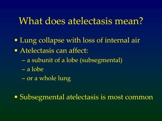
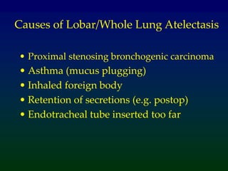
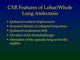
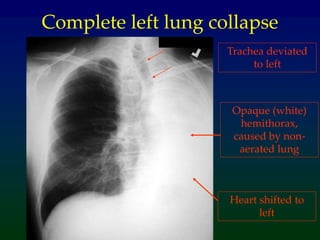
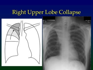
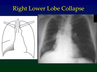
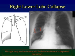
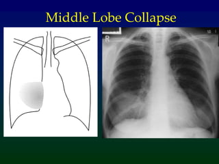
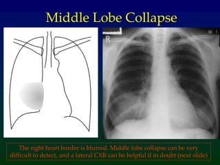
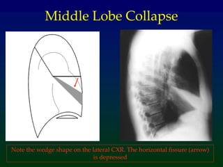
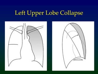
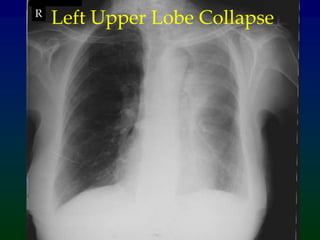
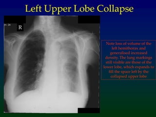
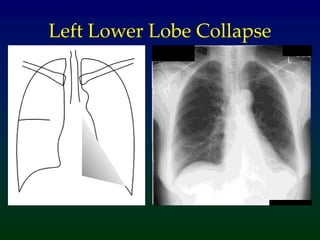
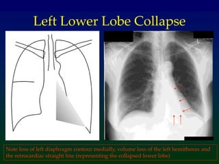
Atelectasis
What does atelectasis mean?
• Lung collapse with loss of internal air
• Atelectasis can affect:
– a subunit of a lobe (subsegmental)
– a lobe
– or a whole lung
• Subsegmental atelectasis is most common
Causes of Lobar/Whole Lung Atelectasis
• Proximal stenosing bronchogenic carcinoma
• Asthma (mucus plugging)
• Inhaled foreign body
• Retention of secretions (e.g. postop)
• Endotracheal tube inserted too far
CXR Features of Lobar/Whole
Lung Atelectasis
• Ipsilateral tracheal displacement
• Increased density of collapsed lung tissue
• Ipsilateral mediastinal shift
• Elevation of the hemidiaphragm
• Herniation of the opposite lung across the
midline
Complete left lung collapse
Trachea deviated
to left
Heart shifted to
left
Opaque (white)
hemithorax,
caused by non-
aerated lung
Patterns of Lobar Collapse
• The left lung has 2 lobes (upper, lower), and
the right lung 3 lobes (upper, middle, and
lower)
• The collapse of each lobe has specific
appearances on a CXR
• Examples of each type of lobar collapse now
follow
• A revision of your lung anatomy now might
be helpful
Surface markings of the lobes
•The lingula is part of the left upper lobe
•Note that the lower lobes extend quite high – above the level of the carina
Right Upper Lobe Collapse
Examples of Lobar Atelectasis
• There now follows examples of each
type of lobar atelectasis.
• Some are difficult to recognise!
• For each one, look for volume loss (e.g.
ribs closer together), and movement of
other structures to occupy the space
created by the atelectasis.
Right Lower Lobe Collapse
The right lung has lost volume and the horizontal fissure is depressed
(red arrow)
Middle Lobe Collapse
The right heart border is blurred. Middle lobe collapse can be very
difficult to detect, and a lateral CXR can be helpful if in doubt (next slide)
Middle Lobe Collapse
Note the wedge shape on the lateral CXR. The horizontal fissure (arrow)
is depressed
Left Upper Lobe Collapse
R
Left Upper Lobe Collapse
Note loss of volume of the
left hemithorax and
generalised increased
density. The lung markings
still visible are those of the
lower lobe, which expands to
fill the space left by the
collapsed upper lobe
R
Left Upper Lobe Collapse
Left Lower Lobe Collapse
Note loss of left diaphragm contour medially, volume loss of the left hemithorax and
the retrocardiac straight line (representing the collapsed lower lobe)
Take Home Points
• The commonest cause of lobar / total
lung collapse in adults is lung cancer
• All patients with lobar or total lung
collapse need further assessment to
determine the underlying cause
• The CXR appearances of collapse vary
according to where the collapse is

atelectasis The Lung Condition and Management

  • 1.
  • 2.
    What does atelectasismean? • Lung collapse with loss of internal air • Atelectasis can affect: – a subunit of a lobe (subsegmental) – a lobe – or a whole lung • Subsegmental atelectasis is most common
  • 3.
    Causes of Lobar/WholeLung Atelectasis • Proximal stenosing bronchogenic carcinoma • Asthma (mucus plugging) • Inhaled foreign body • Retention of secretions (e.g. postop) • Endotracheal tube inserted too far
  • 4.
    CXR Features ofLobar/Whole Lung Atelectasis • Ipsilateral tracheal displacement • Increased density of collapsed lung tissue • Ipsilateral mediastinal shift • Elevation of the hemidiaphragm • Herniation of the opposite lung across the midline
  • 5.
  • 6.
    Complete left lungcollapse Trachea deviated to left Heart shifted to left Opaque (white) hemithorax, caused by non- aerated lung
  • 7.
    Patterns of LobarCollapse • The left lung has 2 lobes (upper, lower), and the right lung 3 lobes (upper, middle, and lower) • The collapse of each lobe has specific appearances on a CXR • Examples of each type of lobar collapse now follow • A revision of your lung anatomy now might be helpful
  • 8.
    Surface markings ofthe lobes •The lingula is part of the left upper lobe •Note that the lower lobes extend quite high – above the level of the carina
  • 9.
  • 10.
    Examples of LobarAtelectasis • There now follows examples of each type of lobar atelectasis. • Some are difficult to recognise! • For each one, look for volume loss (e.g. ribs closer together), and movement of other structures to occupy the space created by the atelectasis.
  • 11.
  • 12.
    Right Lower LobeCollapse The right lung has lost volume and the horizontal fissure is depressed (red arrow)
  • 13.
  • 14.
    Middle Lobe Collapse Theright heart border is blurred. Middle lobe collapse can be very difficult to detect, and a lateral CXR can be helpful if in doubt (next slide)
  • 15.
    Middle Lobe Collapse Notethe wedge shape on the lateral CXR. The horizontal fissure (arrow) is depressed
  • 16.
  • 17.
  • 18.
    Note loss ofvolume of the left hemithorax and generalised increased density. The lung markings still visible are those of the lower lobe, which expands to fill the space left by the collapsed upper lobe R Left Upper Lobe Collapse
  • 19.
  • 20.
    Left Lower LobeCollapse Note loss of left diaphragm contour medially, volume loss of the left hemithorax and the retrocardiac straight line (representing the collapsed lower lobe)
  • 21.
    Take Home Points •The commonest cause of lobar / total lung collapse in adults is lung cancer • All patients with lobar or total lung collapse need further assessment to determine the underlying cause • The CXR appearances of collapse vary according to where the collapse is