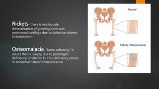
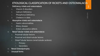
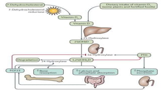
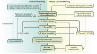
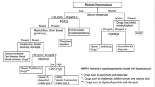
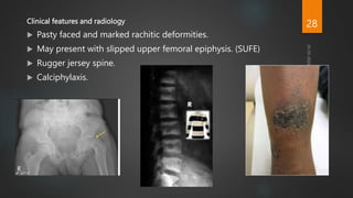
Rickets and Osteomalacia
MODERATOR- DR. SURESH BORA, M.S. , ASSOCIATE PROFESSOR
PRESENTER- DR. SAJID HUSSAIN, PGT
1
Rickets- there is inadequate
mineralization of growing bone and
epiphyseal cartilage due to defective vitamin
D metabolism
Osteomalacia- “bone softening” in
adults that is usually due to prolonged
deficiency of vitamin D. This deficiency results
in abnormal osteoid mineralization.
2
ETIOLOGICAL CLASSIFICATION OF RICKETS AND OSTEOMALACIA
I. Deficiency rickets and osteomalacia
A. Vitamin D disorders
B. Calcium Deficiency
C. Phosphorus Deficiency
D. Chelators in diets
II. Absorptive rickets and osteomalacia
A. Gastric abnormalities
B. Biliary disease
C. Enteric absorptive defects
III. Renal Tubular rickets and osteomalacia
A. Proximal tubular lesions
B. Proximal and distal tubular lesions
C. Distal Tubular lesions (renal tubular acidosis)
1. Primary
2. Secondary
IV. Renal Osteodystrophy
V. Unusual forms
3
4
Office of dietary supplements - vitamin D (no date) NIH Office of Dietary Supplements. Available at:
https://ods.od.nih.gov/factsheets/VitaminD-Consumer/ (Accessed: 16 March 2024).
5
6
7
HISTOLOGY OF GROWTH PLATE
8
 Hypertrophic zone-
 Disordered proliferation of chondrocytes.
 Loss of the columnar arrangement of chondrocytes.
 Thickening and disorganization of the hypertrophic zone.
 Tongue-like projections of cartilage that extend into the spongiosa.
 Irregularity of the limit between the proliferative and hypertrophic
zones.
 Penetration of blood vessels into the hypertrophic zone.
Walker A, El Demellawy D, Davila J. Rickets: Historical, Epidemiological,
Pathophysiological, and Pathological Perspectives. Acad Forensic Pathol.
2017 Jun;7(2):240-262. doi: 10.23907/2017.024. Epub 2017 Jun 1. PMID:
31239976; PMCID: PMC6474539.
9
CLINICAL FEATURES
General
 Failure to thrive
 Protuberant abdomen
 Apathy, listlessness and irritability.
 Proximal muscle weakness.
 Ligament laxity
 Tetany, seizure , laryngospasm.
 Bilateral lamellar cataract.
10
Head
 Craniotabes
 Frontal bossing
 Delayed dentition and tooth caries
 Delayed closure of frontanel
 Craniosynostosis
11
Chest
 Rachitic rosary
 Harrison’s grove
 Pectus carinatum/
Pectus excavatum
12
Limbs and Joints
 Bone pain and tenderness
 Coxa vara
 Genu valgum or varum
 Windswept deformity
 Bowing of long bones
 Rachitic saber shin
 Enlargement of ends of long bones
 String of pearls deformity
 Double malleoli sign
13
Spine
 Scoliosis
 Kyphosis
 Accentuation of lumbar lordosis
14
BIOCHEMISTRY IN VITAMIN D DEFICIENCY RICKETS
 Decreased level of serum phosphate.
 Calcium level is normal and sometimes decreased (if compensatory
mechanism fails).
 Increased serum alkaline phosphate.
 Diminished urinary excretion of calcium.
 Decreased serum level of 25-OH cholecalciferol.
 The “calcium phosphate product” <2.4
15
LABORATORY FINDINGS IN DIFFERENT TYPES OF RICKETS
16
17
Radiological signs
 Generalized osteopenia
 Wide medullary canal and penciling of cortex
 Bowing deformity of long bones
 Widening of growth plate
 Cupping or flaring of metaphysis
 Looser’s zone (Milkmaids pseudofractures or Umbau zones)
 In severe rickets, margins of tarsal and carpal ossification center may disappear.
18
19
 In healing rickets
 Provisional line of calcification (white line)
 Recalcification of spongiosa in the metaphysis
 Dense line at the ends of metaphysis
 Epiphseal shadow defined
 End of shaft and epiphysis becomes clearly differentiated
 Finally, the bone appears to be normal
20
TREATMENT
Vitamin D deficiency rickets
 Adequate intake of vitamin D, calcium, and phosphorus is the mainstay.
 Stoss therapy: 3,00,000-6,00,000 IU of vitamin D administered orally or intramuscularly as 2-4
doses over 1 day.
 Alternate therapy: Vitamin D- 2,000 to 5,000 IU/day over 4-6 weeks.
 Followed by- Vitamin D intake
400IU/day ( for child <1 year of age)
600 IU/day ( for age>1 year of age)
21
RENAL RICKETS
 No radiological healing after 4 weeks of vitamin D therapy, and
compliance confirmed– Rule out renal rickets.
 Renal rickets- Tubular or Glomerular disorders.
 Tubular disorders- Hereditary or Acquired
Proximal or Distal
22
VITAMIN D DEPENDENT RICKETS TYPE 1
 Autosomal recessive mode of inheritance.
 Mutation in 1ɑ-hydroxylase gene.
 Prevents conversion of 25-D into cacitriol.
 Elevated PTH level, Decreased serum calcium, low or undetectable serum concentration
of calcitriol.
23
 Treatment- Calcitriol with initial doses of 0.25-2 mcg/ days, with lower
doses once the rickets has healed, with adequate intake of calcium.
 Target urinary calcium excretion- <4 mg/kg/day.
24
VITAMIN D DEPENDENT RICKETS TYPE 2
 Autosomal recessive disorder.
 End organ resistance to Calcitriol due to gene encoding VDR.
 Levels of calcitriol are extremely elevated.
 3-6 months trial of high dose vitamin D and Oral Calcium.
 If partially functional VDR available, response observed.
 Unresponsive patients- Long term iv Calcium with possible transition to very
high dose oral supplements
25
RICKETS IN CHRONIC RENAL FAILURE AND RENAL OSTEODYSTROPHY
 Rickets/Osteomalacia predominant
 Diminished production of calcitriol.
 Inadequate calcium absorption and secondary
hyperparathyroidism.
26
 Hyperparathyroidism predominant
 Hyperphosphataemia as a result of decreased renal excretion.
 Dominant picture- Secondary hyperparathyroidism
 Osteoporosis Predominant- In older patients, effects of post menopausal osteoporosis
may be superimposed
27
Clinical features and radiology
 Pasty faced and marked rachitic deformities.
 May present with slipped upper femoral epiphysis. (SUFE)
 Rugger jersey spine.
 Calciphylaxis.
28
Treatment
 Calcitriol is the treatment of choice.
 Sevalamer Hydrochloride , a phosphate binder used orally.
 Metabolic acidosis may be corrected with alkalis.
 Calcimimetic drug- Cinacalcet
 Aluminium toxicity redused using Dialysis.
29
HYPOPHOSPHATAEMIC RICKETS
Phosphorus Deficiency
 Inadequate intake- Conditions of prolonged starvation or severe anorexia.
 Isolated phosphorus malabsorption- Long term ingestion of antacids
containing aluminium. Responds to discontinuation of the antacid and
short term phosphorus supplementation.
30
 Role of phosphatonins-
 Phosphatonins viz. FGF-23, FGF-7, SFRP-4 and matrix extracellular phosphoglycoprotein reduce
renal sodium dependent phosphate transport.
 FGF-23 and SFRP-4 inhibit 1,25-dihydroxyvitamin D synthesis, leads to decline in phosphate
absorption and decreased reabsorption of phosphate from kidneys,
31
X-linked Hypophosphatemic Rickets
 aka Familial Vitamin D resistant rickets.
 Defective gene PHEX
 Normally, PHEX regulates FGF-23 produced from FGF-23 gene.
 In absence of normally functioning PHEx enzymatic activity, Osteopontin
accumulates in bone to contribute to the Osteomalacia.
32
 Inv- Hypophostaemia, Decreased 1,25 (OH)2 vit. D3.
 Abnormalities of lower extremity more pronounced as compared to upper
limb.
 Lab Inv- High renal excretion of phosphate, hypophosphataemia, and
increased ALP. Normal PTH and serum Calcium levels.
 Treatment by combination of oral phosphorus and calcitriol. Balance
between both required to prevent complications.
33
Autosomal Dominant Hypophosphatemic Rickets
 Much less common than XLH.
 Autosomal dominant mode of inheritance.
 Gene coding for FGF-23 is mutated.
 Mutated FGF-23 escapes degradation by the proteases.
 Treatment is similar to XLH.
34
Hereditary Hypophosphatemic Rickets with Hypercalciurua
 Isolated renal phosphate wasting disorder leading to low serum
phosphate levels.
 Level of Calcitriol is elevated.
 Loss of function SLC34A3 results primary renal tubular defect and is
compatible with HHRH phenotype.
35
 Normal or low-normal level of FGF-23.
 Presents with rachitic leg abnormalities, muscle weakness and bone pain.
 Kidney stones secondary to hypercalciuria
 Treatment- Oral phosphorus replacement (1-2.5g/ day of elemental
phosphorus in five divided oral doses.)
36
Renal Fanconi’s Syndrome
 Impaired net proximal resorption of amino acids,glucose, phosphates,
bicarbonates and urates.
 Hypophosphataemia secondary to hyperphosphaturia, metabolic acidosis
and impaired synthesis of calcitriol.
 Not all have rickets or osteomalcia
37
Investigations- Hypophosphataemia, normal to low calcium, low calcitriol
and hyperchloremic metabolic acidosis.
 Treatment by administration of large dose of calciferol, or small doses of
dihydrotachysterol or calcitriol.
 Alkali and potassium supplementation
 Thiazide diuretics to reduce intravascular volume and filtered load of
bicarbonates.
38
Overproduction of Phosphatonin
 One of the pathogeneses of Tumor induced osteomalacia (TIO).
 Secrete different phophatonins.
 Produces biochemical phenotype similar to XLH and ADHR.
 Cure can be achieved by resection of tumor.
 If tumor not resectable treatment similar to XLH.
39
FOLLOW UP
 Radiographs should be obtained at 4 weeks and 12 weeks after starting
vitamin D therapy in rickets.
 Serum calcium, phosphate, alkaline phosphatase, serum 25 (OH)D levels
should be performed at 12 weeks after vitamin D therapy to measure
response and toxicity.
 Maintenance dose of vitamin D should be started once complete
healing has been achieved.
 Urine calcium: creatinine ratio and renal ultrasonogram should be done
when there is hypercalcemia or hypervitaminosis.
 In hypophosphatemic rickets height to be monitored every 3 months with
serum P, Ca, ALP, Creatinine and Urinary calcium excretion.
Gupta, P., Dabas, A., Seth, A. et al. Indian Academy of Pediatrics Revised (2021) Guidelines on Prevention and
Treatment of Vitamin D Deficiency and Rickets. Indian Pediatr 59, 142–158 (2022). https://doi.org/10.1007/s13312-
022-2448-y
40
Orthopaedic Treatment
Conservative methods
 Mild deformities correct spontaneously when rickets heals.
 Splints can be used to correct deformities.
Eg- Mermaid splint, Orthopaedic shoes
41
Surgical Management
 In very young children with deformity, treatment of the metabolic defect
supplemented by bracing/splinting may correct the deformity.
 In prepubertal children or adolescent, medical management and bracing
usually not enough.
 Early osteotomy or growth modulation usually indicated.
 Mobilize as early as possible after corrective surgery.
 Control disease metabolically prior to surgery.
42
 Stop vitamin D 3 weeks prior to surgery.
 In older children with severe deformity, without past history of medical
management, go for surgery in less homeostatic but metabolically
compensated state instead of loading the patient with vitamin D and
calcium. ( r/o renal osteodystrophy).
 Role of external fixators for correction to be considered.
 In addition to osteotomy, guided growth modulation with
hemiepiphysiodesis has had promising results.
43
44
OSTEOMALACIA
 In vitamin D deficiency normal serum calcium maintained by mobilizing calcium
from the bones.
 PTH secreted by the parathyroid glands in response to hypocalcemia from
vitamin D deficiency attempts to bring the body back to normal serum calcium
levels.
 Bones- the primary target to recruit calcium, and osteomalacia will ensue by
extracting calcium from the bones.
45
MEDICATIONS CAUSING OSTEOMALACIA
 Antiepileptic drugs, including phenobarbital, phenytoin, and carbamazepine,
enhance catabolism of calcidiol via induction of P-450 activity.
 Isoniazid, rifampicin, and theophylline also precipitate vitamin D deficiency in the
same manner as antiepileptic medications.
 Antifungal agents such as ketoconazole increase vitamin D requirements by
inhibiting 1-alpha-hydroxylase.
 Long-term steroid use also has implications for vitamin D deficiency, possibly by
increasing 24-hydroxylase activity.
46
CRITERIA FOR DIAGNOSIS OF OSTEOMALACIA(Fukumoto et al)
1. Hypophosphatemia or hypocalcemia
2. High bone alkaline phosphatase
3. Muscle weakness or bone pain
4. Less than 80% BMD of the young-adult-mean
5. Multiple uptake zones by bone scintigraphy or radiographic evidence of
Looser zones (pseudofractures)
*Definite osteomalacia is defined as having all of the above findings, numbers 1–5.
5.
*Possible osteomalacia is defined as having the findings of numbers 1, 2, and 2 of
the 3 numbers 3–5 findings described above.
47
X Ray findings in Osteomalacia
 Looser’s Zone
 Triradiate Pelvis
 Protrusio acetabula
 Codfish Vertebrae
48
Looser’s Zone
49
TREATMENT- FOCUSSED ON REVERSING THE UNDERLYING DISORDER
If severe vitamin D deficiency is the underlying cause
 60,000 IU of ergocalciferol (vitamin D2) or cholecalciferol (vitamin D3) orally 1 day per
week for 8 to 12 weeks, followed by
 800 IU–2000 IU of vitamin D3 daily
Follow-up of treatment.
• Serum calcium and urine calcium levels should be monitored, initially after 1 and 3
months.
• and then every 6 to 12 months until 24-hour urine calcium excretion is normal.
• Serum 25(OH)D level can be measured 3 to 4 months after starting therapy.
50
References
 Essential Orthopaedics Principles & Practice (3rd Edition), Varshney
 Textbook of Orthopaedics and Trauma, 2nd Edition, GS Kulkarni
 Campbell’s operative orthopaedics, Fourteenth Edition (International)
 Walker A, El Demellawy D, Davila J. Rickets: Historical, Epidemiological, Pathophysiological,
and Pathological Perspectives. Acad Forensic Pathol. 2017 Jun;7(2):240-262. doi:
10.23907/2017.024. Epub 2017 Jun 1. PMID: 31239976; PMCID: PMC6474539.
 Office of dietary supplements - vitamin D (no date) NIH Office of Dietary Supplements.
Available at: https://ods.od.nih.gov/factsheets/VitaminD-Consumer/
51
52
NEXT SEMINAR
TOPIC- OSTEOPOROSIS
DATE- 28/03/2024
MODERATOR- DR. JISHNU PRAKASH BARUAH(ASSO. PROF.)
PRESENTER- DR. DILDAR HUSSAIN (PGT)
53

Rickets and Osteomalacia presentation- Dr. Sajid

  • 1.
    Rickets and Osteomalacia MODERATOR-DR. SURESH BORA, M.S. , ASSOCIATE PROFESSOR PRESENTER- DR. SAJID HUSSAIN, PGT 1
  • 2.
    Rickets- there isinadequate mineralization of growing bone and epiphyseal cartilage due to defective vitamin D metabolism Osteomalacia- “bone softening” in adults that is usually due to prolonged deficiency of vitamin D. This deficiency results in abnormal osteoid mineralization. 2
  • 3.
    ETIOLOGICAL CLASSIFICATION OFRICKETS AND OSTEOMALACIA I. Deficiency rickets and osteomalacia A. Vitamin D disorders B. Calcium Deficiency C. Phosphorus Deficiency D. Chelators in diets II. Absorptive rickets and osteomalacia A. Gastric abnormalities B. Biliary disease C. Enteric absorptive defects III. Renal Tubular rickets and osteomalacia A. Proximal tubular lesions B. Proximal and distal tubular lesions C. Distal Tubular lesions (renal tubular acidosis) 1. Primary 2. Secondary IV. Renal Osteodystrophy V. Unusual forms 3
  • 4.
  • 5.
    Office of dietarysupplements - vitamin D (no date) NIH Office of Dietary Supplements. Available at: https://ods.od.nih.gov/factsheets/VitaminD-Consumer/ (Accessed: 16 March 2024). 5
  • 6.
  • 7.
  • 8.
  • 9.
     Hypertrophic zone- Disordered proliferation of chondrocytes.  Loss of the columnar arrangement of chondrocytes.  Thickening and disorganization of the hypertrophic zone.  Tongue-like projections of cartilage that extend into the spongiosa.  Irregularity of the limit between the proliferative and hypertrophic zones.  Penetration of blood vessels into the hypertrophic zone. Walker A, El Demellawy D, Davila J. Rickets: Historical, Epidemiological, Pathophysiological, and Pathological Perspectives. Acad Forensic Pathol. 2017 Jun;7(2):240-262. doi: 10.23907/2017.024. Epub 2017 Jun 1. PMID: 31239976; PMCID: PMC6474539. 9
  • 10.
    CLINICAL FEATURES General  Failureto thrive  Protuberant abdomen  Apathy, listlessness and irritability.  Proximal muscle weakness.  Ligament laxity  Tetany, seizure , laryngospasm.  Bilateral lamellar cataract. 10
  • 11.
    Head  Craniotabes  Frontalbossing  Delayed dentition and tooth caries  Delayed closure of frontanel  Craniosynostosis 11
  • 12.
    Chest  Rachitic rosary Harrison’s grove  Pectus carinatum/ Pectus excavatum 12
  • 13.
    Limbs and Joints Bone pain and tenderness  Coxa vara  Genu valgum or varum  Windswept deformity  Bowing of long bones  Rachitic saber shin  Enlargement of ends of long bones  String of pearls deformity  Double malleoli sign 13
  • 14.
    Spine  Scoliosis  Kyphosis Accentuation of lumbar lordosis 14
  • 15.
    BIOCHEMISTRY IN VITAMIND DEFICIENCY RICKETS  Decreased level of serum phosphate.  Calcium level is normal and sometimes decreased (if compensatory mechanism fails).  Increased serum alkaline phosphate.  Diminished urinary excretion of calcium.  Decreased serum level of 25-OH cholecalciferol.  The “calcium phosphate product” <2.4 15
  • 16.
    LABORATORY FINDINGS INDIFFERENT TYPES OF RICKETS 16
  • 17.
  • 18.
    Radiological signs  Generalizedosteopenia  Wide medullary canal and penciling of cortex  Bowing deformity of long bones  Widening of growth plate  Cupping or flaring of metaphysis  Looser’s zone (Milkmaids pseudofractures or Umbau zones)  In severe rickets, margins of tarsal and carpal ossification center may disappear. 18
  • 19.
  • 20.
     In healingrickets  Provisional line of calcification (white line)  Recalcification of spongiosa in the metaphysis  Dense line at the ends of metaphysis  Epiphseal shadow defined  End of shaft and epiphysis becomes clearly differentiated  Finally, the bone appears to be normal 20
  • 21.
    TREATMENT Vitamin D deficiencyrickets  Adequate intake of vitamin D, calcium, and phosphorus is the mainstay.  Stoss therapy: 3,00,000-6,00,000 IU of vitamin D administered orally or intramuscularly as 2-4 doses over 1 day.  Alternate therapy: Vitamin D- 2,000 to 5,000 IU/day over 4-6 weeks.  Followed by- Vitamin D intake 400IU/day ( for child <1 year of age) 600 IU/day ( for age>1 year of age) 21
  • 22.
    RENAL RICKETS  Noradiological healing after 4 weeks of vitamin D therapy, and compliance confirmed– Rule out renal rickets.  Renal rickets- Tubular or Glomerular disorders.  Tubular disorders- Hereditary or Acquired Proximal or Distal 22
  • 23.
    VITAMIN D DEPENDENTRICKETS TYPE 1  Autosomal recessive mode of inheritance.  Mutation in 1ɑ-hydroxylase gene.  Prevents conversion of 25-D into cacitriol.  Elevated PTH level, Decreased serum calcium, low or undetectable serum concentration of calcitriol. 23
  • 24.
     Treatment- Calcitriolwith initial doses of 0.25-2 mcg/ days, with lower doses once the rickets has healed, with adequate intake of calcium.  Target urinary calcium excretion- <4 mg/kg/day. 24
  • 25.
    VITAMIN D DEPENDENTRICKETS TYPE 2  Autosomal recessive disorder.  End organ resistance to Calcitriol due to gene encoding VDR.  Levels of calcitriol are extremely elevated.  3-6 months trial of high dose vitamin D and Oral Calcium.  If partially functional VDR available, response observed.  Unresponsive patients- Long term iv Calcium with possible transition to very high dose oral supplements 25
  • 26.
    RICKETS IN CHRONICRENAL FAILURE AND RENAL OSTEODYSTROPHY  Rickets/Osteomalacia predominant  Diminished production of calcitriol.  Inadequate calcium absorption and secondary hyperparathyroidism. 26
  • 27.
     Hyperparathyroidism predominant Hyperphosphataemia as a result of decreased renal excretion.  Dominant picture- Secondary hyperparathyroidism  Osteoporosis Predominant- In older patients, effects of post menopausal osteoporosis may be superimposed 27
  • 28.
    Clinical features andradiology  Pasty faced and marked rachitic deformities.  May present with slipped upper femoral epiphysis. (SUFE)  Rugger jersey spine.  Calciphylaxis. 28
  • 29.
    Treatment  Calcitriol isthe treatment of choice.  Sevalamer Hydrochloride , a phosphate binder used orally.  Metabolic acidosis may be corrected with alkalis.  Calcimimetic drug- Cinacalcet  Aluminium toxicity redused using Dialysis. 29
  • 30.
    HYPOPHOSPHATAEMIC RICKETS Phosphorus Deficiency Inadequate intake- Conditions of prolonged starvation or severe anorexia.  Isolated phosphorus malabsorption- Long term ingestion of antacids containing aluminium. Responds to discontinuation of the antacid and short term phosphorus supplementation. 30
  • 31.
     Role ofphosphatonins-  Phosphatonins viz. FGF-23, FGF-7, SFRP-4 and matrix extracellular phosphoglycoprotein reduce renal sodium dependent phosphate transport.  FGF-23 and SFRP-4 inhibit 1,25-dihydroxyvitamin D synthesis, leads to decline in phosphate absorption and decreased reabsorption of phosphate from kidneys, 31
  • 32.
    X-linked Hypophosphatemic Rickets aka Familial Vitamin D resistant rickets.  Defective gene PHEX  Normally, PHEX regulates FGF-23 produced from FGF-23 gene.  In absence of normally functioning PHEx enzymatic activity, Osteopontin accumulates in bone to contribute to the Osteomalacia. 32
  • 33.
     Inv- Hypophostaemia,Decreased 1,25 (OH)2 vit. D3.  Abnormalities of lower extremity more pronounced as compared to upper limb.  Lab Inv- High renal excretion of phosphate, hypophosphataemia, and increased ALP. Normal PTH and serum Calcium levels.  Treatment by combination of oral phosphorus and calcitriol. Balance between both required to prevent complications. 33
  • 34.
    Autosomal Dominant HypophosphatemicRickets  Much less common than XLH.  Autosomal dominant mode of inheritance.  Gene coding for FGF-23 is mutated.  Mutated FGF-23 escapes degradation by the proteases.  Treatment is similar to XLH. 34
  • 35.
    Hereditary Hypophosphatemic Ricketswith Hypercalciurua  Isolated renal phosphate wasting disorder leading to low serum phosphate levels.  Level of Calcitriol is elevated.  Loss of function SLC34A3 results primary renal tubular defect and is compatible with HHRH phenotype. 35
  • 36.
     Normal orlow-normal level of FGF-23.  Presents with rachitic leg abnormalities, muscle weakness and bone pain.  Kidney stones secondary to hypercalciuria  Treatment- Oral phosphorus replacement (1-2.5g/ day of elemental phosphorus in five divided oral doses.) 36
  • 37.
    Renal Fanconi’s Syndrome Impaired net proximal resorption of amino acids,glucose, phosphates, bicarbonates and urates.  Hypophosphataemia secondary to hyperphosphaturia, metabolic acidosis and impaired synthesis of calcitriol.  Not all have rickets or osteomalcia 37
  • 38.
    Investigations- Hypophosphataemia, normalto low calcium, low calcitriol and hyperchloremic metabolic acidosis.  Treatment by administration of large dose of calciferol, or small doses of dihydrotachysterol or calcitriol.  Alkali and potassium supplementation  Thiazide diuretics to reduce intravascular volume and filtered load of bicarbonates. 38
  • 39.
    Overproduction of Phosphatonin One of the pathogeneses of Tumor induced osteomalacia (TIO).  Secrete different phophatonins.  Produces biochemical phenotype similar to XLH and ADHR.  Cure can be achieved by resection of tumor.  If tumor not resectable treatment similar to XLH. 39
  • 40.
    FOLLOW UP  Radiographsshould be obtained at 4 weeks and 12 weeks after starting vitamin D therapy in rickets.  Serum calcium, phosphate, alkaline phosphatase, serum 25 (OH)D levels should be performed at 12 weeks after vitamin D therapy to measure response and toxicity.  Maintenance dose of vitamin D should be started once complete healing has been achieved.  Urine calcium: creatinine ratio and renal ultrasonogram should be done when there is hypercalcemia or hypervitaminosis.  In hypophosphatemic rickets height to be monitored every 3 months with serum P, Ca, ALP, Creatinine and Urinary calcium excretion. Gupta, P., Dabas, A., Seth, A. et al. Indian Academy of Pediatrics Revised (2021) Guidelines on Prevention and Treatment of Vitamin D Deficiency and Rickets. Indian Pediatr 59, 142–158 (2022). https://doi.org/10.1007/s13312- 022-2448-y 40
  • 41.
    Orthopaedic Treatment Conservative methods Mild deformities correct spontaneously when rickets heals.  Splints can be used to correct deformities. Eg- Mermaid splint, Orthopaedic shoes 41
  • 42.
    Surgical Management  Invery young children with deformity, treatment of the metabolic defect supplemented by bracing/splinting may correct the deformity.  In prepubertal children or adolescent, medical management and bracing usually not enough.  Early osteotomy or growth modulation usually indicated.  Mobilize as early as possible after corrective surgery.  Control disease metabolically prior to surgery. 42
  • 43.
     Stop vitaminD 3 weeks prior to surgery.  In older children with severe deformity, without past history of medical management, go for surgery in less homeostatic but metabolically compensated state instead of loading the patient with vitamin D and calcium. ( r/o renal osteodystrophy).  Role of external fixators for correction to be considered.  In addition to osteotomy, guided growth modulation with hemiepiphysiodesis has had promising results. 43
  • 44.
  • 45.
    OSTEOMALACIA  In vitaminD deficiency normal serum calcium maintained by mobilizing calcium from the bones.  PTH secreted by the parathyroid glands in response to hypocalcemia from vitamin D deficiency attempts to bring the body back to normal serum calcium levels.  Bones- the primary target to recruit calcium, and osteomalacia will ensue by extracting calcium from the bones. 45
  • 46.
    MEDICATIONS CAUSING OSTEOMALACIA Antiepileptic drugs, including phenobarbital, phenytoin, and carbamazepine, enhance catabolism of calcidiol via induction of P-450 activity.  Isoniazid, rifampicin, and theophylline also precipitate vitamin D deficiency in the same manner as antiepileptic medications.  Antifungal agents such as ketoconazole increase vitamin D requirements by inhibiting 1-alpha-hydroxylase.  Long-term steroid use also has implications for vitamin D deficiency, possibly by increasing 24-hydroxylase activity. 46
  • 47.
    CRITERIA FOR DIAGNOSISOF OSTEOMALACIA(Fukumoto et al) 1. Hypophosphatemia or hypocalcemia 2. High bone alkaline phosphatase 3. Muscle weakness or bone pain 4. Less than 80% BMD of the young-adult-mean 5. Multiple uptake zones by bone scintigraphy or radiographic evidence of Looser zones (pseudofractures) *Definite osteomalacia is defined as having all of the above findings, numbers 1–5. 5. *Possible osteomalacia is defined as having the findings of numbers 1, 2, and 2 of the 3 numbers 3–5 findings described above. 47
  • 48.
    X Ray findingsin Osteomalacia  Looser’s Zone  Triradiate Pelvis  Protrusio acetabula  Codfish Vertebrae 48
  • 49.
  • 50.
    TREATMENT- FOCUSSED ONREVERSING THE UNDERLYING DISORDER If severe vitamin D deficiency is the underlying cause  60,000 IU of ergocalciferol (vitamin D2) or cholecalciferol (vitamin D3) orally 1 day per week for 8 to 12 weeks, followed by  800 IU–2000 IU of vitamin D3 daily Follow-up of treatment. • Serum calcium and urine calcium levels should be monitored, initially after 1 and 3 months. • and then every 6 to 12 months until 24-hour urine calcium excretion is normal. • Serum 25(OH)D level can be measured 3 to 4 months after starting therapy. 50
  • 51.
    References  Essential OrthopaedicsPrinciples & Practice (3rd Edition), Varshney  Textbook of Orthopaedics and Trauma, 2nd Edition, GS Kulkarni  Campbell’s operative orthopaedics, Fourteenth Edition (International)  Walker A, El Demellawy D, Davila J. Rickets: Historical, Epidemiological, Pathophysiological, and Pathological Perspectives. Acad Forensic Pathol. 2017 Jun;7(2):240-262. doi: 10.23907/2017.024. Epub 2017 Jun 1. PMID: 31239976; PMCID: PMC6474539.  Office of dietary supplements - vitamin D (no date) NIH Office of Dietary Supplements. Available at: https://ods.od.nih.gov/factsheets/VitaminD-Consumer/ 51
  • 52.
  • 53.
    NEXT SEMINAR TOPIC- OSTEOPOROSIS DATE-28/03/2024 MODERATOR- DR. JISHNU PRAKASH BARUAH(ASSO. PROF.) PRESENTER- DR. DILDAR HUSSAIN (PGT) 53