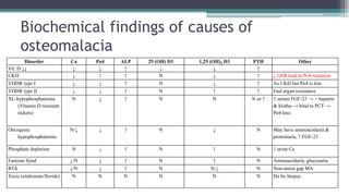
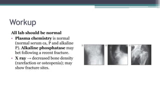
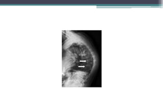
Metabolic bone diseases
Usama Ragab Youssif, MD
Consultant Internal Medicine
Lecturer of Medicine
Zagazig University
Email: usamaragab@medicine.zu.edu.eg
Slideshare: https://www.slideshare.net/dr4spring/
Mobile: 00201000035863
Metabolic bone disease
• Metabolic bone disease, any of
several diseases that cause various abnormalities or
deformities of bone.
• Examples of metabolic bone diseases include
osteoporosis, rickets, osteomalacia, osteogenesis
imperfecta, marble bone disease (osteopetrosis),
Paget disease of bone, and fibrous dysplasia.
Disease of poor women
Introduction
• Osteomalacia is characterized by defective bone
mineralization, bone pain, muscle weakness and pathological
fractures.
• There is failure to replace the turnover of Ca and P in bone matrix
→ bone becomes demineralized and the bony substance
becomes replaced by soft osteoid tissue so it is mainly a
qualitative bone defect.
• There is, unlike osteoporosis, a decline in the ratio of
mineralized bone to matrix.
Synthesis of Vitamin D
Pathogenesis
• Both vitamin D2 (ergocalciferol) from vegetables in the diet, and
D3 (cholecalciferol) from animal tissues and de novo synthesis in
skin, are metabolized in the liver to 25-hydroxyvitamin D and
then in the kidney to 1,25-dihydroxyvitamin D3.
• The latter affects calcium metabolism by acting on the
parathyroid glands (negative-feedback loop on PTH stimulation
of renal vitamin D hydroxylases), GI tract (increased absorption
of calcium and phosphate), and bone (both bone resorption and
osteoblast activation with bone formation). Kidneys; increase
reabsorption of phosphate.
Pathogenesis (cont.)
• The most common cause is vitamin D deficiency, the low levels of
vitamin D causes a reduction of calcium absorption from the intestine.
• The low calcium absorption stimulates parathyroid hormone secretion
which restores serum calcium levels towards normal 1by increasing
bone resorption and 2renal tubular calcium reabsorption. The level of
parathyroid hormone also 3promotes phosphaturia and causes
phosphate depletion.
• It is the combination of calcium loss from bone and phosphate
depletion that leads to impaired bone mineralization.
The classification of osteomalacia
1. Hypocalcemic osteomalacia
• Vitamin D deficiency
• Pseudovitamin D deficiency
2. Hypophosphatemic osteomalacia
3. Osteomalacia with normal bone homeostasis
= Defective mineralization
The classification of osteomalacia
Abnormal vitamin D
metabolism
Reduced availability Poor diet
Inadequate exposure to sun
Malabsorption
Defective metabolism Hepatobiliary disease
Chronic Kidney disease
Anticonvulsant drugs, barbiturate, rifamycin → ↑↑ metabolism
Vitamin D-dependent rickets type I (AR)
X-linked hypophosphatemia (XLD) → Po4 loss
Oncogenic hypophosphatemia→ ↑ FGF-23 → Po4 loss
Receptor defects Vitamin D-dependent rickets type II (AR)
Altered phosphate
homeostasis
Malabsorption Malabsorption syndromes
Renal phosphate loss X-linked hypophosphatemia: phosphate wasting (PHEX)
Fanconi syndrome: loss of Po4, Ca, a.a., sugar...
Defective
mineralization
Aluminum and fluoride toxicity
Bisphosphonate toxicity
Hypophosphatasia
Fibrogenous imperfecta ossium = 1ry collagen defect
VDDR type I: AR, ↓↓ α-hydroxylase → ↓↓ active vitamin D → treated with calcitriol
VDDR type II: AR, end organ resistance if with alopecia type IIa, if without alopecia type IIb, → treated by supraphysiological doses of calcitriol
& calcium.
Clinical features
Osteomalacia is the adult counterpart of rickets
• Skeletal discomfort (from bone and muscle pain), deformities; kyphosis,
triradiate hip i.e. soft hip → this is exaggerated in children → bone bowing.
• Bone tenderness
• Tetany may be manifested: rarely may be severe enough → dysrrhythmia +
convulsions
• Muscular weakness with marked proximal myopathy with waddling gait.
• Rickets: rachitic rosary, Harrison’s groove, delayed dentition.
Causes of waddling gait:
•‫البطة‬
•Myopathy (proximal).
•Osteomalacia ± osteoporosis.
•Bilateral congenital hip dislocation.
•Symphysiotomy.
•Pregnancy.
•Tense ascites
Workup
Bone markers are abnormal
Laboratory + Imaging + Biopsy
A- Laboratory
1. Serum Calcium and phosphorus are decreased; but
phosphorus may be increased in renal failure.
2. Alkaline phosphatase is increased in 90% of cases.
3. Serum FGF-23 is elevated in many people with tumour-
induced osteomalacia, x-linked hypophosphatemic
rickets.
4. Vitamin D levels (25 OH cholecalciferol; calcidiol)
are reduced: normal range = (30 – 100 ng/mL).
A- Laboratory (cont.)
5. Urinary phosphate and low urinary calcium
excretion, low levels of 25-hydroxyvitamin D, and a mild
secondary hyperparathyroidism. The latter may cause a
mild hyperchloremic acidosis due to renal bicarbonate
loss. Severe acidosis suggests a renal tubular defect.
6. Levels of 1,25-dihydroxyvitamin D may be normal
and are, therefore, not helpful. If the serum calcium and
25-hydroxyvitamin D levels are normal as well, then the
defect is likely to be renal handling of phosphate or end-
organ resistance.
Biochemical findings of causes of
osteomalacia
Disorder Ca Po4 ALP 25 (OH) D3 1,25 (OH)2 D3 PTH Other
Vit. D ↓↓ ↓ ↓ ↑ ↓ ↓ ↑
CKD ↓ ↑ ↑ N ↓ ↑ ↓ GFR lead to Po4 retention
VDDR type I ↓ ↓ ↑ N ↓ ↑ As CKD but Po4 is low
VDDR type II ↓ ↓ ↑ N ↑ ↑ End organ resistance
XL-hypophosphatemia
(Vitamin D resistant
rickets)
N ↓ ↑ N N N or ↑ ↑ serum FGF-23 → + heparin
& klotho → bind to PCT →
Po4 loss
Oncogenic
hypophosphatemia
N/↓ ↓ ↑ N ↓ N May have aminoaciduria &
proteinuria, ↑ FGF-23
Phosphate depletion N ↓ ↑ N ↑ N ↑ urine Ca
Fanconi Synd ↓/N ↓ ↑ N ↑ N Aminoaciduria, glucosuria
RTA ↓/N ↓ ↑ N N/↓ N Non-anion gap MA
Toxic (etidronate/floride) N N N N N N Dx by biopsy
B- Imaging
1. X-ray
Bone rarefaction
Radiographic osteopenia
2. Bone scan: can show multiple hot spots in the ribs
and pelvis at the site of fractures and the appearance
may be mistaken for metastases
C- Bone biopsy
• If doubt remains as to the
diagnosis of osteomalacia, a
transiliac bone biopsy can be
taken.
• Tetracycline-labelled bone
biopsy is the gold standard
diagnostic test.
Treatment
1. Treatment of the cause e.g. correct bowel disorder, tumor
resectionm stop offending drug.
2. Sun exposure
3. Diet e.g. milk, cheese or yoghurt
4. Ca supplements: at doses of 800–1000 mg/day for adults.
5. Vitamin D2 (ergocalciferol), or D3 (cholecalciferol)
6. Alfacalcidol especially in cases of renal failure, vitamin D
dependent rickets type I “physiological dose” i.e. 0.5 – 1
mcg/day, vitamin D dependent rickets type II
“supraphysiological dose”, also in hypophosphatemic rickets.
Vitamin D issue
• Goal → 25 (OH) D3 > 30 ng/mL (ideally > 40 ng/mL)
• After replenish → start maintenance vitamin D →
800 – 2000 IU daily of vitamin D or 50,000 IU every
other week.
• After last dose of pharmacological vitamin D →
wait for 2 weeks then measure 25 (OH) vitamin D levels
Vitamin D issue (cont.)
• Recommended vitamin D treatment in age ≥ 18
years:
50,000 IU once weekly for 8 weeks (3 – 12 weeks)
OR
6000 IU once daily for 8 weeks OR
200,000 IU once monthly for 2 – 3 months
Vitamin D issue (cont.)
• Endocrine society (2011) vitamin D
requirements (RDA):
Child 0 – 1 years → 400 IU daily.
Child 1 – 18 years → 600 IU daily.
Adult → 1500 – 2000 IU daily.
Vitamin D in Egyptian Market
• Ossofortin 10,000 & 50,000 IU tab
• Vidrop oral drops 2800 IU/mL (42,000 IU per 15 mL)
• Davalindi 1000 IU tab
• Devarol S 200,000 IU amp
Notes on treatment
• Vitamin D3 is better than vitamin D2 as it ↑↑ serum 25
(OH) vitamin D rapidly
• Patients with CKD or hypoparathyroidism need both
vitamin D2 or D3 + calcitriol as CKD → ↓↓ 1α
hydroxylase and hypoparathyroid patients are unable
to convert 25 (OH) D to 1,25 (OH)2 D in absence of
PTH.
Notes on treatment (cont.)
• With rapid clinical improvement, an elevation in
serum 25(OH)D and a reduction in PTH.
• Serum ALP levels sometimes rise initially as
mineralisation of bone increases, but eventually
fall to within the reference range as the bone
disease heals.
Other causes of osteomalacia
• Aluminium intoxication is now rare due to reduced
use of aluminium containing phosphate binders and
removal of aluminium from the water supplies used in
dialysis. If aluminium intoxication is suspected, the
diagnosis can be confirmed by demonstration of
aluminium at the calcification front in a bone biopsy.
Other causes of osteomalacia
• Osteomalacia due to bisphosphonates has mostly been
described in patients with Paget’s disease receiving etidronate
and high dose pamidronate. It is usually asymptomatic and
healing occurs when treatment is stopped.
• Excessive fluoride intake causes osteomalacia due to
direct inhibition of mineralisation and is common in parts of the
world where there is a high fluoride content in drinking water.
The condition reverses when fluoride intake is reduced.
Osteomalacia vs. Osteoporosis
• osteoMalacia = ↓↓ Mineralization.
• osteoPorosis = ↓↓ Protein matrix.
Osteomalacia vs. Osteoporosis
Osteomalacia Osteoporosis
‫شيبا‬ ‫الرأس‬‫اشتعل‬‫و‬ ‫مني‬ ‫العظم‬‫وهن‬ ‫إني‬‫رب‬ ‫ال‬‫ق‬
Definition
• It is defined as a decrease in the absolute amount of
bone mass leading to enhanced bone fragility with
increased risk of pathological fractures.
• Although the term osteoporosis refers to the reduction
in the amount of bony tissue within the skeleton, this is
generally associated with a loss of structural integrity of
the internal architecture of the bone.
Definition (cont.)
• The combination of both these changes means that
osteoporotic bone is at high risk of fracture, even after
trivial injury = pathological fracture…
• Unlike osteomalacia, the defect in osteoporosis is that
the bone that is present is normally mineralized but is
deficient in quantity, quality and structural integrity i.e.
ratio of matrix to mineral deposit is normal in
osteoporosis.
Epidemiology
• Women outnumber men with osteoporosis with ratio of 4:1
(same as fracture).
• Black women experience hip fracture at approximately half
the rate as white women
• Most women meet diagnostic criteria for osteoporosis at
age of 70s and 80s, more in white than African American
women.
Bone turnover = Bone is dynamic
In order to ensure that bone can
undertake its mechanical and
metabolic functions, it is in a constant
state of turnover
• Osteoclasts, derived from the
hematopoetic stem cell series, resorb
bone.
• Osteoblasts, derived from the
mesenchymal cell precursors, make
bone.
• Osteocytes, buried osteoblasts, sense
mechanical strain and secrete factors
important in bone homeostasis.
Pathogenesis
Many year mismatch between the rate of bone resorption
& bone formation (predominant osteoclast action)
• Osteoporosis may arise from a failure of the body to lay down
sufficient bone during growth and maturation; an earlier bone
loss following maturity; or an increased rate of that loss i.e. early
loss + increased loss.
• Polymorphisms have been identified in several genes that
contribute to the pathogenesis of osteoporosis and many of these
are in the RANK pathways, which play a critical role in regulating
bone turnover
Decreased
bone
formation
Increased
bone
resorption
Pathogenesis (cont.)
Peak bone mass
• Mainly genetically determined:
Racial effects (bone mass higher in Afro-Caribbean and lower in
Caucasians).
Family influence on the risk of osteoporosis—may account for
70% of variation.
• Also influenced by environmental factors:
Exercise—particularly weight-bearing.
Nutrition—especially calcium.
• Exposure to oestrogen is also important: sex protecting effect…
Early menopause or late puberty (in ♂ or ♀) is associated with
increased risk of osteoporosis.
Pathogenesis (cont.)
1- Early onset of loss
• Early menopause.
• Conditions leading to
premature bone loss, e.g.
glucocorticoid therapy.
Pathogenesis (cont.)
2- Increase net loss
• Ageing:
 Decreased intestinal calcium absorption.
 Decrease synthesis of Vitamin D.
 Hyperparathyroidism.
 Increase osteoclast action.
 Declining bone formation.
 Fatty BM; loss of precursor cells & locally generated growth factors.
 Declining renal function.
 Poor nutritional status
• Underlying disease states.
Risk Factors for Low Bone Density and
Osteoporosis
Screening
• A bone density scan uses low dose X-
rays to see how dense (or strong)
bones are.
• You may also hear it called a DEXA scan.
• Bone density scans are often used to
diagnose or assess risk of osteoporosis
DEXA scan
DEXA scan (cont.)
T-score Z- Score
• The T-score is a comparison of a
person's bone density with that of
a healthy 30-year-old of the same
sex.
• It is utilized as standard of care in
postmenopausal women or men >
50 years
• The Z-score is a comparison of a
person's bone density with that of
an average person of the same age
and sex.
• It is used in premenopausal
women or in men < 50 years
Recommendations for Measurement of
Bone Mineral Density
• Women age 65 and older
• The National Osteoporosis Foundation recommends screening men 70
and older, and the Endocrine Society recommends screening men 70
and older and men aged 50–69 who have risk factors such as low body
weight, prior fracture as an adult, or smoking.
• Postmenopausal women and men age 50 to 69, based on risk-factor
profile
• Those who have had a fracture, to determine degree of disease severity
• Radiographic findings suggestive of osteoporosis or vertebral
deformity
• Glucocorticoid therapy for more than 3 months
• Primary hyperparathyroidism
Pitfalls of DEXA scan
• Bone mineral density measurement is specific but not sensitive for identifying patients at
high risk of fracture.
• Almost 50% of postmenopausal women over 50 years old who have an osteoporotic fracture
do not have osteoporosis based on their T-score.
• Conversely, treating all patients based on T-score generally overtreats younger women,
who may be at low risk of bone fracture despite their T-score.
FRAX score
• To address this shortcoming, the World
Health Organization has developed a
Fracture Assessment Tool (FRAX) to
identify patients at highest risk for
osteoporotic fractures. The clinical risk
factors identified by FRAX include:
• Age> 65 years
• Sex
• Current smoker (not history of smoking)
• Alcohol use of greater than 2 drinks daily
• Prior fragility fracture
• Parental history of hip fracture (not
family history of osteoporosis)
• Rheumatoid arthritis
• Secondary osteoporosis (e.g., type 1
diabetes, hypogonadism, premature
menopause, chronic malabsorption,
longstanding hyperthyroidism)
• History of corticosteroid use (≥ 5 mg qd
for ≥ 3 months)
• Low body mass index (weight < 58 kg).
Causes and types
• Primary (Physiologic):-Post menopausal
osteoporosis (Type I), Age related (Type II).
• Idiopathic.
• Juvenile onset.
• Secondary osteoporosis
Conditions and Comorbidities Associated
with Increased Risk for Low Bone
Mass and Osteoporosis
Clinical features
• Usually clinically silent until an acute fracture.
• Typical vertebral fracture
Sudden episode of well-localized pain.
May, or may not, have been related to injury or exertion.
May be radiation of the pain in a girdle distribution. Pain may initially
require bed rest but gradually subsides over 4–8 weeks; even after this
time, there may be residual pain at the fracture site.
Osteoporotic vertebral fractures only rarely lead to neurological
impairment. So, any evidence of spinal cord compression should prompt a
search for malignancy or other underlying cause.
Following vertebral fracture, a patient may be left with persistent back
pain, kyphosis, or height loss.
• Peripheral fractures are also more common in osteoporosis
Notes
• If a bone breaks from a fall from less than standing height,
that represents a low-trauma fracture which might indicate
underlying osteoporosis.
• Osteoporosis does not cause generalized skeletal pain.
• The most common sites of pathological fractures are the
forearm (Colles fracture), spine (vertebral fracture) and
femur (hip fracture).
Workup
All lab should be normal
• Plasma chemistry is normal
(normal serum ca, P and alkaline
P). Alkaline phosphatase may
bet following a recent fracture.
• X ray → decreased bone density
(rarefaction or osteopenia); may
show fracture sites.
Workup (cont.)
• DEXA scan (see before)
• Investigations of the cause
An underlying cause for osteoporosis is present in
approximately 10–30% of women and up to 50% of
men with osteoporosis.
It is helpful to target investigations at those people
who have a significantly lower than expected bone
mass for their age (Z score <–2).
Diagnostic Studies to Evaluate for
Secondary Causes of Osteoporosis
Laboratory evaluation
• Routine Checkup lab * 24h urine calcium
• ALP * Testesterone in Men
• TSH * Calcium, phosphrus
• Vitamin D
Tier 1
• SPEP, UPEP
• Anti TTG and total IgA
• Evaluate for CTD i.e. ANA profile, RF, antiCCPTier 2
• Tryptase
• Prolactin
• 24h urinary cortisol
• Bone biopsy
Tier 3
Workup (cont.)
• Use of fracture risk algorithms is now available to
determine individuals’ fracture risk e.g. FRAX or
Qfracture
• Other methods of bone densitometry
Quantitative US
Quantitative CT scan
• Transiliac bone biopsy
• Bone markers
Treatment
Fall Risk Assessment
Clinical risk factors Environmental risk factors
• Prior falls
• Frailty/deconditioning
• Reduced cognitive function
• Sedating medications
• Poor visual acuity
• Poor balance
• Reduced proprioception
• Weakness/gait disturbance
• Orthostatic hypotension
• Lack of assistive devices in
bathroom
• Loose throw rugs
• Poor lighting
• Obstacles in walking path
• Slippery conditions
A- Prevention of osteoporosis
• Exercise: Lower-impact exercise, e.g. walking outdoors
for 20min 3× weekly, may reduce fracture risk.
• Calcium supplements: 1000 – 1200 mg/day, also dairy
products are recommended e.g. Milk, cheese and Yogurt.
• Vitamin D: 600 – 1000 IU (goal vitamin D: 30 ng/mL)
• Restriction of caffeine intake?
• Stop smoking and alcohol intake.
• Estrogen replacement therapy in early menopause?
Who should be treated
Patients with vertebral or hip fractures Patients with T‐score ≤ ‐2.5
T-score -1 – -2.5 and history of fragility
fracture
T-score -1 – -2.5 and high FRAX:
- ≥3% for hip fracture
- ≥20% for a major osteoporotic fracture
Indication of treatment
Decreased
bone
formation
Increased
bone
resorption
Anabolic
agent
Antiresorpt
ive drugs
Definite treatment
Anabolic agents Antiresorptives
•Teriparatide
•Abaloparatide
•Raloxifene
•Bisphosphonates
•Denosumab
•Calcitonin
•Estrogen‐ progesterone (prevention only)
FDA-Approved Medications for Treatment
and Prevention of Osteoporosis
Hormonal replacement therapy (HRT)
• Because of adverse effects (breast cancer, venous
thromboembolism, coronary disease, and stroke), HRT is
no longer regarded as a primary
treatment for osteoporosis in post-menopausal women.
• When a woman is receiving HRT for climacteric symptoms,
however, there will be a beneficial effect on fracture risk
reduction.
• Skeletal protection is rapidly lost on cessation of HRT.
Raloxifene
• SERM
• Evista or Osteo 60 mg tab
• 60 mg PO qday.
• Similar increase in risk of venous thrombosis as HRT.
• May induce/worsen climacteric symptoms.
• Reduces risk of breast cancer (same as tamoxifene).
• Not indicated for men
Bisphosphonate
Oral agent IV agents
•Alendronate (Fosamax 70 mg tab) weekly
•Risedronate (Actonel 35 mg tab) weekly
•Ibandronate (Bonprove 150 mg tab) monthly
•Ibandronate (Bonprove 3.375 mg/3 mL)
q3months
•Zoledronate (Aclasta 5mg/100 mL) yearly
• Oral bisphosphonates, alendronate and risedronate, are antiresorptive agents
and are generally first-line treatment in postmenopausal women and men over
50 years of age.
• They have been shown to reduce the risk for spine, hip, and nonvertebral
fractures.
Bisphosphonate (cont.)
• Ibandronate, has only shown efficacy in reducing vertebral
fractures (see later).
• In glucocorticoid-induced osteoporosis with moderate to high
fracture risk, oral bisphosphonates are recommended as first-line
therapy in adult men and women regardless of age.
• Intravenous formulation are an option if patients experience
upper gastrointestinal symptoms or have difficulty taking the
medication as directed.
Dose Recommendations for Bisphosphonates
Bisphosphonate Prophylactic Dose Treatment Dose
CrCl
Recommendation
Alendronate 5 mg PO once daily or 35 mg
PO once weekly
10 mg PO once daily or 70
mg PO once weekly
≥ 35 mL/min
Risedronate (IR) 5 mg PO once daily or 35 mg
once weekly
5 mg PO once daily or 35 mg
PO once weekly or 150 mg
PO once monthly
≥ 30 mL/min
Zoledronic acid 5 mg IV every 2 years 5 mg IV once yearly ≥ 35 mL/min
Ibandronate 2.5 mg PO once daily or 150
mg PO once monthly
2.5 mg PO once daily or 150
mg PO once monthly or 3
mg IV every 3 months
≥ 30 mL/min
Bisphosphonate (cont.)
• It inhibits bone resorption by
preventing formation of osteoclast
ruffled border
• Prolonged Lifespan in Bone
• Attracted to sites of rapid turnover
• Not metabolized
• Inactive, buried in bone for up to
10 years
• Activated by osteoclastic bone
resorption
Bisphosphonate Side effects
Oral agent IV agents
•GERD/Upper GI •Acute phase reaction
•Hypocalcemia
•Renal toxicity
•Osteonecrosis of the jaw
•Atypical femur fracture
•Conflicting risk of atrial fibrillation
•Hypomagnesemia, hypophosphatemia
Bisphosphonate (cont.)
Side effects
• Osteonecrosis of the jaw is a very rare side effect of
bisphosphonates in doses given to treat osteoporosis
(<0.5%). It may be treated with teriparatide.
• There is a possible association between atypical
subtrochanteric femoral fractures in patients who have
taken several years of bisphosphonates
Bisphosphonate (cont.)
A drug holiday
• Can be considered in postmenopausal women who are not at high
fracture risk after 3 years (intravenous) to 5 years (oral) of
bisphosphonate treatment.
• In postmenopausal women at high risk due to a T-score -3.5 or below,
previous osteoporotic fracture, or who sustain a fracture while on
therapy, continuation of treatment for up to 10 years (oral) or 6 years
(intravenous) should be considered.
• During a drug holiday, you may use another agent e.g. denosumab or
anabolic agent
Bisphosphonate drug holiday
Calcitonin (Miacalcic)
Very rarely used for this indication nowadays
• Parenteral administration (miaclacic 50 – 100 IU amp), or
intranasal spray (miaclacic 50 – 200 IU nasal spray).
• It has an analgesic effect and is useful for acute fracture
pain (e.g. 50–100 mg SC 3 times a week for 6 weeks).
• Side effects
1. Flushing.
2. Nausea and diarrhea.
3. Nasal irritation (nostril alteration every day).
Monoclonal antibodies to RANK-lignad
• Denosumab is a monoclonal antibody
that neutralizes the effects of RANKL
• It is administered SC q6 months
• It is a powerful inhibitor of bone
resorption and reduces the risk of hip
fractures by 40%, vertebral fractures
by 70% and other nonvertebral
fractures by 20%
• Effective for at least 6 years.
• The effects of denosumab are not
sustained when treatment is stopped.
Denosumab
• It may be preferred in patients with stage 4 CKD and in
those intolerant of or incompletely responding to
bisphosphonate therapy.
• Uses other than osteoporosis: Hypercalcemia of
malignancy, giant cell tumor (but with different dose
and marketing as Xgeva 120 mg SC q4weeks with 120
mg SC at day 8 and day 15).
Denosumab (cont.)
• Profound hypocalcemia (esp if CKD)
• Risk for vertebral fractures if therapy stopped or
interrupted (no drug holiday)
• Osteonecrosis of the jaw (ONJ)
• Atypical femur fracture (AFF)
• Hypersensitivity
• Immune effects: Increased risk of cellulitis
Anabolic agents
• Bases on: when PTH intermittently injected it can paradoxically
increase bone density and fracture risk decreases. It initially
causes release of bone then increase growth factos IGF-1 and
TGF-β → ++ osteoblast bone formation
• Teriparatide, recombinant fragment of human PTH “rhPTH”
(1-34)
• Abaloparatide, rhPTHrP (1-34)
• Daily SQ injection for 18‐24 months
• Teriparatide – follow with an anti‐resorptive to maintain BMD
gains
Anabolic agents (cont.)
Contraindications Side effects
1. Hypercalcaemia
2. Renal impairment (relative CI)
3. Unexplained elevation of alkaline
phosphatase—Paget’s disease,
prior irradiation.
4. History of osteosarcoma
5. Shouldn’t coadminstrated with
bisphosphonate (but followed by
it)
1. Risk of hypercalcaemia but is not
great, and no specific monitoring
of treatment is recommended,
however if occurred → stop it.
2. Nausea, vomiting.
3. Leg cramps.
4. Increased risk of osteosarcoma
seen in rats given teriparatide for
most of their life.
5. Hyperuricemia (3%)
How to choose osteoporosis drug
• Efficacy against vertebral
vs non‐vertebral
fractures
• Ease of administration
• Adverse effects
• Long‐term safety
• Non‐skeletal effects
• Cost and insurance
coverage
Monitoring of therapy
• Routine serial DEXA measurements of BMD are not
indicated for follow-up of low-risk patients who do not
have osteoporosis.
Subsequent BMD testing depends on baseline
BMD.
Repeating after 15 years may be reasonable if the
hip T-score is normal (>-1), while retesting at 2
years may be considered if the hip T-score is -2 to
-2.4
Monitoring of therapy (cont.)
• The primary reason for repeating BMD testing in patients
taking antiresorptive agents is to detect treatment failure.
• Declining BMD, indicated by a statistically significant
percent drop in g/cm2 of bone (not declining T-scores in
subsequent DEXA scans) or a fracture while on treatment,
raises concern for an unrecognized secondary cause,
nonadherence, or insufficient response that necessitates
reevaluation.
Monitoring of therapy (cont.)
• ACP recommends against monitoring of BMD during treatment
benefit of antiresorptive reduce fractures even if BMD did not
increase.
• Instead, follow-up management should include review of indication
for treatment, monitoring of adherence to treatment, and
reinforcement of lifestyle measures to prevent fractures, minimize
bone loss, and avoid frailty.
• Drug holiday from antiresorptive therapy usually involves
measurement of BMD to establish a baseline and repeated
measurement in 2 to 3 years.
Corticosteroid-induced osteoporosis
• This is an important cause of osteoporosis that relates to dose
and duration of corticosteroid therapy.
• Although there is no ‘safe’ dose of corticosteroid, the risk
increases when the dose of prednisolone exceeds ≥5 mg daily and
is continued for ≥3 months (may occur in as low as 2.5 mg dose).
May occur in improper use of inhaled steroids.
• Corticosteroids have adverse effects on calcium metabolism and
bone cell function.
Corticosteroid-induced osteoporosis
1. Direct inhibitory effect on osteoblast function and
2. steroid induced osteoblast and osteocyte apoptosis.
3. Corticosteroids also inhibit intestinal calcium absorption and
4. cause a renal leak of calcium, and this tends to reduce serum
calcium, leading to secondary hyperparathyroidism with
increased osteoclastic bone resorption.
5. Hypogonadism may also occur with highdose steroids.
6. Also it increases PTH
7. It may contribute through decrease muscle mass
Pregnancy-associated osteoporosis
• This is a rare condition that typically presents with
back pain and multiple vertebral fractures during the
second or third trimester.
• The cause is unknown but may relate to an
exaggeration of the bone loss that normally occurs
during pregnancy, in patients with preexisting low
bone mass.
• May occur with prolonged use of heparin
Definition & pathology
• Paget’s disease is the result of greatly increased local bone
turnover, which occurs particularly in the elderly but can
affect younger people.
• The 1ry abnormality in Paget’s disease is gross overactivity
of the osteoclasts, resulting in greatly increased bone
resorption.
• This secondarily results in increased osteoblastic activity.
The new bone is laid down in a highly disorganized manner
and leads to the characteristic pagetic abnormality, with
irregular packets of woven bone.
Site
• Paget’s disease can affect any bone in the skeleton but
is most frequently found in the pelvis, vertebral
column, femur, skull, and tibia.
• In most patients, it affects several sites, but, in about
20% of cases, a single bone is affected (monostotic
disease).
• Typically, the disease will start in one end of a long
bone and spread along the bone at a rate of about 1cm
per year
Pathophysiology (cont.)
• Pagetic bones are more likely to bend under normal
physiological
loads and are thus liable to fracture.
• This can take the form of complete fractures, which
tend to be transverse, rather than the more common
spiral fractures of long bones.
• More frequently, fissure or incremental fractures are
seen on the convex surface of bowed pagetic bones.
Pathophysiology (cont.)
• These may be painful in their own right but are also
liable to proceed to complete fracture.
• Pagetic bones are also larger than their normal
counterparts. This can lead to increased arthritis at
adjacent joints and to pressure on nerves, leading to
neurological compression syndromes and, when it
occurs in the skull base, sensorineural deafness.
Clinical Picture
• 90% asymptomatic.
• Most notable feature is pain. This is frequently
multifactorial:
Increased metabolic activity of the bone.
Changes in bone shape.
Fissure fractures.
Nerve compression.
Arthritis.
Clinical Picture (cont.)
• Pagetic bones tend to increase in size or become bowed
(16% cases): bowing can be so severe as to interfere
with function.
• Fractures (either complete or fissure) present in 10%.
• Risk of osteosarcoma is increased in active Paget’s
disease but is a very rare finding.
Workup
The diagnosis of Paget’s disease is primarily radiological
• Xray
Early disease—primarily lytic
Combined phase (mixed lytic and sclerotic
Late phase—primarily sclerotic
• An isotope bone scan is frequently helpful in assessing the extent.
• In active disease, plasma alkaline phosphatase activity is usually
(85%) elevated. An exception to this is in monostotic disease
when there may be insufficient bone involved to raise the
enzyme.
Complications
• Deafness is present in up to half of cases of skull base Paget’s.
• Other neurological complications are rare. These can
include:
Compression of other cranial nerves with skull base disease.
Spinal cord compression. Most common with involvement of
the thoracic spine and is thought to result as much from a
vascular steal syndrome as from physical compression. It
frequently responds to medical therapy without need for
surgical decompression.
Platybasia which can lead to an obstructive hydrocephalus
that may require surgical drainage.
Complications (cont.)
• Osteogenic sarcoma:
Very rare complication of Paget’s disease.
Rarely amenable to treatment.
Presents with i pain/radiological evidence of tumour, a
mass, and very elevated alkaline phosphatase.
• Any increase of pain in a patient with Paget’s disease
should arouse suspicion of sarcomatous degeneration. A
more common cause, however, is resumption of activity of
disease.
Treatment
• Bisphosphonates have become the
mainstay of treatment. IV
zoledronate is the drug of choice
and results in long-term
normalization of alkaline
phosphatase in the majority of
patients.
• Calcitonin and plicamycin are no
longer used.
• Goals of treatment
 Minimize symptoms.
 Prevent long-term
complications.
 Normalize bone turnover.
 Alkaline phosphatase in normal
range.
 No actual evidence that
treatment achieves this.
Treatment (cont.)
Typical indications for treating Paget’s disease Monitoring therapy
• Pain likely due to Paget’s.
• Neurological complications (e.g.
deafness, spinal cord compression).
• Disease in weight-bearing bones.
• Disease in periarticular location.
• Prevention of long-term
complications (e.g. bone deformation,
osteoarthritis).
• Young patients.
• In preparation for surgery.
• Hypercalcaemia.
• Following fracture.
• Plasma alkaline phosphatase every
6–12 months.
• Clinical assessment.
Metabolic bone diseases for students

Metabolic bone diseases for students

  • 1.
    Metabolic bone diseases UsamaRagab Youssif, MD Consultant Internal Medicine Lecturer of Medicine Zagazig University Email: usamaragab@medicine.zu.edu.eg Slideshare: https://www.slideshare.net/dr4spring/ Mobile: 00201000035863
  • 2.
    Metabolic bone disease •Metabolic bone disease, any of several diseases that cause various abnormalities or deformities of bone. • Examples of metabolic bone diseases include osteoporosis, rickets, osteomalacia, osteogenesis imperfecta, marble bone disease (osteopetrosis), Paget disease of bone, and fibrous dysplasia.
  • 3.
  • 4.
    Introduction • Osteomalacia ischaracterized by defective bone mineralization, bone pain, muscle weakness and pathological fractures. • There is failure to replace the turnover of Ca and P in bone matrix → bone becomes demineralized and the bony substance becomes replaced by soft osteoid tissue so it is mainly a qualitative bone defect. • There is, unlike osteoporosis, a decline in the ratio of mineralized bone to matrix.
  • 5.
  • 6.
    Pathogenesis • Both vitaminD2 (ergocalciferol) from vegetables in the diet, and D3 (cholecalciferol) from animal tissues and de novo synthesis in skin, are metabolized in the liver to 25-hydroxyvitamin D and then in the kidney to 1,25-dihydroxyvitamin D3. • The latter affects calcium metabolism by acting on the parathyroid glands (negative-feedback loop on PTH stimulation of renal vitamin D hydroxylases), GI tract (increased absorption of calcium and phosphate), and bone (both bone resorption and osteoblast activation with bone formation). Kidneys; increase reabsorption of phosphate.
  • 7.
    Pathogenesis (cont.) • Themost common cause is vitamin D deficiency, the low levels of vitamin D causes a reduction of calcium absorption from the intestine. • The low calcium absorption stimulates parathyroid hormone secretion which restores serum calcium levels towards normal 1by increasing bone resorption and 2renal tubular calcium reabsorption. The level of parathyroid hormone also 3promotes phosphaturia and causes phosphate depletion. • It is the combination of calcium loss from bone and phosphate depletion that leads to impaired bone mineralization.
  • 8.
    The classification ofosteomalacia 1. Hypocalcemic osteomalacia • Vitamin D deficiency • Pseudovitamin D deficiency 2. Hypophosphatemic osteomalacia 3. Osteomalacia with normal bone homeostasis = Defective mineralization
  • 9.
    The classification ofosteomalacia Abnormal vitamin D metabolism Reduced availability Poor diet Inadequate exposure to sun Malabsorption Defective metabolism Hepatobiliary disease Chronic Kidney disease Anticonvulsant drugs, barbiturate, rifamycin → ↑↑ metabolism Vitamin D-dependent rickets type I (AR) X-linked hypophosphatemia (XLD) → Po4 loss Oncogenic hypophosphatemia→ ↑ FGF-23 → Po4 loss Receptor defects Vitamin D-dependent rickets type II (AR) Altered phosphate homeostasis Malabsorption Malabsorption syndromes Renal phosphate loss X-linked hypophosphatemia: phosphate wasting (PHEX) Fanconi syndrome: loss of Po4, Ca, a.a., sugar... Defective mineralization Aluminum and fluoride toxicity Bisphosphonate toxicity Hypophosphatasia Fibrogenous imperfecta ossium = 1ry collagen defect VDDR type I: AR, ↓↓ α-hydroxylase → ↓↓ active vitamin D → treated with calcitriol VDDR type II: AR, end organ resistance if with alopecia type IIa, if without alopecia type IIb, → treated by supraphysiological doses of calcitriol & calcium.
  • 10.
    Clinical features Osteomalacia isthe adult counterpart of rickets • Skeletal discomfort (from bone and muscle pain), deformities; kyphosis, triradiate hip i.e. soft hip → this is exaggerated in children → bone bowing. • Bone tenderness • Tetany may be manifested: rarely may be severe enough → dysrrhythmia + convulsions • Muscular weakness with marked proximal myopathy with waddling gait. • Rickets: rachitic rosary, Harrison’s groove, delayed dentition. Causes of waddling gait: •‫البطة‬ •Myopathy (proximal). •Osteomalacia ± osteoporosis. •Bilateral congenital hip dislocation. •Symphysiotomy. •Pregnancy. •Tense ascites
  • 11.
    Workup Bone markers areabnormal Laboratory + Imaging + Biopsy
  • 12.
    A- Laboratory 1. SerumCalcium and phosphorus are decreased; but phosphorus may be increased in renal failure. 2. Alkaline phosphatase is increased in 90% of cases. 3. Serum FGF-23 is elevated in many people with tumour- induced osteomalacia, x-linked hypophosphatemic rickets. 4. Vitamin D levels (25 OH cholecalciferol; calcidiol) are reduced: normal range = (30 – 100 ng/mL).
  • 13.
    A- Laboratory (cont.) 5.Urinary phosphate and low urinary calcium excretion, low levels of 25-hydroxyvitamin D, and a mild secondary hyperparathyroidism. The latter may cause a mild hyperchloremic acidosis due to renal bicarbonate loss. Severe acidosis suggests a renal tubular defect. 6. Levels of 1,25-dihydroxyvitamin D may be normal and are, therefore, not helpful. If the serum calcium and 25-hydroxyvitamin D levels are normal as well, then the defect is likely to be renal handling of phosphate or end- organ resistance.
  • 14.
    Biochemical findings ofcauses of osteomalacia Disorder Ca Po4 ALP 25 (OH) D3 1,25 (OH)2 D3 PTH Other Vit. D ↓↓ ↓ ↓ ↑ ↓ ↓ ↑ CKD ↓ ↑ ↑ N ↓ ↑ ↓ GFR lead to Po4 retention VDDR type I ↓ ↓ ↑ N ↓ ↑ As CKD but Po4 is low VDDR type II ↓ ↓ ↑ N ↑ ↑ End organ resistance XL-hypophosphatemia (Vitamin D resistant rickets) N ↓ ↑ N N N or ↑ ↑ serum FGF-23 → + heparin & klotho → bind to PCT → Po4 loss Oncogenic hypophosphatemia N/↓ ↓ ↑ N ↓ N May have aminoaciduria & proteinuria, ↑ FGF-23 Phosphate depletion N ↓ ↑ N ↑ N ↑ urine Ca Fanconi Synd ↓/N ↓ ↑ N ↑ N Aminoaciduria, glucosuria RTA ↓/N ↓ ↑ N N/↓ N Non-anion gap MA Toxic (etidronate/floride) N N N N N N Dx by biopsy
  • 15.
    B- Imaging 1. X-ray Bonerarefaction Radiographic osteopenia 2. Bone scan: can show multiple hot spots in the ribs and pelvis at the site of fractures and the appearance may be mistaken for metastases
  • 17.
    C- Bone biopsy •If doubt remains as to the diagnosis of osteomalacia, a transiliac bone biopsy can be taken. • Tetracycline-labelled bone biopsy is the gold standard diagnostic test.
  • 18.
    Treatment 1. Treatment ofthe cause e.g. correct bowel disorder, tumor resectionm stop offending drug. 2. Sun exposure 3. Diet e.g. milk, cheese or yoghurt 4. Ca supplements: at doses of 800–1000 mg/day for adults. 5. Vitamin D2 (ergocalciferol), or D3 (cholecalciferol) 6. Alfacalcidol especially in cases of renal failure, vitamin D dependent rickets type I “physiological dose” i.e. 0.5 – 1 mcg/day, vitamin D dependent rickets type II “supraphysiological dose”, also in hypophosphatemic rickets.
  • 19.
    Vitamin D issue •Goal → 25 (OH) D3 > 30 ng/mL (ideally > 40 ng/mL) • After replenish → start maintenance vitamin D → 800 – 2000 IU daily of vitamin D or 50,000 IU every other week. • After last dose of pharmacological vitamin D → wait for 2 weeks then measure 25 (OH) vitamin D levels
  • 20.
    Vitamin D issue(cont.) • Recommended vitamin D treatment in age ≥ 18 years: 50,000 IU once weekly for 8 weeks (3 – 12 weeks) OR 6000 IU once daily for 8 weeks OR 200,000 IU once monthly for 2 – 3 months
  • 21.
    Vitamin D issue(cont.) • Endocrine society (2011) vitamin D requirements (RDA): Child 0 – 1 years → 400 IU daily. Child 1 – 18 years → 600 IU daily. Adult → 1500 – 2000 IU daily.
  • 22.
    Vitamin D inEgyptian Market • Ossofortin 10,000 & 50,000 IU tab • Vidrop oral drops 2800 IU/mL (42,000 IU per 15 mL) • Davalindi 1000 IU tab • Devarol S 200,000 IU amp
  • 23.
    Notes on treatment •Vitamin D3 is better than vitamin D2 as it ↑↑ serum 25 (OH) vitamin D rapidly • Patients with CKD or hypoparathyroidism need both vitamin D2 or D3 + calcitriol as CKD → ↓↓ 1α hydroxylase and hypoparathyroid patients are unable to convert 25 (OH) D to 1,25 (OH)2 D in absence of PTH.
  • 24.
    Notes on treatment(cont.) • With rapid clinical improvement, an elevation in serum 25(OH)D and a reduction in PTH. • Serum ALP levels sometimes rise initially as mineralisation of bone increases, but eventually fall to within the reference range as the bone disease heals.
  • 25.
    Other causes ofosteomalacia • Aluminium intoxication is now rare due to reduced use of aluminium containing phosphate binders and removal of aluminium from the water supplies used in dialysis. If aluminium intoxication is suspected, the diagnosis can be confirmed by demonstration of aluminium at the calcification front in a bone biopsy.
  • 26.
    Other causes ofosteomalacia • Osteomalacia due to bisphosphonates has mostly been described in patients with Paget’s disease receiving etidronate and high dose pamidronate. It is usually asymptomatic and healing occurs when treatment is stopped. • Excessive fluoride intake causes osteomalacia due to direct inhibition of mineralisation and is common in parts of the world where there is a high fluoride content in drinking water. The condition reverses when fluoride intake is reduced.
  • 27.
    Osteomalacia vs. Osteoporosis •osteoMalacia = ↓↓ Mineralization. • osteoPorosis = ↓↓ Protein matrix.
  • 28.
  • 29.
    ‫شيبا‬ ‫الرأس‬‫اشتعل‬‫و‬ ‫مني‬‫العظم‬‫وهن‬ ‫إني‬‫رب‬ ‫ال‬‫ق‬
  • 30.
    Definition • It isdefined as a decrease in the absolute amount of bone mass leading to enhanced bone fragility with increased risk of pathological fractures. • Although the term osteoporosis refers to the reduction in the amount of bony tissue within the skeleton, this is generally associated with a loss of structural integrity of the internal architecture of the bone.
  • 31.
    Definition (cont.) • Thecombination of both these changes means that osteoporotic bone is at high risk of fracture, even after trivial injury = pathological fracture… • Unlike osteomalacia, the defect in osteoporosis is that the bone that is present is normally mineralized but is deficient in quantity, quality and structural integrity i.e. ratio of matrix to mineral deposit is normal in osteoporosis.
  • 33.
    Epidemiology • Women outnumbermen with osteoporosis with ratio of 4:1 (same as fracture). • Black women experience hip fracture at approximately half the rate as white women • Most women meet diagnostic criteria for osteoporosis at age of 70s and 80s, more in white than African American women.
  • 34.
    Bone turnover =Bone is dynamic In order to ensure that bone can undertake its mechanical and metabolic functions, it is in a constant state of turnover • Osteoclasts, derived from the hematopoetic stem cell series, resorb bone. • Osteoblasts, derived from the mesenchymal cell precursors, make bone. • Osteocytes, buried osteoblasts, sense mechanical strain and secrete factors important in bone homeostasis.
  • 36.
    Pathogenesis Many year mismatchbetween the rate of bone resorption & bone formation (predominant osteoclast action) • Osteoporosis may arise from a failure of the body to lay down sufficient bone during growth and maturation; an earlier bone loss following maturity; or an increased rate of that loss i.e. early loss + increased loss. • Polymorphisms have been identified in several genes that contribute to the pathogenesis of osteoporosis and many of these are in the RANK pathways, which play a critical role in regulating bone turnover
  • 37.
  • 38.
    Pathogenesis (cont.) Peak bonemass • Mainly genetically determined: Racial effects (bone mass higher in Afro-Caribbean and lower in Caucasians). Family influence on the risk of osteoporosis—may account for 70% of variation. • Also influenced by environmental factors: Exercise—particularly weight-bearing. Nutrition—especially calcium. • Exposure to oestrogen is also important: sex protecting effect… Early menopause or late puberty (in ♂ or ♀) is associated with increased risk of osteoporosis.
  • 39.
    Pathogenesis (cont.) 1- Earlyonset of loss • Early menopause. • Conditions leading to premature bone loss, e.g. glucocorticoid therapy.
  • 40.
    Pathogenesis (cont.) 2- Increasenet loss • Ageing:  Decreased intestinal calcium absorption.  Decrease synthesis of Vitamin D.  Hyperparathyroidism.  Increase osteoclast action.  Declining bone formation.  Fatty BM; loss of precursor cells & locally generated growth factors.  Declining renal function.  Poor nutritional status • Underlying disease states.
  • 41.
    Risk Factors forLow Bone Density and Osteoporosis
  • 42.
    Screening • A bonedensity scan uses low dose X- rays to see how dense (or strong) bones are. • You may also hear it called a DEXA scan. • Bone density scans are often used to diagnose or assess risk of osteoporosis
  • 43.
  • 44.
    DEXA scan (cont.) T-scoreZ- Score • The T-score is a comparison of a person's bone density with that of a healthy 30-year-old of the same sex. • It is utilized as standard of care in postmenopausal women or men > 50 years • The Z-score is a comparison of a person's bone density with that of an average person of the same age and sex. • It is used in premenopausal women or in men < 50 years
  • 45.
    Recommendations for Measurementof Bone Mineral Density • Women age 65 and older • The National Osteoporosis Foundation recommends screening men 70 and older, and the Endocrine Society recommends screening men 70 and older and men aged 50–69 who have risk factors such as low body weight, prior fracture as an adult, or smoking. • Postmenopausal women and men age 50 to 69, based on risk-factor profile • Those who have had a fracture, to determine degree of disease severity • Radiographic findings suggestive of osteoporosis or vertebral deformity • Glucocorticoid therapy for more than 3 months • Primary hyperparathyroidism
  • 46.
    Pitfalls of DEXAscan • Bone mineral density measurement is specific but not sensitive for identifying patients at high risk of fracture. • Almost 50% of postmenopausal women over 50 years old who have an osteoporotic fracture do not have osteoporosis based on their T-score. • Conversely, treating all patients based on T-score generally overtreats younger women, who may be at low risk of bone fracture despite their T-score.
  • 47.
    FRAX score • Toaddress this shortcoming, the World Health Organization has developed a Fracture Assessment Tool (FRAX) to identify patients at highest risk for osteoporotic fractures. The clinical risk factors identified by FRAX include: • Age> 65 years • Sex • Current smoker (not history of smoking) • Alcohol use of greater than 2 drinks daily • Prior fragility fracture • Parental history of hip fracture (not family history of osteoporosis) • Rheumatoid arthritis • Secondary osteoporosis (e.g., type 1 diabetes, hypogonadism, premature menopause, chronic malabsorption, longstanding hyperthyroidism) • History of corticosteroid use (≥ 5 mg qd for ≥ 3 months) • Low body mass index (weight < 58 kg).
  • 49.
    Causes and types •Primary (Physiologic):-Post menopausal osteoporosis (Type I), Age related (Type II). • Idiopathic. • Juvenile onset. • Secondary osteoporosis
  • 50.
    Conditions and ComorbiditiesAssociated with Increased Risk for Low Bone Mass and Osteoporosis
  • 51.
    Clinical features • Usuallyclinically silent until an acute fracture. • Typical vertebral fracture Sudden episode of well-localized pain. May, or may not, have been related to injury or exertion. May be radiation of the pain in a girdle distribution. Pain may initially require bed rest but gradually subsides over 4–8 weeks; even after this time, there may be residual pain at the fracture site. Osteoporotic vertebral fractures only rarely lead to neurological impairment. So, any evidence of spinal cord compression should prompt a search for malignancy or other underlying cause. Following vertebral fracture, a patient may be left with persistent back pain, kyphosis, or height loss. • Peripheral fractures are also more common in osteoporosis
  • 52.
    Notes • If abone breaks from a fall from less than standing height, that represents a low-trauma fracture which might indicate underlying osteoporosis. • Osteoporosis does not cause generalized skeletal pain. • The most common sites of pathological fractures are the forearm (Colles fracture), spine (vertebral fracture) and femur (hip fracture).
  • 53.
    Workup All lab shouldbe normal • Plasma chemistry is normal (normal serum ca, P and alkaline P). Alkaline phosphatase may bet following a recent fracture. • X ray → decreased bone density (rarefaction or osteopenia); may show fracture sites.
  • 55.
    Workup (cont.) • DEXAscan (see before) • Investigations of the cause An underlying cause for osteoporosis is present in approximately 10–30% of women and up to 50% of men with osteoporosis. It is helpful to target investigations at those people who have a significantly lower than expected bone mass for their age (Z score <–2).
  • 56.
    Diagnostic Studies toEvaluate for Secondary Causes of Osteoporosis
  • 57.
    Laboratory evaluation • RoutineCheckup lab * 24h urine calcium • ALP * Testesterone in Men • TSH * Calcium, phosphrus • Vitamin D Tier 1 • SPEP, UPEP • Anti TTG and total IgA • Evaluate for CTD i.e. ANA profile, RF, antiCCPTier 2 • Tryptase • Prolactin • 24h urinary cortisol • Bone biopsy Tier 3
  • 58.
    Workup (cont.) • Useof fracture risk algorithms is now available to determine individuals’ fracture risk e.g. FRAX or Qfracture • Other methods of bone densitometry Quantitative US Quantitative CT scan • Transiliac bone biopsy • Bone markers
  • 59.
  • 60.
    Fall Risk Assessment Clinicalrisk factors Environmental risk factors • Prior falls • Frailty/deconditioning • Reduced cognitive function • Sedating medications • Poor visual acuity • Poor balance • Reduced proprioception • Weakness/gait disturbance • Orthostatic hypotension • Lack of assistive devices in bathroom • Loose throw rugs • Poor lighting • Obstacles in walking path • Slippery conditions
  • 61.
    A- Prevention ofosteoporosis • Exercise: Lower-impact exercise, e.g. walking outdoors for 20min 3× weekly, may reduce fracture risk. • Calcium supplements: 1000 – 1200 mg/day, also dairy products are recommended e.g. Milk, cheese and Yogurt. • Vitamin D: 600 – 1000 IU (goal vitamin D: 30 ng/mL) • Restriction of caffeine intake? • Stop smoking and alcohol intake. • Estrogen replacement therapy in early menopause?
  • 62.
    Who should betreated Patients with vertebral or hip fractures Patients with T‐score ≤ ‐2.5 T-score -1 – -2.5 and history of fragility fracture T-score -1 – -2.5 and high FRAX: - ≥3% for hip fracture - ≥20% for a major osteoporotic fracture Indication of treatment
  • 63.
  • 64.
    Definite treatment Anabolic agentsAntiresorptives •Teriparatide •Abaloparatide •Raloxifene •Bisphosphonates •Denosumab •Calcitonin •Estrogen‐ progesterone (prevention only)
  • 65.
    FDA-Approved Medications forTreatment and Prevention of Osteoporosis
  • 66.
    Hormonal replacement therapy(HRT) • Because of adverse effects (breast cancer, venous thromboembolism, coronary disease, and stroke), HRT is no longer regarded as a primary treatment for osteoporosis in post-menopausal women. • When a woman is receiving HRT for climacteric symptoms, however, there will be a beneficial effect on fracture risk reduction. • Skeletal protection is rapidly lost on cessation of HRT.
  • 67.
    Raloxifene • SERM • Evistaor Osteo 60 mg tab • 60 mg PO qday. • Similar increase in risk of venous thrombosis as HRT. • May induce/worsen climacteric symptoms. • Reduces risk of breast cancer (same as tamoxifene). • Not indicated for men
  • 68.
    Bisphosphonate Oral agent IVagents •Alendronate (Fosamax 70 mg tab) weekly •Risedronate (Actonel 35 mg tab) weekly •Ibandronate (Bonprove 150 mg tab) monthly •Ibandronate (Bonprove 3.375 mg/3 mL) q3months •Zoledronate (Aclasta 5mg/100 mL) yearly • Oral bisphosphonates, alendronate and risedronate, are antiresorptive agents and are generally first-line treatment in postmenopausal women and men over 50 years of age. • They have been shown to reduce the risk for spine, hip, and nonvertebral fractures.
  • 69.
    Bisphosphonate (cont.) • Ibandronate,has only shown efficacy in reducing vertebral fractures (see later). • In glucocorticoid-induced osteoporosis with moderate to high fracture risk, oral bisphosphonates are recommended as first-line therapy in adult men and women regardless of age. • Intravenous formulation are an option if patients experience upper gastrointestinal symptoms or have difficulty taking the medication as directed.
  • 70.
    Dose Recommendations forBisphosphonates Bisphosphonate Prophylactic Dose Treatment Dose CrCl Recommendation Alendronate 5 mg PO once daily or 35 mg PO once weekly 10 mg PO once daily or 70 mg PO once weekly ≥ 35 mL/min Risedronate (IR) 5 mg PO once daily or 35 mg once weekly 5 mg PO once daily or 35 mg PO once weekly or 150 mg PO once monthly ≥ 30 mL/min Zoledronic acid 5 mg IV every 2 years 5 mg IV once yearly ≥ 35 mL/min Ibandronate 2.5 mg PO once daily or 150 mg PO once monthly 2.5 mg PO once daily or 150 mg PO once monthly or 3 mg IV every 3 months ≥ 30 mL/min
  • 71.
    Bisphosphonate (cont.) • Itinhibits bone resorption by preventing formation of osteoclast ruffled border • Prolonged Lifespan in Bone • Attracted to sites of rapid turnover • Not metabolized • Inactive, buried in bone for up to 10 years • Activated by osteoclastic bone resorption
  • 72.
    Bisphosphonate Side effects Oralagent IV agents •GERD/Upper GI •Acute phase reaction •Hypocalcemia •Renal toxicity •Osteonecrosis of the jaw •Atypical femur fracture •Conflicting risk of atrial fibrillation •Hypomagnesemia, hypophosphatemia
  • 73.
    Bisphosphonate (cont.) Side effects •Osteonecrosis of the jaw is a very rare side effect of bisphosphonates in doses given to treat osteoporosis (<0.5%). It may be treated with teriparatide. • There is a possible association between atypical subtrochanteric femoral fractures in patients who have taken several years of bisphosphonates
  • 74.
    Bisphosphonate (cont.) A drugholiday • Can be considered in postmenopausal women who are not at high fracture risk after 3 years (intravenous) to 5 years (oral) of bisphosphonate treatment. • In postmenopausal women at high risk due to a T-score -3.5 or below, previous osteoporotic fracture, or who sustain a fracture while on therapy, continuation of treatment for up to 10 years (oral) or 6 years (intravenous) should be considered. • During a drug holiday, you may use another agent e.g. denosumab or anabolic agent
  • 75.
  • 76.
    Calcitonin (Miacalcic) Very rarelyused for this indication nowadays • Parenteral administration (miaclacic 50 – 100 IU amp), or intranasal spray (miaclacic 50 – 200 IU nasal spray). • It has an analgesic effect and is useful for acute fracture pain (e.g. 50–100 mg SC 3 times a week for 6 weeks). • Side effects 1. Flushing. 2. Nausea and diarrhea. 3. Nasal irritation (nostril alteration every day).
  • 77.
    Monoclonal antibodies toRANK-lignad • Denosumab is a monoclonal antibody that neutralizes the effects of RANKL • It is administered SC q6 months • It is a powerful inhibitor of bone resorption and reduces the risk of hip fractures by 40%, vertebral fractures by 70% and other nonvertebral fractures by 20% • Effective for at least 6 years. • The effects of denosumab are not sustained when treatment is stopped.
  • 78.
    Denosumab • It maybe preferred in patients with stage 4 CKD and in those intolerant of or incompletely responding to bisphosphonate therapy. • Uses other than osteoporosis: Hypercalcemia of malignancy, giant cell tumor (but with different dose and marketing as Xgeva 120 mg SC q4weeks with 120 mg SC at day 8 and day 15).
  • 79.
    Denosumab (cont.) • Profoundhypocalcemia (esp if CKD) • Risk for vertebral fractures if therapy stopped or interrupted (no drug holiday) • Osteonecrosis of the jaw (ONJ) • Atypical femur fracture (AFF) • Hypersensitivity • Immune effects: Increased risk of cellulitis
  • 80.
    Anabolic agents • Baseson: when PTH intermittently injected it can paradoxically increase bone density and fracture risk decreases. It initially causes release of bone then increase growth factos IGF-1 and TGF-β → ++ osteoblast bone formation • Teriparatide, recombinant fragment of human PTH “rhPTH” (1-34) • Abaloparatide, rhPTHrP (1-34) • Daily SQ injection for 18‐24 months • Teriparatide – follow with an anti‐resorptive to maintain BMD gains
  • 81.
    Anabolic agents (cont.) ContraindicationsSide effects 1. Hypercalcaemia 2. Renal impairment (relative CI) 3. Unexplained elevation of alkaline phosphatase—Paget’s disease, prior irradiation. 4. History of osteosarcoma 5. Shouldn’t coadminstrated with bisphosphonate (but followed by it) 1. Risk of hypercalcaemia but is not great, and no specific monitoring of treatment is recommended, however if occurred → stop it. 2. Nausea, vomiting. 3. Leg cramps. 4. Increased risk of osteosarcoma seen in rats given teriparatide for most of their life. 5. Hyperuricemia (3%)
  • 82.
    How to chooseosteoporosis drug • Efficacy against vertebral vs non‐vertebral fractures • Ease of administration • Adverse effects • Long‐term safety • Non‐skeletal effects • Cost and insurance coverage
  • 83.
    Monitoring of therapy •Routine serial DEXA measurements of BMD are not indicated for follow-up of low-risk patients who do not have osteoporosis. Subsequent BMD testing depends on baseline BMD. Repeating after 15 years may be reasonable if the hip T-score is normal (>-1), while retesting at 2 years may be considered if the hip T-score is -2 to -2.4
  • 84.
    Monitoring of therapy(cont.) • The primary reason for repeating BMD testing in patients taking antiresorptive agents is to detect treatment failure. • Declining BMD, indicated by a statistically significant percent drop in g/cm2 of bone (not declining T-scores in subsequent DEXA scans) or a fracture while on treatment, raises concern for an unrecognized secondary cause, nonadherence, or insufficient response that necessitates reevaluation.
  • 85.
    Monitoring of therapy(cont.) • ACP recommends against monitoring of BMD during treatment benefit of antiresorptive reduce fractures even if BMD did not increase. • Instead, follow-up management should include review of indication for treatment, monitoring of adherence to treatment, and reinforcement of lifestyle measures to prevent fractures, minimize bone loss, and avoid frailty. • Drug holiday from antiresorptive therapy usually involves measurement of BMD to establish a baseline and repeated measurement in 2 to 3 years.
  • 87.
    Corticosteroid-induced osteoporosis • Thisis an important cause of osteoporosis that relates to dose and duration of corticosteroid therapy. • Although there is no ‘safe’ dose of corticosteroid, the risk increases when the dose of prednisolone exceeds ≥5 mg daily and is continued for ≥3 months (may occur in as low as 2.5 mg dose). May occur in improper use of inhaled steroids. • Corticosteroids have adverse effects on calcium metabolism and bone cell function.
  • 88.
    Corticosteroid-induced osteoporosis 1. Directinhibitory effect on osteoblast function and 2. steroid induced osteoblast and osteocyte apoptosis. 3. Corticosteroids also inhibit intestinal calcium absorption and 4. cause a renal leak of calcium, and this tends to reduce serum calcium, leading to secondary hyperparathyroidism with increased osteoclastic bone resorption. 5. Hypogonadism may also occur with highdose steroids. 6. Also it increases PTH 7. It may contribute through decrease muscle mass
  • 89.
    Pregnancy-associated osteoporosis • Thisis a rare condition that typically presents with back pain and multiple vertebral fractures during the second or third trimester. • The cause is unknown but may relate to an exaggeration of the bone loss that normally occurs during pregnancy, in patients with preexisting low bone mass. • May occur with prolonged use of heparin
  • 91.
    Definition & pathology •Paget’s disease is the result of greatly increased local bone turnover, which occurs particularly in the elderly but can affect younger people. • The 1ry abnormality in Paget’s disease is gross overactivity of the osteoclasts, resulting in greatly increased bone resorption. • This secondarily results in increased osteoblastic activity. The new bone is laid down in a highly disorganized manner and leads to the characteristic pagetic abnormality, with irregular packets of woven bone.
  • 92.
    Site • Paget’s diseasecan affect any bone in the skeleton but is most frequently found in the pelvis, vertebral column, femur, skull, and tibia. • In most patients, it affects several sites, but, in about 20% of cases, a single bone is affected (monostotic disease). • Typically, the disease will start in one end of a long bone and spread along the bone at a rate of about 1cm per year
  • 93.
    Pathophysiology (cont.) • Pageticbones are more likely to bend under normal physiological loads and are thus liable to fracture. • This can take the form of complete fractures, which tend to be transverse, rather than the more common spiral fractures of long bones. • More frequently, fissure or incremental fractures are seen on the convex surface of bowed pagetic bones.
  • 94.
    Pathophysiology (cont.) • Thesemay be painful in their own right but are also liable to proceed to complete fracture. • Pagetic bones are also larger than their normal counterparts. This can lead to increased arthritis at adjacent joints and to pressure on nerves, leading to neurological compression syndromes and, when it occurs in the skull base, sensorineural deafness.
  • 95.
    Clinical Picture • 90%asymptomatic. • Most notable feature is pain. This is frequently multifactorial: Increased metabolic activity of the bone. Changes in bone shape. Fissure fractures. Nerve compression. Arthritis.
  • 96.
    Clinical Picture (cont.) •Pagetic bones tend to increase in size or become bowed (16% cases): bowing can be so severe as to interfere with function. • Fractures (either complete or fissure) present in 10%. • Risk of osteosarcoma is increased in active Paget’s disease but is a very rare finding.
  • 97.
    Workup The diagnosis ofPaget’s disease is primarily radiological • Xray Early disease—primarily lytic Combined phase (mixed lytic and sclerotic Late phase—primarily sclerotic • An isotope bone scan is frequently helpful in assessing the extent. • In active disease, plasma alkaline phosphatase activity is usually (85%) elevated. An exception to this is in monostotic disease when there may be insufficient bone involved to raise the enzyme.
  • 99.
    Complications • Deafness ispresent in up to half of cases of skull base Paget’s. • Other neurological complications are rare. These can include: Compression of other cranial nerves with skull base disease. Spinal cord compression. Most common with involvement of the thoracic spine and is thought to result as much from a vascular steal syndrome as from physical compression. It frequently responds to medical therapy without need for surgical decompression. Platybasia which can lead to an obstructive hydrocephalus that may require surgical drainage.
  • 100.
    Complications (cont.) • Osteogenicsarcoma: Very rare complication of Paget’s disease. Rarely amenable to treatment. Presents with i pain/radiological evidence of tumour, a mass, and very elevated alkaline phosphatase. • Any increase of pain in a patient with Paget’s disease should arouse suspicion of sarcomatous degeneration. A more common cause, however, is resumption of activity of disease.
  • 101.
    Treatment • Bisphosphonates havebecome the mainstay of treatment. IV zoledronate is the drug of choice and results in long-term normalization of alkaline phosphatase in the majority of patients. • Calcitonin and plicamycin are no longer used. • Goals of treatment  Minimize symptoms.  Prevent long-term complications.  Normalize bone turnover.  Alkaline phosphatase in normal range.  No actual evidence that treatment achieves this.
  • 102.
    Treatment (cont.) Typical indicationsfor treating Paget’s disease Monitoring therapy • Pain likely due to Paget’s. • Neurological complications (e.g. deafness, spinal cord compression). • Disease in weight-bearing bones. • Disease in periarticular location. • Prevention of long-term complications (e.g. bone deformation, osteoarthritis). • Young patients. • In preparation for surgery. • Hypercalcaemia. • Following fracture. • Plasma alkaline phosphatase every 6–12 months. • Clinical assessment.

Editor's Notes

  • #49 Pharmaceutical treatment is cost‐ effective if risk of fracture in 10 years is: • 3% or greater for hip fracture • 20% or greater for major osteoporotic fracture ---------- Limitations of FRAX® • Intended for use in post‐menopausal women and men>50 • Not validated for patients currently or previously treated for osteoporosis • Relies on Femoral Neck BMD Does not include fall risk as a clinical factor • Underestimates risk in patients with multiple osteoporosis‐related fractures • Underestimates risk in patients with low spine BMD but relatively preserved hip BMD
  • #60 The U.S. National Osteoporosis Foundation recommends pharmacologic treatment for patients with osteoporosis-related hip or spine fractures, those with a BMD T-score of -2.5 or less, and those with a BMD T-score between -1 and -2.5 with a 10-year risk of 3% or greater for hip fracture or risk of 20% or greater for major osteoporosis-related fracture as estimated by the Fracture Risk Assessment Tool (FRAX). The American College of Rheumatology recommends treatment for glucocortieoideinduced osteoporosis based on age, gender, and fracture risk.
  • #63 Fragility fractures are those occurring spontaneously or from minor trauma, such as a fall from a standing height or less Skull, cervical spine, hands, feet, and ankles, are not associated with fragility fractures. Stress fractures are also not considered fragility fractures
  • #65 • Raloxifene • Bisphosphonates • Denosumab • Calcitonin • Estrogen‐ progesterone (prevention only)
  • #73 An acute-phase response reaction including pyrexia and myalgia may occur after first administration. Oral preparations require patients to follow strict dosing instructions. Oral bisphosphonates should be taken on an empty stomach, and 30–60min (dependent on drug) should pass prior to additional oral intake. Avoid in severe GERD Contraindicated in patients with reduced kidney function GFR <35 mL/min) Correct hypocalcemia and vitamin D.
  • #78 Does not reside in bone
  • #83 Efficacy against vertebral vs non‐vertebral fractures • Ease of administration • Adverse effects • Long‐term safety • Non‐skeletal effects • Cost and insurance coverage
  • #86 ACP recommends against monitoring of BMD during treatment because data from several studies showed that women treated with antiresorptive treatment benefited from reduced fractures even if BMD did not increase. -------------- Although this approach would apparently inform subsequent treatment decisions, it has not been validated. Effective treatment = modest rise in bone density (about 5%) Biochemical bone markers may be useful: P1NP & urinary NTX.
  • #101 Pagetic sarcomas are most frequently found in the humerus or femur but can affect any bone involved with Paget’s disease.
  • #103 Re-treat if symptoms recur with objective evidence of disease recurrence (alkaline phosphatase or +ve isotope scan). There is no evidence that treating a raised alkaline phosphatase in the absence of symptoms affects outcome in Paget’s.