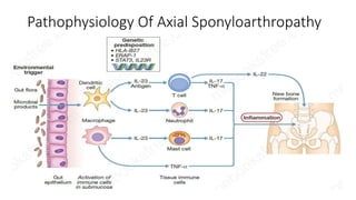
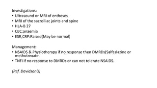
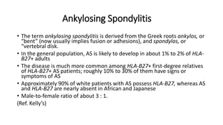
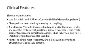
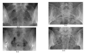
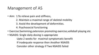
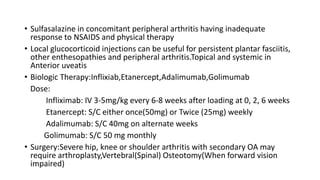
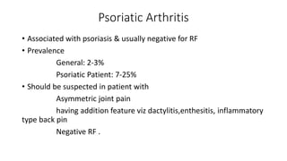
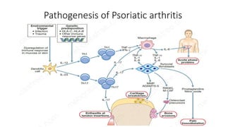
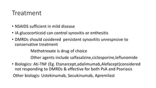
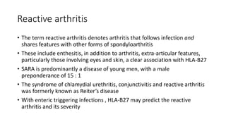
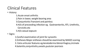
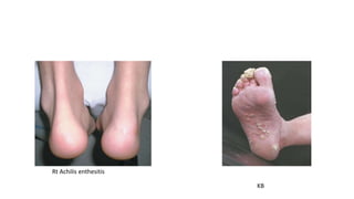
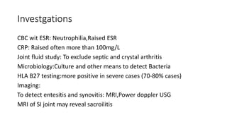
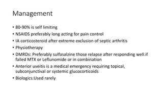
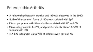
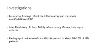
Spondyloarthropathy
Dr. Md. Rafiqul islam
Registrar(Medicine)
M Abdur Rahim Medical College Hospital,
Dinajpur.
Defination & Types
Spondyloarthropathies (SpAs) comprise a group of related
inflammatory musculoskeletal diseases that show overlap in their
clinical features and have a shared immunogenetic association with HLA-B27.
It Includes:
• axial spondyloarthritis
• ankylosing spondylitis
• reactive arthritis
• psoriatic arthritis
• arthritis with inflammatory bowel disease (enteropathic spondyloarthritis).
(Ref. Davidson’s)
Clinical Characteristics of Spondyloarthritis
• Typical pattern of arthritis—predominantly of lower limb, asymmetric
• Absence of rheumatoid factor
• Absence of subcutaneous nodules and other extra-articular features of
rheumatoid arthritis
• Overlapping extra-articular features characteristic of the group
(e.g.anterior uveitis)
• Significant familial aggregation
• Association with HLA-B27
• Enthesitis (e.g. gluteus medius insertion, plantar fascia origin)
(Ref. kelley’s & Davidson’s)
Aspects of Inflammatory Back Pain in
Ankylosing Spondylitis and Axial Spondyloarthritis
• Onset of complaints before age 45
• Duration of symptoms more than 3 months’ (chronic pain)
• Located at the lower back
• Alternating buttock pain
• Awaking due to back pain during the second half of the night
• Morning stiffness for at least 30 min
• Insidious onset of complaints
• Improvement with exercises
• No improvement of back pain with rest
• Improvement with use of nonsteroidal agents
(Ref. Kelley’s)
ASAS Criteria for axial Spondyloarthritis
ASAS Criteria for Peripheral Spondyloarthritis
Arthritis or Enthesitis
Plus
One or more of the following:
a.Uveitis
b.Psoriasis
c.Crohn’s disease or ulcerative colitis
OR two or more of the following:
a.Arthritis
b.Enthesitis
c.Dactylitis
d.Inflammatory back pain ever
e.Family history for SpA
Axial Spndyloarthropathy
• Includes classical ankylosing Spondylitis(AS) & Axial Spondyloarthritis
• Inflammatory change in entire axial skeleton
• Structural alteration (Syndesmophytes,Ankylosis etc) latter
• Not all patient will develop AS from axSpA
(Ref. Davidson’s)
Ref. Kelly’s
Pathophysiology Of Axial Sponyloarthropathy
Investigations:
• Ultrasound or MRI of entheses
• MRI of the sacroiliac joints and spine
• HLA-B 27
• CBC:anaemia
• ESR,CRP:Raised(May be normal)
Management:
• NSAIDS & Physiotherapy if no response then DMRDs(Salfaslazine or
methotrexate.
• TNFi if no response to DMRDs or can not tolerate NSAIDS.
(Ref. Davidson’s)
Ankylosing Spondylitis
• The term ankylosing spondylitis is derived from the Greek roots ankylos, or
“bent” (now usually implies fusion or adhesions), and spondylos, or
“vertebral disk.
• In the general population, AS is likely to develop in about 1% to 2% of HLA-
B27+ adults
• The disease is much more common among HLA-B27+ first-degree relatives
of HLA-B27+ AS patients; roughly 10% to 30% of them have signs or
symptoms of AS
• Approximately 90% of white patients with AS possess HLA-B27, whereas AS
and HLA-B27 are nearly absent in African and Japanese
• Male-to-female ratio of about 3 : 1.
(Ref. Kelly’s)
Clinical Features
Skeletal manifetation:
• Low Back Pain and Stiffness:Common(80% of General population)
• Chest pain: accentuated by sneezing or coughing
• Tentderness: These lesions are due to enthesitis. Common tender
sites are the costosternal junctions, spinous processes, iliac crests,
greater trochanters, ischial tuberosities, tibial tubercles, and heels
(Achilles tendinitis or plantar fasciitis)
• Joint: The girdle most frequently,Knee joint with intermittent
effusion.TMJ(about 10% patients.
Extraskeletal Manifestaion
• Fatigue,wt loss,low grade fever commonly
• Eye:Acute anterior uveitis or iridocyclitis most common extrarticular
manifetation(25%-35%),
• CVS: Ascending aortitis,aortic valve incompetence,conduction
abnormalities,cardiomegally,pericarditis,increased prevalence of MI.
• Pulmonary:Rare and late manifestaion.Slowly progressive fibrosis of
upper lobe of lungs,VC and TLC reduced but RV normal.
• Neurologic involvement: can be cuased by fracture, instability,
compression or inflammation.
• Renal invovement:IgA nephropathy,Microscopic hematuria and
proteinuria,amyloidosis(rare),Prostatis(80% men),urthritis
• GIT:Inflammatory bowel disease (up to 50% have IBD lesions)
Physical findings
Spinal mobility:
Limitation of movement of lumber sipne elicited by forward
flexion(schober’s Test),Hyperextension,lateral flexion.
• Chest expansion: Reduction below 5 cm in young
persons with an insidious onset of chronic,
inflammatory low back pain strongly suggests AS.
• Enthesitis:MASES provides a helpful list of 13
major entheses: first and fourth costochondral joints,
L + R; anterior and posterior iliac spines, L +R;
iliac crest, L + R; fifth lumbar spinous process; and
Achilles tendon insertion, L + R, which should be
checked.Missing from this list but readily
examined is the calcaneal plantar fascia insertion
• Examination of SI joint for Sacroilitis: Direct
Pressure on SI joint May elicit pain
• Posture:
Forward slope evidened by Flesche test (WOT)
Loss of lumber lordosis and kyphosis
Inestigations:
• Routine blood tests are not helpful.
• ESR,CRP raised upto 75%( but may be normal)
• Mild normocytic normochromic anemia may be present in 15% cases
• Autoantibodies, such as RF, ACPA and ANA, are negative.
Imaging
• Xray SI joint:
irregularity and loss of cortical margins
widening of the joint space
Subsequently sclerosis, joint space narrowing and fusion
• Lateral thoracolumbar spine X-rays : Findings may be
1. Suarring of verterae
2.Shiny corners(Romanus lesion)
3.Spondylodiscitis(Anderson lesion)
4. Syndesmophytes with partial or complete fusion
5.In advanced disease, ossification of the anterior longitudinal ligament and facet joint
fusion may also be visible
6.Bamboo spine appearance(Combination of mentioned feature)
Grading of Sacroiliitis: New York Criteria
• Grade 0: normal
• Grade 1: suspicious -some blurring of the joint margins
• Grade 2: Minimal abnormality with small areas of erosions or
sclerosis without alteration in the joint width
• Grade 3: Unequivocal abnormality - moderate or advanced sacroiliitis
consisting of erosion, sclerosis, widening, narrowing, and/or partial
joint fusion (ankylosis)
• Grade 4: Complete ankylosis
G-0
G-1
G-3
G-2
G-5
Syndesmophytes
Bamboo spine with Ankylosis SI
Squaring Romanus lesion
Anderson lesion
Romanus lesion MRI
Diagnosis
Modified New York, 1984
• Criteria
1. L ow back pain of at least 3 months’ duration improved by exercise and
not relieved by rest
2. Limitation of lumbar spine in sagittal and frontal planes
3. Chest expansion decreased relative to normal values for age and sex
4. Bilateral sacroiliitis grade 2 to 4
5. Unilateral sacroiliitis grade 3 or 4
• Definite Ankylosing Spondylitis
Unilateral grade 3 or 4, or bilateral grade 2 to 4 sacroiliitis and any other
clinical criterion
The patients with at least 5 points out of above 13 points have definite diagnosis of AS.
Management of AS
• Aim: 1.To relieve pain and stiffness,
2. Maintain a maximal range of skeletal mobility
3. Avoid the development of deformities.
4. Psychosocial functioning
• Exercise:Swimming,extension promoting exercise,volleball playing etc
• NSAIDS: Single daily dosing is appropriate
Upto 2 weeks for maximal symptomatic benefit
If inadequate response then Another NSAIDS
Consider other strategy if Two NSAIDS failed
• Sulfasalazine in concomitant peripheral arthritis having inadequate
response to NSAIDS and physical therapy
• Local glucocorticoid injections can be useful for persistent plantar fasciitis,
other enthesopathies and peripheral arthritis.Topical and systemic in
Anterior uveatis
• Biologic Therapy:Inflixiab,Etanercept,Adalimumab,Golimumab
Dose:
Infliximab: IV 3-5mg/kg every 6-8 weeks after loading at 0, 2, 6 weeks
Etanercept: S/C either once(50mg) or Twice (25mg) weekly
Adalimumab: S/C 40mg on alternate weeks
Golimumab: S/C 50 mg monthly
• Surgery:Severe hip, knee or shoulder arthritis with secondary OA may
require arthroplasty,Vertebral(Spinal) Osteotomy(When forward vision
impaired)
ASAS/EULAR recommendations for the
management of AS
Psoriatic Arthritis
• Associated with psoriasis & usually negative for RF
• Prevalence
General: 2-3%
Psoriatic Patient: 7-25%
• Should be suspected in patient with
Asymmetric joint pain
having addition feature viz dactylitis,enthesitis, inflammatory
type back pin
Negative RF .
Pathogenesis of Psoriatic arthritis
Clinical Features
• Plaque psoriasis is the most common skin phenotype
• Arthritis in established psoriasis usually,but aaprox, in 15% cases psoriasis
develops latter
• 5 clinical pattern of arthritis
1.Asymetric oligoarthritis
2.Symetric polyarthritis
3.Predominant DIP involvement
4.Predominant Spondyloarthritis
5.Destructive (mutilans) arthritis
• Dactylitis,Enthesitis,Nail pitting ,onycholysis,Subangual hyperkeratosis,
uveitis(bilateral)
Plaque Psoriasis
Plaque Psoriasis Scalp
Submammary & Umbelical
Guttate psoriasis Erythodermic psoriasis in trunk upper limb Pustular psoriasis in Foot
Koebner Phenomena in surgical Wound
Sausage Finger
Finger And Nail Change in PsA
Pitting
Pitting With Onycholysis
Onycholysis Pustule formation and nail loss
Dactylitis
H0rizontal line
Predominant DIPDIP Involvement
Toe dactylitis
Arthritis Mutilans
Investigations
• No diagnostic laboratory test are known.
• RF is negative usually (may be positive in 5%-16% cases)
• Anti-CCP: Usually neggative (may be positive in 5% cases)
• ESR,CRP:May be raised less commonly and lesser degree than RA
• Plain Radiograph:May be Normal.Features which may be found
1.Involvent of interphalngeal joint of fingers and toes
2.Features of bony erosion and resorption(Pencil-in-cup
deformity)
3.Joint space narrowinng and involvement of entheseal sites
• MSUS:Features include
Entheseal Thickening
Hypoechoic change
Increased vascularity(Power doppler)
Tenosynovitis
Bony erosion or enthesophyte formation
• MRI:Based on entheseal-realated bone marrow edema
• CT: Superseded by MRI
Imaging in Psoriatic arthritis
3rd digit arthritis with new bone formation
Bone Scan in same patient Right sided sacroilitis
Arthritis mutilans Pencil-in-cup deformity
MRI in Psoriasis
Tlonavicular disease with bone edema
Synovial enhancement and suprapatellar effusion
SI
DIAGNOSIS
The CASPAR criteria for psoriatic arthritis
Inflammatory articular disease (joint, spine or enthesis) with ≥ 3 points from
the following (1 point each unless stated):
• Current psoriasis (scores 2 points)
• History of psoriasis in first- or second-degree relative
• Psoriatic nail dystrophy
• Negative IgM rheumatoid factor
• Current dactylitis
• History of dactylitis (Recorded by Rheumatologist)
• Juxta-articular new bone formation: ill defined ossification near joint
margin on plain xray of hand or foot
Treatment
• NSAIDS sufficient in mild disease
• IA glucocorticoid can control synovitis or enthesitis
• DMRDs should cosidered persistent synovitits unrespnsive to
conservative treatment
Methotrexate is drug of choice
Other agents include salfasalzine,ciclosporine,leflunomide
• Biologics: Ati-TNF (Eg. Etanarcept,adalimumab,Alefacept)considered
not responding to DMRDs & effective for both PsA and Psoriasis
Other biologis: Ustekinumab, Secukinumab, Apremilast
Reactive arthritis
• The term reactive arthritis denotes arthritis that follows infection and
shares features with other forms of spondyloarthritis
• These include enthesitis, in addition to arthritis, extra-articular features,
particularly those involving eyes and skin, a clear association with HLA-B27
• SARA is predominantly a disease of young men, with a male
preponderance of 15 : 1
• The syndrome of chlamydial urethritis, conjunctivitis and reactive arthritis
was formerly known as Reiter’s disease
• With enteric triggering infections , HLA-B27 may predict the reactive
arthritis and its severity
Organisms Associated with Reactive Arthritis
• Common
Gastrointestinal Pathogens
Salmonella species
Campylobacter jejuni and Campylobacter coli
Yersinia enterocolitica and Yersinia pseudotuberculosis
Shigella flexneri; less commonly, Shigella sonnei or Shigella dysenteriae
Clostridium difficile
Genitourinary Pathogens
Chlamydia trachomatis
?Mycoplasma species
Respiratory Pathogens
Chlamydia pneumoniae
• Reported
Mycobacterium bovis bacillus Calmette-Guérin
Enterotoxigenic Escherichia coli and many others in small numbers of case reports
Clinical Features
• History:
1.Acute onset arthritis
2.Pain in lower, weight bearing area
3.Conjuntivitis:Transient and painless
4.H/o of preceeding infection eg. Gastroenteritis, RTI, Urethritis,
Cervicitis etc
5.H/o sexual expsure
• Signs:
1.Careful examination of joint for synovitis
2.Entheses:Major entheses should be examined by MASES scoring
3. Extra articular features eg.keratoderma blenorrhagica,circinate
4.balanitis,conjuntivitis,uveatis,pustular psoriasis
KB
Rt Achilis enthesitis
Investgations
CBC wit ESR: Neutrophilia,Raised ESR
CRP: Raised often more than 100mg/L
Joint fluid study: To exclude septic and crystal arthritis
Microbiology:Culture and other means to detect Bacteria
HLA B27 testing:more positive in severe cases (70-80% cases)
Imaging:
To detect entesitis and synovitis: MRI,Power doppler USG
MRI of SI joint may reveal sacroilitis
Management
• 80-90% is self limiting
• NSAIDS preferably long acting for pain control
• IA corticosteroid after extreme exclusion of septic arthritis
• Physiotherapy
• DMRDs: Preferably sulfasalzine those relapse after responding well.if
failed MTX or Leflunomide or in combination
• Anterior uveitis is a medical emergency requiring topical,
subconjunctival or systemic glucocorticoids
• Biologics:Used rarely
Enteropathic Arthritis
• A relationship between arthritis and IBD was observed in the 1930s
• Both of the common forms of IBD are associated with SpA
• AS and peripheral arthritis are both associated with UC and CD
• AS was diagnosed in 1–10%, and peripheral arthritis in 10–50% of
patients with IBD
• HLA-B27 is found in up to 70% of patients with IBD and AS
Clinical Features
• AS associated with IBD is clinically indistinguishable from idiopathic AS.
• Peripheral arthritis may also begin before onset of overt bowel disease
• The patterns of joint involvement are similar in UC and CD.
• In general, erosions and deformities are infrequent in IBD-associated
peripheral arthritis
• Dactylitis and enthesopathy are occasionally found
• Upto 20% of IBD patients with SpA, a comparable percentage have
arthralgias or fibromyalgia symptoms.
• Other extraintestinal manifestations of IBD including uveitis, pyoderma
gangrenosum, erythema nodosum, and finger clubbing
Pyoderma Gangrenosum Erythema nodosum
Investigations
• Laboratory findings reflect the inflammatory and metabolic
manifestations of IBD
• Joint Fluid study: At least Mildly Inflammatory.Mus exclude septic
arthritis.
• Radiographic evidence of sacroiliitis is present in about 20–25% of IBD
patients
Mangement
• Arthritis improves with effective treatment of the bowel disease.
• NSAIDS should be avoided.
• judicious use of glucocorticoids, sulfasalazine and methotrexate may be
considered
• Liason is necceary between gastroenterologist & Rheumatolgist for
appropriate thearpy.
• Biologics:infliximab results in remission of gastrointestinal manifestations
in close to 60% of patients
• adalimumab, sme action as infliximab
• etanercept, which is not beneficial for intestinal manifestation, seems to be
effective for spinal and peripheral involvement(better to avoid)
Thank You

Spondyloarthropathy:An update

  • 1.
    Spondyloarthropathy Dr. Md. Rafiqulislam Registrar(Medicine) M Abdur Rahim Medical College Hospital, Dinajpur.
  • 2.
    Defination & Types Spondyloarthropathies(SpAs) comprise a group of related inflammatory musculoskeletal diseases that show overlap in their clinical features and have a shared immunogenetic association with HLA-B27. It Includes: • axial spondyloarthritis • ankylosing spondylitis • reactive arthritis • psoriatic arthritis • arthritis with inflammatory bowel disease (enteropathic spondyloarthritis). (Ref. Davidson’s)
  • 3.
    Clinical Characteristics ofSpondyloarthritis • Typical pattern of arthritis—predominantly of lower limb, asymmetric • Absence of rheumatoid factor • Absence of subcutaneous nodules and other extra-articular features of rheumatoid arthritis • Overlapping extra-articular features characteristic of the group (e.g.anterior uveitis) • Significant familial aggregation • Association with HLA-B27 • Enthesitis (e.g. gluteus medius insertion, plantar fascia origin) (Ref. kelley’s & Davidson’s)
  • 4.
    Aspects of InflammatoryBack Pain in Ankylosing Spondylitis and Axial Spondyloarthritis • Onset of complaints before age 45 • Duration of symptoms more than 3 months’ (chronic pain) • Located at the lower back • Alternating buttock pain • Awaking due to back pain during the second half of the night • Morning stiffness for at least 30 min • Insidious onset of complaints • Improvement with exercises • No improvement of back pain with rest • Improvement with use of nonsteroidal agents (Ref. Kelley’s)
  • 6.
    ASAS Criteria foraxial Spondyloarthritis
  • 7.
    ASAS Criteria forPeripheral Spondyloarthritis Arthritis or Enthesitis Plus One or more of the following: a.Uveitis b.Psoriasis c.Crohn’s disease or ulcerative colitis OR two or more of the following: a.Arthritis b.Enthesitis c.Dactylitis d.Inflammatory back pain ever e.Family history for SpA
  • 8.
    Axial Spndyloarthropathy • Includesclassical ankylosing Spondylitis(AS) & Axial Spondyloarthritis • Inflammatory change in entire axial skeleton • Structural alteration (Syndesmophytes,Ankylosis etc) latter • Not all patient will develop AS from axSpA (Ref. Davidson’s)
  • 9.
  • 10.
    Pathophysiology Of AxialSponyloarthropathy
  • 11.
    Investigations: • Ultrasound orMRI of entheses • MRI of the sacroiliac joints and spine • HLA-B 27 • CBC:anaemia • ESR,CRP:Raised(May be normal) Management: • NSAIDS & Physiotherapy if no response then DMRDs(Salfaslazine or methotrexate. • TNFi if no response to DMRDs or can not tolerate NSAIDS. (Ref. Davidson’s)
  • 13.
    Ankylosing Spondylitis • Theterm ankylosing spondylitis is derived from the Greek roots ankylos, or “bent” (now usually implies fusion or adhesions), and spondylos, or “vertebral disk. • In the general population, AS is likely to develop in about 1% to 2% of HLA- B27+ adults • The disease is much more common among HLA-B27+ first-degree relatives of HLA-B27+ AS patients; roughly 10% to 30% of them have signs or symptoms of AS • Approximately 90% of white patients with AS possess HLA-B27, whereas AS and HLA-B27 are nearly absent in African and Japanese • Male-to-female ratio of about 3 : 1. (Ref. Kelly’s)
  • 14.
    Clinical Features Skeletal manifetation: •Low Back Pain and Stiffness:Common(80% of General population) • Chest pain: accentuated by sneezing or coughing • Tentderness: These lesions are due to enthesitis. Common tender sites are the costosternal junctions, spinous processes, iliac crests, greater trochanters, ischial tuberosities, tibial tubercles, and heels (Achilles tendinitis or plantar fasciitis) • Joint: The girdle most frequently,Knee joint with intermittent effusion.TMJ(about 10% patients.
  • 15.
    Extraskeletal Manifestaion • Fatigue,wtloss,low grade fever commonly • Eye:Acute anterior uveitis or iridocyclitis most common extrarticular manifetation(25%-35%), • CVS: Ascending aortitis,aortic valve incompetence,conduction abnormalities,cardiomegally,pericarditis,increased prevalence of MI. • Pulmonary:Rare and late manifestaion.Slowly progressive fibrosis of upper lobe of lungs,VC and TLC reduced but RV normal. • Neurologic involvement: can be cuased by fracture, instability, compression or inflammation. • Renal invovement:IgA nephropathy,Microscopic hematuria and proteinuria,amyloidosis(rare),Prostatis(80% men),urthritis • GIT:Inflammatory bowel disease (up to 50% have IBD lesions)
  • 16.
    Physical findings Spinal mobility: Limitationof movement of lumber sipne elicited by forward flexion(schober’s Test),Hyperextension,lateral flexion.
  • 17.
    • Chest expansion:Reduction below 5 cm in young persons with an insidious onset of chronic, inflammatory low back pain strongly suggests AS. • Enthesitis:MASES provides a helpful list of 13 major entheses: first and fourth costochondral joints, L + R; anterior and posterior iliac spines, L +R; iliac crest, L + R; fifth lumbar spinous process; and Achilles tendon insertion, L + R, which should be checked.Missing from this list but readily examined is the calcaneal plantar fascia insertion
  • 18.
    • Examination ofSI joint for Sacroilitis: Direct Pressure on SI joint May elicit pain • Posture: Forward slope evidened by Flesche test (WOT) Loss of lumber lordosis and kyphosis
  • 19.
    Inestigations: • Routine bloodtests are not helpful. • ESR,CRP raised upto 75%( but may be normal) • Mild normocytic normochromic anemia may be present in 15% cases • Autoantibodies, such as RF, ACPA and ANA, are negative.
  • 20.
    Imaging • Xray SIjoint: irregularity and loss of cortical margins widening of the joint space Subsequently sclerosis, joint space narrowing and fusion • Lateral thoracolumbar spine X-rays : Findings may be 1. Suarring of verterae 2.Shiny corners(Romanus lesion) 3.Spondylodiscitis(Anderson lesion) 4. Syndesmophytes with partial or complete fusion 5.In advanced disease, ossification of the anterior longitudinal ligament and facet joint fusion may also be visible 6.Bamboo spine appearance(Combination of mentioned feature)
  • 21.
    Grading of Sacroiliitis:New York Criteria • Grade 0: normal • Grade 1: suspicious -some blurring of the joint margins • Grade 2: Minimal abnormality with small areas of erosions or sclerosis without alteration in the joint width • Grade 3: Unequivocal abnormality - moderate or advanced sacroiliitis consisting of erosion, sclerosis, widening, narrowing, and/or partial joint fusion (ankylosis) • Grade 4: Complete ankylosis
  • 22.
  • 23.
  • 24.
    Syndesmophytes Bamboo spine withAnkylosis SI Squaring Romanus lesion Anderson lesion Romanus lesion MRI
  • 25.
    Diagnosis Modified New York,1984 • Criteria 1. L ow back pain of at least 3 months’ duration improved by exercise and not relieved by rest 2. Limitation of lumbar spine in sagittal and frontal planes 3. Chest expansion decreased relative to normal values for age and sex 4. Bilateral sacroiliitis grade 2 to 4 5. Unilateral sacroiliitis grade 3 or 4 • Definite Ankylosing Spondylitis Unilateral grade 3 or 4, or bilateral grade 2 to 4 sacroiliitis and any other clinical criterion
  • 26.
    The patients withat least 5 points out of above 13 points have definite diagnosis of AS.
  • 27.
    Management of AS •Aim: 1.To relieve pain and stiffness, 2. Maintain a maximal range of skeletal mobility 3. Avoid the development of deformities. 4. Psychosocial functioning • Exercise:Swimming,extension promoting exercise,volleball playing etc • NSAIDS: Single daily dosing is appropriate Upto 2 weeks for maximal symptomatic benefit If inadequate response then Another NSAIDS Consider other strategy if Two NSAIDS failed
  • 28.
    • Sulfasalazine inconcomitant peripheral arthritis having inadequate response to NSAIDS and physical therapy • Local glucocorticoid injections can be useful for persistent plantar fasciitis, other enthesopathies and peripheral arthritis.Topical and systemic in Anterior uveatis • Biologic Therapy:Inflixiab,Etanercept,Adalimumab,Golimumab Dose: Infliximab: IV 3-5mg/kg every 6-8 weeks after loading at 0, 2, 6 weeks Etanercept: S/C either once(50mg) or Twice (25mg) weekly Adalimumab: S/C 40mg on alternate weeks Golimumab: S/C 50 mg monthly • Surgery:Severe hip, knee or shoulder arthritis with secondary OA may require arthroplasty,Vertebral(Spinal) Osteotomy(When forward vision impaired)
  • 29.
    ASAS/EULAR recommendations forthe management of AS
  • 30.
    Psoriatic Arthritis • Associatedwith psoriasis & usually negative for RF • Prevalence General: 2-3% Psoriatic Patient: 7-25% • Should be suspected in patient with Asymmetric joint pain having addition feature viz dactylitis,enthesitis, inflammatory type back pin Negative RF .
  • 31.
  • 32.
    Clinical Features • Plaquepsoriasis is the most common skin phenotype • Arthritis in established psoriasis usually,but aaprox, in 15% cases psoriasis develops latter • 5 clinical pattern of arthritis 1.Asymetric oligoarthritis 2.Symetric polyarthritis 3.Predominant DIP involvement 4.Predominant Spondyloarthritis 5.Destructive (mutilans) arthritis • Dactylitis,Enthesitis,Nail pitting ,onycholysis,Subangual hyperkeratosis, uveitis(bilateral)
  • 34.
    Plaque Psoriasis Plaque PsoriasisScalp Submammary & Umbelical Guttate psoriasis Erythodermic psoriasis in trunk upper limb Pustular psoriasis in Foot
  • 35.
    Koebner Phenomena insurgical Wound Sausage Finger
  • 36.
    Finger And NailChange in PsA Pitting Pitting With Onycholysis Onycholysis Pustule formation and nail loss Dactylitis
  • 37.
    H0rizontal line Predominant DIPDIPInvolvement Toe dactylitis Arthritis Mutilans
  • 38.
    Investigations • No diagnosticlaboratory test are known. • RF is negative usually (may be positive in 5%-16% cases) • Anti-CCP: Usually neggative (may be positive in 5% cases) • ESR,CRP:May be raised less commonly and lesser degree than RA • Plain Radiograph:May be Normal.Features which may be found 1.Involvent of interphalngeal joint of fingers and toes 2.Features of bony erosion and resorption(Pencil-in-cup deformity) 3.Joint space narrowinng and involvement of entheseal sites
  • 39.
    • MSUS:Features include EnthesealThickening Hypoechoic change Increased vascularity(Power doppler) Tenosynovitis Bony erosion or enthesophyte formation • MRI:Based on entheseal-realated bone marrow edema • CT: Superseded by MRI
  • 40.
    Imaging in Psoriaticarthritis 3rd digit arthritis with new bone formation Bone Scan in same patient Right sided sacroilitis Arthritis mutilans Pencil-in-cup deformity
  • 41.
    MRI in Psoriasis Tlonaviculardisease with bone edema Synovial enhancement and suprapatellar effusion SI
  • 42.
    DIAGNOSIS The CASPAR criteriafor psoriatic arthritis Inflammatory articular disease (joint, spine or enthesis) with ≥ 3 points from the following (1 point each unless stated): • Current psoriasis (scores 2 points) • History of psoriasis in first- or second-degree relative • Psoriatic nail dystrophy • Negative IgM rheumatoid factor • Current dactylitis • History of dactylitis (Recorded by Rheumatologist) • Juxta-articular new bone formation: ill defined ossification near joint margin on plain xray of hand or foot
  • 43.
    Treatment • NSAIDS sufficientin mild disease • IA glucocorticoid can control synovitis or enthesitis • DMRDs should cosidered persistent synovitits unrespnsive to conservative treatment Methotrexate is drug of choice Other agents include salfasalzine,ciclosporine,leflunomide • Biologics: Ati-TNF (Eg. Etanarcept,adalimumab,Alefacept)considered not responding to DMRDs & effective for both PsA and Psoriasis Other biologis: Ustekinumab, Secukinumab, Apremilast
  • 44.
    Reactive arthritis • Theterm reactive arthritis denotes arthritis that follows infection and shares features with other forms of spondyloarthritis • These include enthesitis, in addition to arthritis, extra-articular features, particularly those involving eyes and skin, a clear association with HLA-B27 • SARA is predominantly a disease of young men, with a male preponderance of 15 : 1 • The syndrome of chlamydial urethritis, conjunctivitis and reactive arthritis was formerly known as Reiter’s disease • With enteric triggering infections , HLA-B27 may predict the reactive arthritis and its severity
  • 45.
    Organisms Associated withReactive Arthritis • Common Gastrointestinal Pathogens Salmonella species Campylobacter jejuni and Campylobacter coli Yersinia enterocolitica and Yersinia pseudotuberculosis Shigella flexneri; less commonly, Shigella sonnei or Shigella dysenteriae Clostridium difficile Genitourinary Pathogens Chlamydia trachomatis ?Mycoplasma species Respiratory Pathogens Chlamydia pneumoniae • Reported Mycobacterium bovis bacillus Calmette-Guérin Enterotoxigenic Escherichia coli and many others in small numbers of case reports
  • 46.
    Clinical Features • History: 1.Acuteonset arthritis 2.Pain in lower, weight bearing area 3.Conjuntivitis:Transient and painless 4.H/o of preceeding infection eg. Gastroenteritis, RTI, Urethritis, Cervicitis etc 5.H/o sexual expsure • Signs: 1.Careful examination of joint for synovitis 2.Entheses:Major entheses should be examined by MASES scoring 3. Extra articular features eg.keratoderma blenorrhagica,circinate 4.balanitis,conjuntivitis,uveatis,pustular psoriasis
  • 47.
  • 48.
    Investgations CBC wit ESR:Neutrophilia,Raised ESR CRP: Raised often more than 100mg/L Joint fluid study: To exclude septic and crystal arthritis Microbiology:Culture and other means to detect Bacteria HLA B27 testing:more positive in severe cases (70-80% cases) Imaging: To detect entesitis and synovitis: MRI,Power doppler USG MRI of SI joint may reveal sacroilitis
  • 49.
    Management • 80-90% isself limiting • NSAIDS preferably long acting for pain control • IA corticosteroid after extreme exclusion of septic arthritis • Physiotherapy • DMRDs: Preferably sulfasalzine those relapse after responding well.if failed MTX or Leflunomide or in combination • Anterior uveitis is a medical emergency requiring topical, subconjunctival or systemic glucocorticoids • Biologics:Used rarely
  • 50.
    Enteropathic Arthritis • Arelationship between arthritis and IBD was observed in the 1930s • Both of the common forms of IBD are associated with SpA • AS and peripheral arthritis are both associated with UC and CD • AS was diagnosed in 1–10%, and peripheral arthritis in 10–50% of patients with IBD • HLA-B27 is found in up to 70% of patients with IBD and AS
  • 51.
    Clinical Features • ASassociated with IBD is clinically indistinguishable from idiopathic AS. • Peripheral arthritis may also begin before onset of overt bowel disease • The patterns of joint involvement are similar in UC and CD. • In general, erosions and deformities are infrequent in IBD-associated peripheral arthritis • Dactylitis and enthesopathy are occasionally found • Upto 20% of IBD patients with SpA, a comparable percentage have arthralgias or fibromyalgia symptoms. • Other extraintestinal manifestations of IBD including uveitis, pyoderma gangrenosum, erythema nodosum, and finger clubbing
  • 52.
  • 53.
    Investigations • Laboratory findingsreflect the inflammatory and metabolic manifestations of IBD • Joint Fluid study: At least Mildly Inflammatory.Mus exclude septic arthritis. • Radiographic evidence of sacroiliitis is present in about 20–25% of IBD patients
  • 54.
    Mangement • Arthritis improveswith effective treatment of the bowel disease. • NSAIDS should be avoided. • judicious use of glucocorticoids, sulfasalazine and methotrexate may be considered • Liason is necceary between gastroenterologist & Rheumatolgist for appropriate thearpy. • Biologics:infliximab results in remission of gastrointestinal manifestations in close to 60% of patients • adalimumab, sme action as infliximab • etanercept, which is not beneficial for intestinal manifestation, seems to be effective for spinal and peripheral involvement(better to avoid)
  • 55.