Downloaded 691 times


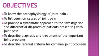


![ Inflammatory arthritis is characterized by inflammation affecting
joint structures, such as the synovium, synovial cavity, and
enthuses.
No inflammatory arthritis is joint disease resulting primarily from
alterations in the structure or mechanics of the joint.
Arthralgia is characterized by joint tenderness, but abnormalities
of the joint cannot be identified.
Such patients may have a syndrome of altered pain sensation (eg,
fibromyalgia) or an early rheumatic syndrome whose clinical signs
are not yet apparent or are too subtle for detection (eg, arthralgia
of systemic lupus erythematosus [SLE]).](https://image.slidesharecdn.com/seminarapproachtojointpain-180517160335/85/Seminar-approach-to-joint-pain-18-320.jpg)


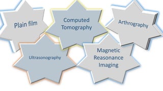
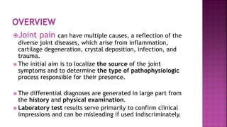
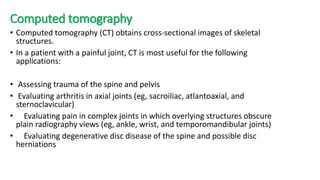
![Plain films
• Plain radiography is the least expensive imaging
modality and is most useful for clarifying the nature
of joint abnormalities already noted during the
physical examination.
• The appearance of joints on plain radiographs is
often distinctive for various forms of arthritis though
these characteristic changes may not be apparent
early in the disease course.
• Plain radiographs are also useful for monitoring the
progression of chronic arthritides (eg:osteoarthritis
and rheumatoid arthritis [RA]).](https://image.slidesharecdn.com/seminarapproachtojointpain-180517160335/85/Seminar-approach-to-joint-pain-56-320.jpg)


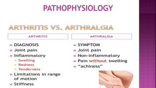
![:indicationSlow-Acting antirheumatica drugs
1-persistent synovitis more than 6 wk.
2-sever extra-articular disease [ vasculitise ,scleritise renal
involvment].
3-steroid-sparing effect [polymyalgia rheumatica resistant to low-dose
Corticosteroid].
4- Inflammatory myositis.](https://image.slidesharecdn.com/seminarapproachtojointpain-180517160335/85/Seminar-approach-to-joint-pain-86-320.jpg)


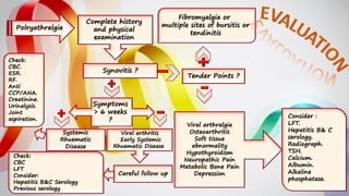
This document provides an overview of the pathophysiology, diagnosis, and treatment of joint pain. It discusses the various causes of joint pain including inflammation, cartilage degeneration, crystal deposition, infection, and trauma. The document outlines the approach to evaluating a patient with joint pain, including obtaining a thorough history regarding symptoms, physical examination of the joints, and initial laboratory tests. Common differential diagnoses are also reviewed depending on characteristics such as number of involved joints, symmetry, and distribution of pain.

![[Int. med] approach to joint pain from SIMS Lahore](https://cdn.slidesharecdn.com/ss_thumbnails/064ftjyatacrqjgurlzo-signature-b01672da1ecf8b94befb115319b147a085de390b8cb403389bce6c156545fbb5-poli-150815171713-lva1-app6891-thumbnail.jpg?width=640&height=640&fit=bounds)