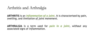
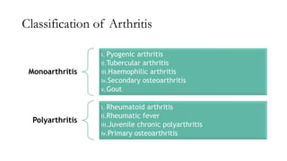
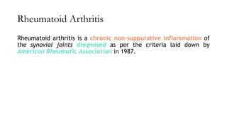
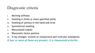
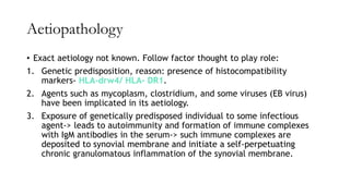
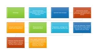
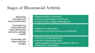
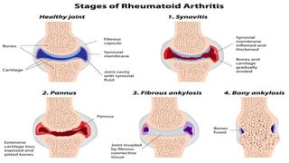
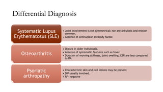
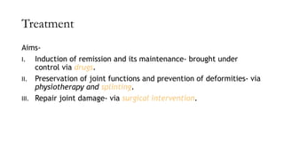
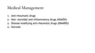
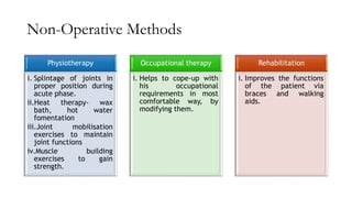
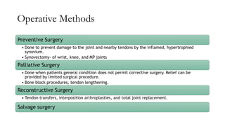
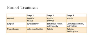
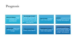
Rheumatoid Arthritis
Presented by: Rutvi Raualji (MPT)
Contents
❖ Arthritis and Arthralgia
❖ Classification of arthritis
❖ Rheumatoid arthritis
❖ Diagnostic criteria
❖ Aetiopathology
❖ Stages of RA
❖ Dignosis
❖ Differntial Diagnosis
❖ Treatment
Arthritis and Arthralgia
ARTHRITIS is an inflammation of a joint. It is characterised by pain,
swelling, and limitation of joint movement.
ARTHRALGIA is a term used for pain in a joint, without any
associated signs of inflammation.
Classification of Arthritis
Monoarthritis
i. Pyogenic arthritis
ii.Tubercular arthritis
iii.Haemophilic arthritis
iv.Secondary osteoarthritis
v.Gout
Polyarthritis
i. Rheumatoid arthritis
ii.Rheumatic fever
iii.Juvenile chronic polyarthritis
iv.Primary osteoarthritis
Rheumatoid Arthritis
Rheumatoid arthritis is a chronic non-suppurative inflammation of
the synovial joints diagnosed as per the criteria laid down by
American Rheumatic Association in 1987.
Diagnostic criteria
i. Morning stiffness
ii. Swelling in three or more specified joints
iii. Swelling of joint(s) in the hand and wrist
iv. Symmetrical swelling
v. Rheumatoid nodule
vi. Rheumatic factor positive
vii. X-ray changes- erosion or unequivocal peri-articular osteopenia
If four or more of these are present, it is rheumatoid arthritis.
Aetiopathology
• Exact aetiology not known. Follow factor thought to play role:
1. Genetic predisposition, reason: presence of histocompatibility
markers- HLA-drw4/ HLA- DR1.
2. Agents such as mycoplasm, clostridium, and some viruses (EB virus)
have been implicated in its aetiology.
3. Exposure of genetically predisposed individual to some infectious
agent-> leads to autoimmunity and formation of immune complexes
with IgM antibodies in the serum-> such immune complexes are
deposited to synovial membrane and initiate a self-perpetuating
chronic granulomatous inflammation of the synovial membrane.
Pathology:
Synovium becomes
oedematous, filled with fibrin
exudates and cellular
infiltrates.
Synovial fluid increases
Inflammation persists,
synovium hypertrophies and
surrounding periphery forms a
pannus
Articular surface loses its
smooth shiny appearance.
Pannus burrows in
subchondral bone.
Progression- cartilage worn
off and surface becomes raw.
Joints get deformed, initially
due to severe muscle spasm,
and later due to fibrosis of
capsules and joint tissues.
Adhesions develop between
apposing layers of pannus,
leading to fibrous ankylosis,
and later bony ankylosis.
Joint capsule distended due
to hypertrophied synovium
and synovial fluid, ligaments
supporting joints are
stretched- resulting in
subluxation of joint.
• Osteoporosis develop in bones
adjacent to the affected joint.
• Peri-articular structures becomes
oedematous.
• Course of disease: varies from
patient to patient; mild which
totally recovers to severe resulting
into deformities.
Stages of Rheumatoid Arthritis
Potentially
reversible soft
tissue proliferation
• Disease limited to synovium.
• Synovium hypertrophy and effusion.
• No destructive changes radiologically.
Controlled but
irreversible soft
tissue destruction
and early cartilage
erosion
• Reduction in joint space.
• Outlines of articular surface are maintained.
Irreversible soft
tissue and bony
changes
• Articular cartilage destroyed.
• Erosion of subchondral bone.
• Joint ankylosed in deformed position- later on
dislocated or subluxated.
Diagnosis
CLINICAL PRESENTATION
✓ Age: 20 to 50 years
✓ Female: Male ratio- 3:1
✓ Presentation- acute, symmetrical polyarthritis
✓ Pain and stiffness in multiple joints, particularly in morning (beginning of
the disease)
✓ Commonly affected joints: MP joints of hands, PIP of fingers, wrist,
knees, elbow, ankles.
✓ Less common: Hip, temporomandibular joint
✓ Uncommon: atlanto-axial and facet joints of cervical spine
✓ In some cases, onset may be with fever or visceral manifestations such as
pneumonitis, rheumatoid nodules, etc.
EXAMINATION:
✓ Swollen boggy joints due to intra-articular effusion, synovial hypertrophy
and oedema in peri-articular structures.
✓ Severe muscle spasm
✓ Limited range of motion
✓ Subluxation or dislocation of joints.
Deformities
a) Hand:
Ulnar drift of the hand, Boutonniere
deformity, Swan neck deformity.
b) Elbow:
Flexion deformity
c) Knee:
Early- flexion deformity, later-triple
subluxation
d) Ankle:
Equinus deformity
e) Foot
Hallux valgus, hammer toe
Extra-articular Manifestations
i. Vasculitis:
Digital arteritis, Raynaud’s
phenomenon, fever, skin lesions,
chronic leg ulcers, peripheral neuritis
ii. Rheumatoid nodules:
Olecranon, dorsal surface of forearm,
tendo-achilles
iii. Serositis:
Lung and pleura- pleurisy, honey comb
lung; Heart- pericarditis,
cardiomyopathy; Eye- iridocyclitis;
Nervous system- peripheral neuritis,
CTS
iv. Others:
Fever, Amyloidosis, Sjogren’s
syndrome
INVESTIGATIONS
I. Radiological examination: consist of X-rays of affected joints. Findings:
Reduced joint space
Erosion of articular cysts
Subchondral cysts
Juxta-articular rarefaction
Soft tissue shadow at the level of joint (joint effusion or synovial
hypertrophy)
Deformities of hand and fingers
II. Blood:
Elevated ESR
Low haemoglobin value
Rheumatoid factor- auto antibody directed against Fc fragment of
immunoglobulin G (IgG) [Can be checked via Latex fixation test or Rose-
Waaler test]
Note: all the patients with RF do not have RA and vice versa
III. Synovial fluid examination
IV. Synovial biopsy
Differential Diagnosis
• Joint involvement is not symmetrical; nor are ankylosis and erosion
common.
• Absence of antinuclear antibody factor.
Systematic Lupus
Erythematosus (SLE)
• Occurs in older individuals.
• Absence of systematic features such as fever.
• Duration of morning stiffness, joint swelling, ESR are less compared
to RA.
Osteoarthritis
• Characteristic skin and nail lesions may be present
• DIP usually involved.
• RF- negative
Psoriatic
arthropathy
Treatment
Aims-
I. Induction of remission and its maintenance- brought under
control via drugs.
II. Preservation of joint functions and prevention of deformities- via
physiotherapy and splinting.
III. Repair joint damage- via surgical intervention.
Medical Management
1) Anti-rheumatic drugs
2) Non- steroidal anti-inflammatory drugs (NSAIDS)
3) Disease modifying anti-rheumatic drugs (DMARDS)
4) Steroids
Non-Operative Methods
Physiotherapy
i. Splintage of joints in
proper position during
acute phase.
ii.Heat therapy- wax
bath, hot water
fomentation
iii.Joint mobilisation
exercises to maintain
joint functions
iv.Muscle building
exercises to gain
strength.
Occupational therapy
i. Helps to cope-up with
his occupational
requirements in most
comfortable way, by
modifying them.
Rehabilitation
i. Improves the functions
of the patient via
braces and walking
aids.
Operative Methods
Preventive Surgery
• Done to prevent damage to the joint and nearby tendons by the inflamed, hypertrophied
synovium.
• Synovectomy- of wrist, knee, and MP joints
Palliative Surgery
• Done when patients general condition does not permit corrective surgery. Relief can be
provided by limited surgical procedure.
• Bone block procedures, tendon lengthening.
Reconstructive Surgery
• Tendon transfers, interposition arthroplasties, and total joint replacement.
Salvage surgery
Plan of Treatment
Stage 1 Stage 2 Stage 3
Medical DMARDs,
NSAIDs
NSAIDs,
DMARDs
NSAIDs
Surgical Synovectomy Soft tissue repair,
Arthroplasty
Joint replacement,
Arthrodesis
Physiotherapy Joint mobilization Splints Splints,
Walking aids
Prognosis
Nature of History: Varies
from fulminant to more
remissions and
exacerbations.
Sex and age of onset:
Women in child-bearing
age with predominant UE
involvement; Males under
age of 30, with sparing UE
– bad prognosis.
Type of onset: Insidious
onset- bad prognosis.
Anaemia: unresponsiveness
of anaemia to oral iron
therapy-bad prognosis
ESR and CRP: High levels
indicate more erosive
changes.
Rheumatoid Factor:
present- bad prognosis.
Radiological erosions:
Present within 2 years of
onset- bad prognosis.
Histopathological changes:
Synovial proliferation,
with increased number of
synovial cells with DR
antigen- bad prognosis.
Reference
Essential Orthopaedics- 5th Edition- Maheshwari and Mhaskar

Rheumatoid Arthritis.pdf

  • 1.
  • 2.
    Contents ❖ Arthritis andArthralgia ❖ Classification of arthritis ❖ Rheumatoid arthritis ❖ Diagnostic criteria ❖ Aetiopathology ❖ Stages of RA ❖ Dignosis ❖ Differntial Diagnosis ❖ Treatment
  • 3.
    Arthritis and Arthralgia ARTHRITISis an inflammation of a joint. It is characterised by pain, swelling, and limitation of joint movement. ARTHRALGIA is a term used for pain in a joint, without any associated signs of inflammation.
  • 4.
    Classification of Arthritis Monoarthritis i.Pyogenic arthritis ii.Tubercular arthritis iii.Haemophilic arthritis iv.Secondary osteoarthritis v.Gout Polyarthritis i. Rheumatoid arthritis ii.Rheumatic fever iii.Juvenile chronic polyarthritis iv.Primary osteoarthritis
  • 5.
    Rheumatoid Arthritis Rheumatoid arthritisis a chronic non-suppurative inflammation of the synovial joints diagnosed as per the criteria laid down by American Rheumatic Association in 1987.
  • 6.
    Diagnostic criteria i. Morningstiffness ii. Swelling in three or more specified joints iii. Swelling of joint(s) in the hand and wrist iv. Symmetrical swelling v. Rheumatoid nodule vi. Rheumatic factor positive vii. X-ray changes- erosion or unequivocal peri-articular osteopenia If four or more of these are present, it is rheumatoid arthritis.
  • 7.
    Aetiopathology • Exact aetiologynot known. Follow factor thought to play role: 1. Genetic predisposition, reason: presence of histocompatibility markers- HLA-drw4/ HLA- DR1. 2. Agents such as mycoplasm, clostridium, and some viruses (EB virus) have been implicated in its aetiology. 3. Exposure of genetically predisposed individual to some infectious agent-> leads to autoimmunity and formation of immune complexes with IgM antibodies in the serum-> such immune complexes are deposited to synovial membrane and initiate a self-perpetuating chronic granulomatous inflammation of the synovial membrane.
  • 8.
    Pathology: Synovium becomes oedematous, filledwith fibrin exudates and cellular infiltrates. Synovial fluid increases Inflammation persists, synovium hypertrophies and surrounding periphery forms a pannus Articular surface loses its smooth shiny appearance. Pannus burrows in subchondral bone. Progression- cartilage worn off and surface becomes raw. Joints get deformed, initially due to severe muscle spasm, and later due to fibrosis of capsules and joint tissues. Adhesions develop between apposing layers of pannus, leading to fibrous ankylosis, and later bony ankylosis. Joint capsule distended due to hypertrophied synovium and synovial fluid, ligaments supporting joints are stretched- resulting in subluxation of joint.
  • 9.
    • Osteoporosis developin bones adjacent to the affected joint. • Peri-articular structures becomes oedematous. • Course of disease: varies from patient to patient; mild which totally recovers to severe resulting into deformities.
  • 10.
    Stages of RheumatoidArthritis Potentially reversible soft tissue proliferation • Disease limited to synovium. • Synovium hypertrophy and effusion. • No destructive changes radiologically. Controlled but irreversible soft tissue destruction and early cartilage erosion • Reduction in joint space. • Outlines of articular surface are maintained. Irreversible soft tissue and bony changes • Articular cartilage destroyed. • Erosion of subchondral bone. • Joint ankylosed in deformed position- later on dislocated or subluxated.
  • 12.
    Diagnosis CLINICAL PRESENTATION ✓ Age:20 to 50 years ✓ Female: Male ratio- 3:1 ✓ Presentation- acute, symmetrical polyarthritis ✓ Pain and stiffness in multiple joints, particularly in morning (beginning of the disease) ✓ Commonly affected joints: MP joints of hands, PIP of fingers, wrist, knees, elbow, ankles. ✓ Less common: Hip, temporomandibular joint ✓ Uncommon: atlanto-axial and facet joints of cervical spine ✓ In some cases, onset may be with fever or visceral manifestations such as pneumonitis, rheumatoid nodules, etc.
  • 13.
    EXAMINATION: ✓ Swollen boggyjoints due to intra-articular effusion, synovial hypertrophy and oedema in peri-articular structures. ✓ Severe muscle spasm ✓ Limited range of motion ✓ Subluxation or dislocation of joints.
  • 14.
    Deformities a) Hand: Ulnar driftof the hand, Boutonniere deformity, Swan neck deformity. b) Elbow: Flexion deformity c) Knee: Early- flexion deformity, later-triple subluxation d) Ankle: Equinus deformity e) Foot Hallux valgus, hammer toe Extra-articular Manifestations i. Vasculitis: Digital arteritis, Raynaud’s phenomenon, fever, skin lesions, chronic leg ulcers, peripheral neuritis ii. Rheumatoid nodules: Olecranon, dorsal surface of forearm, tendo-achilles iii. Serositis: Lung and pleura- pleurisy, honey comb lung; Heart- pericarditis, cardiomyopathy; Eye- iridocyclitis; Nervous system- peripheral neuritis, CTS iv. Others: Fever, Amyloidosis, Sjogren’s syndrome
  • 15.
    INVESTIGATIONS I. Radiological examination:consist of X-rays of affected joints. Findings: Reduced joint space Erosion of articular cysts Subchondral cysts Juxta-articular rarefaction Soft tissue shadow at the level of joint (joint effusion or synovial hypertrophy) Deformities of hand and fingers II. Blood: Elevated ESR Low haemoglobin value Rheumatoid factor- auto antibody directed against Fc fragment of immunoglobulin G (IgG) [Can be checked via Latex fixation test or Rose- Waaler test] Note: all the patients with RF do not have RA and vice versa III. Synovial fluid examination IV. Synovial biopsy
  • 17.
    Differential Diagnosis • Jointinvolvement is not symmetrical; nor are ankylosis and erosion common. • Absence of antinuclear antibody factor. Systematic Lupus Erythematosus (SLE) • Occurs in older individuals. • Absence of systematic features such as fever. • Duration of morning stiffness, joint swelling, ESR are less compared to RA. Osteoarthritis • Characteristic skin and nail lesions may be present • DIP usually involved. • RF- negative Psoriatic arthropathy
  • 18.
    Treatment Aims- I. Induction ofremission and its maintenance- brought under control via drugs. II. Preservation of joint functions and prevention of deformities- via physiotherapy and splinting. III. Repair joint damage- via surgical intervention.
  • 19.
    Medical Management 1) Anti-rheumaticdrugs 2) Non- steroidal anti-inflammatory drugs (NSAIDS) 3) Disease modifying anti-rheumatic drugs (DMARDS) 4) Steroids
  • 20.
    Non-Operative Methods Physiotherapy i. Splintageof joints in proper position during acute phase. ii.Heat therapy- wax bath, hot water fomentation iii.Joint mobilisation exercises to maintain joint functions iv.Muscle building exercises to gain strength. Occupational therapy i. Helps to cope-up with his occupational requirements in most comfortable way, by modifying them. Rehabilitation i. Improves the functions of the patient via braces and walking aids.
  • 21.
    Operative Methods Preventive Surgery •Done to prevent damage to the joint and nearby tendons by the inflamed, hypertrophied synovium. • Synovectomy- of wrist, knee, and MP joints Palliative Surgery • Done when patients general condition does not permit corrective surgery. Relief can be provided by limited surgical procedure. • Bone block procedures, tendon lengthening. Reconstructive Surgery • Tendon transfers, interposition arthroplasties, and total joint replacement. Salvage surgery
  • 22.
    Plan of Treatment Stage1 Stage 2 Stage 3 Medical DMARDs, NSAIDs NSAIDs, DMARDs NSAIDs Surgical Synovectomy Soft tissue repair, Arthroplasty Joint replacement, Arthrodesis Physiotherapy Joint mobilization Splints Splints, Walking aids
  • 23.
    Prognosis Nature of History:Varies from fulminant to more remissions and exacerbations. Sex and age of onset: Women in child-bearing age with predominant UE involvement; Males under age of 30, with sparing UE – bad prognosis. Type of onset: Insidious onset- bad prognosis. Anaemia: unresponsiveness of anaemia to oral iron therapy-bad prognosis ESR and CRP: High levels indicate more erosive changes. Rheumatoid Factor: present- bad prognosis. Radiological erosions: Present within 2 years of onset- bad prognosis. Histopathological changes: Synovial proliferation, with increased number of synovial cells with DR antigen- bad prognosis.
  • 24.
    Reference Essential Orthopaedics- 5thEdition- Maheshwari and Mhaskar