SlideShare a Scribd company logo
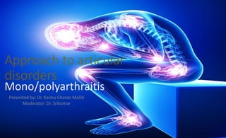
Approach to articular
disorders
Mono/polyarthraitis
Presented by: Dr. Kanhu Charan Mallik
Moderator: Dr. Srikumar
Evaluation of Patients With
Musculoskeletal Complaints
Goals
• Accurate diagnosis
• Timely provision of therapy
• Avoidance of unnecessary diagnostic testing
Approach
 Anatomic localization of complaint (articular vs. nonarticular)
 Determination of the nature of the pathologic process
 (inflammatory vs. noninflammatory)
 Determination of the extent of involvement (monoarticular,
 polyarticular, focal, widespread)
 Determination of chronology (acute vs. chronic)
 Consider the most common disorders first
 Formulation of a differential diagnosis
 What is the impact of the condition on the patient’s life?
 Articular Vs. Non-articular
Must discriminate the anatomic origin(s) & pt.s complaint
Requires a careful & detailed examination
Articular
 Articular structures- the synovium,
synovial fluid, articular cartilage,
intra-articular lig., joint capsule and
juxta-articular bone.
 Articular disorders may be
characterised by deep or diffuse
pain, pain or limited ROM on
active and passive movement,
and swelling( caused by synovial
proliferation, effusion, or bony
enlargement), crepitation,
instability, “locking” or deformity.
Non-articular
 Non-articular( Peri-articular) structures-
supportive extra-articular lig.s,
tendons, bursae, muscles, fascia,
bone, nerve and overlying skin.
 By contrast non-articular disorders
tend to be painful on active, but not
on passive(or assisted) ROM. Non-
articular joints seldom demonstrate
swelling, crepitation's, instability or
deformity of jt. itself.
 Peri-articular conditions often
demonstrate joint or focal tenderness
in regions adjacent to articular
structures and have physical findings
remote from the joint capsule
Distinguishing Inflammatory vs
Noninflammatory Joint Disease by Features
Feature Inflammatory Noninflammatory
Systemic symptoms Prominent, including
fatigue
Unusual
Onset Insidious
Usually affecting multiple
joints
Gradual
1 joint or a few joints
Morning stiffness > 1 h < 30 min
Worst time of day Morning As day progresses
Effect of activity on
symptoms (joint pain and
stiffness)
Lessen with activity
Worse after periods of rest
May also have pain with
use
Worsen with activity
Lessen with rest
Impact of the condition on the patient
 Understanding the impact of the disease on the patient is crucial to
negotiating a suitable management plan.
 Ask open questions about functional problems and difficulty in doing
things.
 It may be easiest to get the patient to describe a typical day, from
getting out of bed to washing, dressing, toileting etc.
 Potentially sensitive areas, such as hygiene or sexual activity, should
be approached with simple, direct, open questions.
 The impact of the disease on the patient’s employment will be
important.
 A patient’s needs and aspirations are an important part of the
equation and will influence their ability to adapt to the condition.
Drug-Induced Musculoskeletal Conditions
Arthralgias
Quinidine, cimetidine, quinolones, chronic acyclovir, interferon, IL-2, nicardipine, vaccines, rifabutin, aromatase and
HIVprotease inhibitors
Myalgias/myopathy
Glucocorticoids, penicillamine, hydroxychloroquine, AZT, lovastatin, simvastatin, pravastatin, clofibrate, interferon, IL-2,
alcohol, cocaine, taxol, docetaxel, colchicine, quinolones, cyclosporine, protease inhibitors
Tendon rupture/tendinitis
Quinolones, glucocorticoids, isotretinoin
Gout
Diuretics, aspirin, cytotoxics, cyclosporine, alcohol, moonshine, ethambutol
Drug-induced lupus
Hydralazine, procainamide, quinidine, phenytoin, carbamazepine, methyldopa, isoniazid, chlorpromazine,
lithium, penicillamine, tetracyclines, TNF inhibitors, ACE inhibitors, ticlopidine
Osteonecrosis
Glucocorticoids, alcohol, radiation, bisphosphonates
Osteopenia
Glucocorticoids, chronic heparin, phenytoin, methotrexate
Scleroderma
Vinyl chloride, bleomycin, pentazocine, organic solvents, carbidopa, tryptophan, rapeseed oil
Vasculitis
Allopurinol, amphetamines, cocaine, thiazides, penicillamine, propylthiouracil, montelukast, TNF inhibitors,
Clinical history
 Aspects of the pt.s profile( Age, sex, race considerations)
 Complaints chronology
 Extent of joint involvement
 Precipitating factors
 Age groups( Age prevalence of different conditions)
 Sex
 Racial
Young Middle age Elderly
SLE
Reactive arthritis
Fibromyalgia
RA
OA
Polymyalgia
rheumatica
Male Female
Gout
Spondyloarthropathies(e.g. AS)
RA
Fibromyalgia
Lupus
 Familial aggregation- as in AS, gout,Heberden’s nodule of OA
 Chronology of the Complains- imp. Diagnostic feature
- divided into onset, evolution, duration
 Onset:
 Evolution
 Duration
Abrupt Indolent
Septic arthritis
Gout
OA
RA
Fibromyalgia
Chronic Intermittent Migratory Additive
OA Crystal or Lyme’s
disease
Rheumatic fever
Gonnococcal or
Viral Arthritis
RA
Psoriatic
arthritis
Acute ( < 6 wk ) Chronic ( > 6 wk )
Infectious
Crystal induced
-non-inflammatory or immunologic arthrides (
e.g. OA, RA ) & non-articular disorders ( e.g.
fibrmyalgia)
Precipitating Events
 Trauma( Osteonecrosis, meniscal tear)
 Drug
 Antecedent or intercurrent illness( Rheumatic fever, Reactive arthritis,
Hepatitis)
Co-morbidities predisposing to musculoskeletal complaints
 DM-CTS
 Renal insufficiency- Gout
 Psoriasis- psoriatic arthritis
 Myeloma- low back pain
 Cancer- myositis
 Drugs-
 Glucocorticoids- osteonecrosis, septic arthritis
 Diuretics
 Chemotherapy
Symptoms
• Pain
• Stiffness
• Limitation of motion
• Swelling
• Weakness
• Fatigue
Pain
 Localize pain anatomically.
 Ask the pt. to point to the area of pain with one finger
 Pain in the jt.- most probably articular disorder
 Pain between jt.s- bone or muscle disorders or referred pain
 Diffuse pain, variable, poorly described or unrelated to
anatomic structures- Fibromyalgia, malingering or
psychogenic problems.
 Severity of pain- 1 t 10
 Jt. Pain at rest and mov.- inflammatory process.
 Pain primarily during activity- Mechanical
Stiffness
 Discomfort perceived when the pt. attempts to move jt.s after a
period of inactivity.
 When it occurs stiffness or gelling, usually develops after several
hours of inactivity.
 Morning stiffness- RA
Morning stiffness a/w non-inflammatory jt. Dis. Is
always of short duration( usually less than 30 min) & of less severity than
the stiffness of the inflammatory jt. Dis.
 Absence of morning stiffness though does not exclude systemic
inflammatory diseases, it’s absence is uncommon.
Limitation of Motion
 It must be differentiated from stiffness because stiffness is usually
transient but true limitation of motion is fixed & not variable from hour
to hour.
 Determination of the extent of disability resulting from lack of motion is
imp.
 Ascertain- the length of time of limitation of ROM,
whether both active & passive ROM are restricted,
whether it began abruptly( may suggest mechanical
derangement e.g. a tendon rupture) or gradually suggesting
inflammatory jt. Dis.
Swelling
 True joint swelling narrows the D.D.
 Ask where & when the swelling occurs
 A description of the exact location of the swelling
 Onset, persistence & factors that influence the joint are also imp.
 Discomfort with use of swollen part may indicate synovitis or bursitis
Weakness
o when it is present, a loss of motor power or muscle strength is nearly
always objectively demonstrable on physical examination
o Examiner must determine whether there is “true weakness” or “give
way” weakness.
 In musculoskeletal disorders- weakness is persistent rather than
intermittent
 Initially good strength with subsequent weakness- clue to
neuromuscular disorders
 Inflammatory myopathies- proximal weakness
 Distal weakness- neurologic disorders or inclusion body myositis
Fatigue
 Defined as inclination to rest even though pain & weakness are not
limiting factors
 It is a normal phenomena after various degrees of activity but should
resolve after rest.
 In rheumatic disease fatigue may be prominent even after rest.
 Malaise commonly occurs with fatigue.
 Malaise is an indefinite feeling of lack of health.
 Stiffness is a discomfort during movement & weakness is an inability to
move normally, esp. against resistance.
Important physical signs of arthritis
• Swelling
• Tenderness
• Limitation of motion
• Crepitation
• Deformity
• Instability
Swelling
 Swelling around a joint may be caused by intra-articular effusion, synovial
thickening, periarticular soft tissue inflammation( such as tendinitis or bursitis),
bony enlargements or extra-articular fat pads.
 A joint effusion is often visible, compare jt. Of one side with the opp. Side for
symmetry or asymmetry
 Palpable fluid in a jt. Without recent trauma suggests synovitis.
 Thickened synovial membrane- chr. Inflammatory arthrides such as RA, may have
a “doughy” or “ boggy” consistency.
Tenderness
 Is an unusual sensitivity to touch or pressure
 Localization of tenderness by palpation helps to determine whether
the pathologic site is intra- or peri-articular, such as fat pad, tendon
attachment, ligament, bursa or muscle or in the skin.
 Also palpate the non-involved str.s to help asses the significance of
tenderness.
Limitation of Motion
 Normal type & ROM of each jt. Must be kept in
mind
 Comparison with an unaffected jt. Of opp.
Extremity should be done
 Limitations in ROM may be d/t limitation in the jt.
Itself or in the periarticular strs.
 In pt. with jt. Dis. Passive ROM is often greater
than the active type, possibly because of pain,
weakness or the state of articular or peri-
articular stress.
 The pt. must be relaxed.
Crepitation
 It is a palpable grating or crunching sensation produced by motion.
 May or may not be accompanied by pain.
 Fine creps on palpation over jt.s- Chronic inflammatory arthritis
 Coarse creps- Inflammatory or non-inflammatory
 Bone-on-bone creps- a palpable or audible “squeak” of higher frequency.
Deformity
 Is the mal-alignment of the joints
 Manifested by a bony enlargement, articular sublaxation,
contracture or ankyloses in non-anatomical positions.
 Deformed joints:-
1) Do not function normally
2) Frequently restrict activities
3) May give rise to pain esp. hen put to stressful use
4) May be of cosmetic concern
Instability
 Is present when the joint has greater than normal movement in any
plane
 Subluxation
 Dislocation
 Again pt. must be relaxed.
Rheumatic review of systems
Systemic features:-
o Fever
o Rash
o Nail abnormalities
o Myalgias
o Weakness
Involvement of organs
 Eyes ( Behcet’s disease, Sarcoidosis, Spondyloarthritis)
 GIT ( Scleroderma, IBD)
 GUT ( Reactive arthritis, gonococcemia)
 Nervous system( Lyme’s disease, vasculitis)
Red flags:
 These red flags should always prompt consideration of serious pathology and
can be indicative of any inflammatory, infective or neoplastic process
 Erythema, warmth, effusion, and decreased range of motion
 Fever with acute joint pain
 Acute joint pain in a sexually active young adult
 Skin breaks with signs of cellulitis adjacent to the affected joint
 Underlying bleeding disorder or use of anticoagulants
 Systemic or extra-articular symptoms
 Weight loss
 Night pain
 Single joint involvement
 Neurological symptoms and signs
Recording of joint Examinations
The “S-T-L “ system records:-
Degree of Swelling (S)
Tenderness (T)
Limitation of movement (M)
on a basis of a quantitative estimation of gradation.
grade ranges from 0(normal) to 4(highly abnormal)
In limitation of motion:
Grade 1= 25% loss of motion
2= 50% loss of motion
3= 75% loss of motion
4= Ankylosis
 Performing a regional examination of the musculoskeletal system
(‘REMS’) -
 Regional examination of the musculoskeletal system refers to the
more detailed examination that should be carried out once an
abnormality has been detected either through the history or
through the screening examination.
 REMS involves the examination of a group of joints which are linked
by function, and may require a detailed neurological and vascular
examination.
 five key stages:-
 Introduce yourself.
 Look at the joint(s).
 Feel the joint(s).
 Move the joint(s).
 Assess the function of the joint(s).
Introduction
 It is important to introduce yourself, explain to the patient what you
are going to do, gain verbal consent to examine, and ask the
patient to let you know if you cause them any pain or discomfort at
any time.
 In all cases it is important to make the patient feel comfortable
about being examined. A good musculoskeletal examination relies
on patient cooperation, in order for them to relax their muscles, if
important clinical signs are not to be missed.
Look
 The examination should always start with a visual inspection of the
exposed area at rest.
 Compare one side with the other, checking for symmetry. You
should look specifically for skin changes, muscle bulk, and swelling in
and around the joint.
 Look also for deformity in terms of alignment and posture of the joint.
Feel
 Using the back of your hand, feel for skin temperature across the
joint line and at relevant neighbouring sites.
 Any swellings should be assessed for fluctuance and mobility. The
hard bony swellings of osteoarthritis should be distinguished from the
soft, rubbery swellings of inflammatory joint disease.
 Tenderness is an important clinical sign to elicit – both in and around
the joint.
 Identifying inflammation of a joint (synovitis) relies on detecting the
triad of warmth, swelling and tenderness.
Move
 The full range of movement of the joint should be assessed.
 Compare one side with the other. As a general rule both active movements
(where the patient moves the joint themselves) and passive movements (where
the examiner moves the joint) should be performed.
 If there is a loss of active movement, but passive movement is unaffected, this
may suggest a problem with the muscles, tendons or nerves rather than in the
joints, or it may be an effect of pain in the joints.
 In certain instances joints may move further than expected – this is called
hypermobility. It is important to elicit a loss of full flexion or a loss of full extension
as either may affect function.
 A loss of movement should be recorded as mild, moderate or severe. The quality
of movement should be recorded, with reference to abnormalities such as
increased muscle tone or the presence of crepitus.
Function
 It is important to make a functional assessment of the joint – for
example, in the case of limited elbow flexion, does this make it
difficult for the patient to bring their hands to their mouth? In the
case of the lower limbs, function mainly involves gait and the
patient’s ability to get out of a chair.
 one group of joints may need to be examined in conjunction with
another group (e.g. the shoulder and cervical spine).
RECORDING OF FINDINGS FROM THE REGIONAL
EXAMINATION
 The positive and significant negative
findings of the REMS examination are
usually documented longhand in the
notes.
 If no abnormality is found then ‘REMS
normal’ is sufficient.
 You may find it helpful to document joint
involvement on a homunculus such as
the one shown in fig.
 The total number of tender and swollen
joints can be used for calculating disease
activity scores – these are useful in
monitoring disease severity and response
to treatment over time.
LAB. INVESTIGATIONS
 Majority of musculoskeletal disorders can be easily diagnosed by a complete
history & physical examination.
 An additional objective of the initial counter is to determine whether additional
investigations or immediate therapy is required.
Indications for additional evaluation:-
 Monoarticular conditions
 Conditions accompanied by neurologic changes or systemic manifestations of
serious disease
 In individual with Chronic symptoms(>6 wk.), esp. when there’s lack of response
to symptomatic measures.
Investigations
 CBC
 ESR
 CRP
 Serum Uric acid
 RF
 Anti-CCP
 ANA
 Complement levels
 ASO titre
 Lymes and ANCA
Only if there is clinical evidence to
suggestion
of associated diagnosis.
ESR, CRP
 Ac. Phase reactants
 Useful in discriminating inflammatory Vs. non-
inflammatory
 Elevated in- infection, inflammation, auto-immune
disorders, neoplasia, pregnancy, renal insufficiency,
advanced age, hyperlipidemia.
 Extreme elevation- serious illness, such as( sepsis,
pleuropericarditis, polymyalgia rheumatic, giant cell
arteritis, adult Still’s disease)
S. Uric acid
 end product of purine metabolism, excreted in urine
 Useful in diagnosis of gout & in monitoring the response
to urate-lowering therapy
 level is a/w gout, nephrolithiasis, but levels may not
correlate with severity of the disease.
 level ( hence risk of gout)
 Inborn errors of metabolism
 Disease states( renal insufficiency, myeloproliferative disease,
psoriasis)
 Drugs( Alcohol, cytotoxic therapy, thiazides)
IgM RF
 Autoantibodies against the Fc portion of IgG
 Found in 80% of pt.s with RA
 Sensitivity= 70% & specificity= 80%
 Also seen in low titres in pt.s with
 Chronic infections( tuberculosis, leprosy, hepatitis)
 Other auto-immune diseases( SLE, Sjogren’s syndrome)
 Chronic pulmonary, hepatic and renal diseases
 When considering RA, both Serum RF and Anti-CCP
antibodies should be obtained as these are
complementary.
Anti-CCP
 Now Anti-CCP-3
 Method of estimation- ELISA
 Arbitary unit
 Higher value- higher specificity
 Specificity
 In healthy controls= 99%
 In diseased population= 95%
 In RA presence of both anti-CCP & RF antibodies may indicate a
greater risk for more severe, erosive polyarthritis.
ANAs
 Found in all pt.s ith SLE
 Also seen in pt.s with other auto-immune diseases(
polymyositis, scleroderma, anti-phospholipid syndrome,
Sjogren’s syndrome), drug induced lupus(d/t
Hydralazine, procainamide, quinidine, tetracyclines, TNF
inhibitors), Chronic liver or renal disorders and advanced
age.
 The interpretation of a positive ANA test may depend on
the magnitude of the titre & the pattern of IF
microscopy.
 Indirect IF is the Gold standard.
Aspiration &
Analysis of Synovial Fluid
.
Crystals
Crystals In polarised microscopy
In Gout • Long, needle-shaped,
negatively birefringent
• (Monosodium urate
crystals), usually intra-
cellular
In Calcium Pyrophosphate
Deposition (CPPD) (formerly
called Pseudogout)
& Chondrocalcinosis
• Short, rhomboid shaped
and positively birefringent
• Calcium pyrophosphate
dehydrate crystals
The negatively birefringent crystals appear
yellow when they lie parallel to the optical
axis of the compensator and blue when
perpendicular to it.
Diagnostic Imaging in Joint diseases
Conventional radiograph
 Plain x-rays are most appropriate when there is a history
of trauma, suspected chronic infection, progressive
disability, or monoarticular involvement; when
therapeutic alterations are considered; or when a
baseline assessment is desired for what appears to be a
chronic process.
 However, in acute inflammatory arthritis, early
radiography is rarely helpful in establishing a diagnosis
and may only reveal soft tissue swelling or juxtaarticular
demineralization
Diagnostic Imaging Techniques for Musculoskeletal Disorders
Method Imaging Time, h Costa Current Indications
Ultrasoundb <1 ++ Synovial cysts, Rotator cuff tears, Tendon injury
Radionuclide
scintigraphy
99mTc 1–4 ++ Metastatic bone survey
Evaluation of Paget's disease
Acute and chronic osteomyelitis
111In-WBC 24 +++ Acute infection, Prosthetic infection, Acute
osteomyelitis
67Ga 24–48 ++++ Acute and chronic infection, Acute osteomyelitis
Computed
tomography
<1 +++ Herniated intervertebral disk, Sacroiliitis ,Spinal
stenosis, Spinal trauma, Osteoid osteoma, Stress
fracture
Magnetic resonance
imaging
1/2–2 ++++ Avascular necrosis, Osteomyelitis, Intraarticular
derangement and soft tissue injury, Derangements of
axial skeleton and spinal cord, Herniated
intervertebral disk, Pigmented villonodular synovitis,
Inflammatory and metabolic muscle pathology
Summary
 Clinical history is by far the most important diagnostic tool together
with the power of physical examination.
 Appropriate Lab. & radiological inv.s should only be ordered based
on the strong clinical findings and in difficult & doubtful cases.
 Recognising the Red Flag signs.
Approach to articular disorders( Mono/Poly Arthritis)

More Related Content

What's hot

Rheumatology
RheumatologyRheumatology
Rheumatology
DJ CrissCross
 
Approach to case of arthritis
Approach to case of arthritisApproach to case of arthritis
Approach to case of arthritis
Sarath Menon
 
Approach to arthritis
Approach to arthritisApproach to arthritis
Approach to arthritis
Shivshankar Badole
 
4 Rheumatoid Arthriis 2010
4 Rheumatoid Arthriis 20104 Rheumatoid Arthriis 2010
4 Rheumatoid Arthriis 2010
NorthTec
 
Psoriatic arthritis
Psoriatic arthritisPsoriatic arthritis
Psoriatic arthritis
hamidreza227
 
Approach to the patient with arthritis
Approach to the patient with arthritisApproach to the patient with arthritis
Approach to the patient with arthritisdattasrisaila
 
Psoriatic arthropathy
Psoriatic arthropathyPsoriatic arthropathy
Psoriatic arthropathy
Dr Daulatram Dhaked
 
Approach To A Patient With Polyarthritis
Approach To A Patient With PolyarthritisApproach To A Patient With Polyarthritis
Approach To A Patient With PolyarthritisPramod Mahender
 
Polyarthritis (clinical approach)
Polyarthritis (clinical approach)Polyarthritis (clinical approach)
Polyarthritis (clinical approach)
ankita0809
 
Rheumatic disorders summary
Rheumatic disorders summaryRheumatic disorders summary
Rheumatic disorders summaryRasha Dabbagh
 
Spondyloarthritis a brief
Spondyloarthritis a briefSpondyloarthritis a brief
Spondyloarthritis a brief
Chandra Shekar
 
Rheumatoid arthritis
Rheumatoid arthritisRheumatoid arthritis
Rheumatoid arthritis
Pratap Tiwari
 
Septic Arthritis
Septic ArthritisSeptic Arthritis
Septic Arthritis
yuyuricci
 
Septic arthritis
Septic arthritisSeptic arthritis
Septic arthritisorthoprince
 
Septic arthritis
Septic arthritisSeptic arthritis
Septic arthritisgroup7usmkk
 
Rheumatoid arthritis
Rheumatoid arthritisRheumatoid arthritis
Rheumatoid arthritis
Syed Muhammad Ali Shah
 

What's hot (20)

Rheumatology
RheumatologyRheumatology
Rheumatology
 
Approach to case of arthritis
Approach to case of arthritisApproach to case of arthritis
Approach to case of arthritis
 
Approach to arthritis
Approach to arthritisApproach to arthritis
Approach to arthritis
 
4 Rheumatoid Arthriis 2010
4 Rheumatoid Arthriis 20104 Rheumatoid Arthriis 2010
4 Rheumatoid Arthriis 2010
 
Psoriatic arthritis
Psoriatic arthritisPsoriatic arthritis
Psoriatic arthritis
 
Reactive Arthritis
Reactive  ArthritisReactive  Arthritis
Reactive Arthritis
 
Approach to the patient with arthritis
Approach to the patient with arthritisApproach to the patient with arthritis
Approach to the patient with arthritis
 
Polymyalgia rheumatica
Polymyalgia rheumaticaPolymyalgia rheumatica
Polymyalgia rheumatica
 
Psoriatic arthropathy
Psoriatic arthropathyPsoriatic arthropathy
Psoriatic arthropathy
 
Approach To A Patient With Polyarthritis
Approach To A Patient With PolyarthritisApproach To A Patient With Polyarthritis
Approach To A Patient With Polyarthritis
 
Polyarthritis (clinical approach)
Polyarthritis (clinical approach)Polyarthritis (clinical approach)
Polyarthritis (clinical approach)
 
Rheumatic disorders summary
Rheumatic disorders summaryRheumatic disorders summary
Rheumatic disorders summary
 
Spondyloarthropathy
SpondyloarthropathySpondyloarthropathy
Spondyloarthropathy
 
Inflammatory arthritis
Inflammatory arthritisInflammatory arthritis
Inflammatory arthritis
 
Spondyloarthritis a brief
Spondyloarthritis a briefSpondyloarthritis a brief
Spondyloarthritis a brief
 
Rheumatoid arthritis
Rheumatoid arthritisRheumatoid arthritis
Rheumatoid arthritis
 
Septic Arthritis
Septic ArthritisSeptic Arthritis
Septic Arthritis
 
Septic arthritis
Septic arthritisSeptic arthritis
Septic arthritis
 
Septic arthritis
Septic arthritisSeptic arthritis
Septic arthritis
 
Rheumatoid arthritis
Rheumatoid arthritisRheumatoid arthritis
Rheumatoid arthritis
 

Viewers also liked

Topic review approach_arthritis
Topic review approach_arthritisTopic review approach_arthritis
Topic review approach_arthritisSorawit Boonyathee
 
Effective Treatment For Gout
Effective Treatment For GoutEffective Treatment For Gout
Effective Treatment For Gout
CureGout
 
Hypothyroidism in SLE
Hypothyroidism in SLEHypothyroidism in SLE
Hypothyroidism in SLEDarls
 
Treatment For Gout
Treatment For GoutTreatment For Gout
Treatment For Gout
CureGout
 
Joint Disorders
Joint DisordersJoint Disorders
Joint Disorders
Dr. Prabhakar Manu
 
Drug Treatment Of Gout
Drug Treatment Of GoutDrug Treatment Of Gout
Drug Treatment Of Gout
Dr Shah Murad
 
Idiopathic inflammatory myopathy
Idiopathic inflammatory myopathyIdiopathic inflammatory myopathy
Idiopathic inflammatory myopathydattasrisaila
 
Lupus update for rhem fellows wramc 2014 talk
Lupus update for rhem fellows wramc 2014  talkLupus update for rhem fellows wramc 2014  talk
Lupus update for rhem fellows wramc 2014 talk
Donald Thomas
 
Meningitis and Encephalitis
Meningitis and EncephalitisMeningitis and Encephalitis
Meningitis and EncephalitisTondy Rimando
 
[Int. med] approach to joint pain from SIMS Lahore
[Int. med] approach to joint pain from SIMS Lahore[Int. med] approach to joint pain from SIMS Lahore
[Int. med] approach to joint pain from SIMS Lahore
Muhammad Ahmad
 
Approach to diagnosis of arthritis
Approach to diagnosis of arthritis Approach to diagnosis of arthritis
Approach to diagnosis of arthritis
Panganani Njobvu
 
Systemic lupus erythematosus
Systemic lupus erythematosusSystemic lupus erythematosus
Systemic lupus erythematosus
Hirdesh Chawla
 
2014 GSS Updates on Gout
2014 GSS Updates on Gout2014 GSS Updates on Gout
2014 GSS Updates on Gout
Sidney Erwin Manahan
 
Gout.
Gout.Gout.
Gout.
Shaikhani.
 
Rheumatoid arthritis for undergraduates
Rheumatoid arthritis for undergraduatesRheumatoid arthritis for undergraduates
Rheumatoid arthritis for undergraduatesDhananjaya Sabat
 
Arthritis
ArthritisArthritis
Arthritis
Paudel Sushil
 
Systemic lupus erythematosus
Systemic lupus erythematosusSystemic lupus erythematosus
Systemic lupus erythematosus
Furqan Khan
 
RHEUMATOID ARTHRITIS
RHEUMATOID ARTHRITISRHEUMATOID ARTHRITIS
RHEUMATOID ARTHRITISshruti87
 

Viewers also liked (20)

Topic review approach_arthritis
Topic review approach_arthritisTopic review approach_arthritis
Topic review approach_arthritis
 
Effective Treatment For Gout
Effective Treatment For GoutEffective Treatment For Gout
Effective Treatment For Gout
 
Hypothyroidism in SLE
Hypothyroidism in SLEHypothyroidism in SLE
Hypothyroidism in SLE
 
Treatment For Gout
Treatment For GoutTreatment For Gout
Treatment For Gout
 
Joint Disorders
Joint DisordersJoint Disorders
Joint Disorders
 
Drug Treatment Of Gout
Drug Treatment Of GoutDrug Treatment Of Gout
Drug Treatment Of Gout
 
Systemic lupus erythematosus overview
Systemic lupus erythematosus   overviewSystemic lupus erythematosus   overview
Systemic lupus erythematosus overview
 
Idiopathic inflammatory myopathy
Idiopathic inflammatory myopathyIdiopathic inflammatory myopathy
Idiopathic inflammatory myopathy
 
Lupus update for rhem fellows wramc 2014 talk
Lupus update for rhem fellows wramc 2014  talkLupus update for rhem fellows wramc 2014  talk
Lupus update for rhem fellows wramc 2014 talk
 
Meningitis and Encephalitis
Meningitis and EncephalitisMeningitis and Encephalitis
Meningitis and Encephalitis
 
2009 gout pharmacology
2009 gout pharmacology2009 gout pharmacology
2009 gout pharmacology
 
[Int. med] approach to joint pain from SIMS Lahore
[Int. med] approach to joint pain from SIMS Lahore[Int. med] approach to joint pain from SIMS Lahore
[Int. med] approach to joint pain from SIMS Lahore
 
Approach to diagnosis of arthritis
Approach to diagnosis of arthritis Approach to diagnosis of arthritis
Approach to diagnosis of arthritis
 
Systemic lupus erythematosus
Systemic lupus erythematosusSystemic lupus erythematosus
Systemic lupus erythematosus
 
2014 GSS Updates on Gout
2014 GSS Updates on Gout2014 GSS Updates on Gout
2014 GSS Updates on Gout
 
Gout.
Gout.Gout.
Gout.
 
Rheumatoid arthritis for undergraduates
Rheumatoid arthritis for undergraduatesRheumatoid arthritis for undergraduates
Rheumatoid arthritis for undergraduates
 
Arthritis
ArthritisArthritis
Arthritis
 
Systemic lupus erythematosus
Systemic lupus erythematosusSystemic lupus erythematosus
Systemic lupus erythematosus
 
RHEUMATOID ARTHRITIS
RHEUMATOID ARTHRITISRHEUMATOID ARTHRITIS
RHEUMATOID ARTHRITIS
 

Similar to Approach to articular disorders( Mono/Poly Arthritis)

Rhematoid arthritis uploaded
Rhematoid                 arthritis uploadedRhematoid                 arthritis uploaded
Rhematoid arthritis uploaded
GAUTAMI TIRPUDE
 
ATHRITIS Presentation, diqgnosis an.pptx
ATHRITIS Presentation, diqgnosis an.pptxATHRITIS Presentation, diqgnosis an.pptx
ATHRITIS Presentation, diqgnosis an.pptx
okumuatanas1
 
Rhematoid arthiritis
Rhematoid arthiritisRhematoid arthiritis
Rhematoid arthiritis
jasleenbrar03
 
Musculoskeletal System Trauma
Musculoskeletal System TraumaMusculoskeletal System Trauma
Musculoskeletal System Trauma
Jofred Martinez
 
connective tissue diseases.pptx
connective tissue diseases.pptxconnective tissue diseases.pptx
connective tissue diseases.pptx
MohammedAbdela7
 
Physiotherapy management for rheumatoid arthritis
Physiotherapy management for rheumatoid arthritisPhysiotherapy management for rheumatoid arthritis
Physiotherapy management for rheumatoid arthritis
senphysio
 
Rheumatoid arthritis
Rheumatoid arthritisRheumatoid arthritis
Rheumatoid arthritis
Rounak Bhandari
 
Ra conference may 2017
Ra conference may 2017Ra conference may 2017
Ra conference may 2017
Gregorio Cortes-Maisonet, MD, CHCP
 
Diagnosis 2014 june DR.HAFFIZ UR REHMAN MOHAMMAD
Diagnosis  2014   june DR.HAFFIZ UR REHMAN MOHAMMADDiagnosis  2014   june DR.HAFFIZ UR REHMAN MOHAMMAD
Diagnosis 2014 june DR.HAFFIZ UR REHMAN MOHAMMADKarachi
 
Chronic pain syndromes
Chronic pain syndromes Chronic pain syndromes
Chronic pain syndromes
Aftab Hussain
 
Clinical evaluation of the patient with rheumatic disease
Clinical evaluation of the patient with rheumatic diseaseClinical evaluation of the patient with rheumatic disease
Clinical evaluation of the patient with rheumatic disease
Faculty of Medicine, Ain Shams University
 
Degenerative joint disorders of temporomandibular joint
Degenerative joint disorders of temporomandibular jointDegenerative joint disorders of temporomandibular joint
Degenerative joint disorders of temporomandibular joint
Shibani Sarangi
 
Disesases of locomotor system
Disesases of locomotor systemDisesases of locomotor system
Disesases of locomotor system
DrRavi Jain
 
Diagnosis of TMDs and General therapy.pptx
Diagnosis of TMDs and General therapy.pptxDiagnosis of TMDs and General therapy.pptx
Diagnosis of TMDs and General therapy.pptx
Vishaltrivedi62
 
Rheumatic pain indramayu 29june2013
Rheumatic pain indramayu 29june2013Rheumatic pain indramayu 29june2013
Rheumatic pain indramayu 29june2013dhoan Evridho
 
Rheumatic pain management
Rheumatic pain managementRheumatic pain management
Rheumatic pain management
Rachmat Gunadi Wachjudi
 
CASE STUDY ON OSTEOARTHRITIS
CASE STUDY ON OSTEOARTHRITISCASE STUDY ON OSTEOARTHRITIS
CASE STUDY ON OSTEOARTHRITIS
AnilDhakal14
 
RHS joint pain, degeneration presentation.pptx
RHS  joint pain, degeneration presentation.pptxRHS  joint pain, degeneration presentation.pptx
RHS joint pain, degeneration presentation.pptx
jhobarry
 
Seropositive arthritis ppt
Seropositive arthritis pptSeropositive arthritis ppt
Seropositive arthritis ppt
Saurabh Chahar
 

Similar to Approach to articular disorders( Mono/Poly Arthritis) (20)

Rhematoid arthritis uploaded
Rhematoid                 arthritis uploadedRhematoid                 arthritis uploaded
Rhematoid arthritis uploaded
 
ATHRITIS Presentation, diqgnosis an.pptx
ATHRITIS Presentation, diqgnosis an.pptxATHRITIS Presentation, diqgnosis an.pptx
ATHRITIS Presentation, diqgnosis an.pptx
 
Rhematoid arthiritis
Rhematoid arthiritisRhematoid arthiritis
Rhematoid arthiritis
 
Musculoskeletal System Trauma
Musculoskeletal System TraumaMusculoskeletal System Trauma
Musculoskeletal System Trauma
 
connective tissue diseases.pptx
connective tissue diseases.pptxconnective tissue diseases.pptx
connective tissue diseases.pptx
 
Physiotherapy management for rheumatoid arthritis
Physiotherapy management for rheumatoid arthritisPhysiotherapy management for rheumatoid arthritis
Physiotherapy management for rheumatoid arthritis
 
Rheumatoid arthritis
Rheumatoid arthritisRheumatoid arthritis
Rheumatoid arthritis
 
Osteoarthritis
OsteoarthritisOsteoarthritis
Osteoarthritis
 
Ra conference may 2017
Ra conference may 2017Ra conference may 2017
Ra conference may 2017
 
Diagnosis 2014 june DR.HAFFIZ UR REHMAN MOHAMMAD
Diagnosis  2014   june DR.HAFFIZ UR REHMAN MOHAMMADDiagnosis  2014   june DR.HAFFIZ UR REHMAN MOHAMMAD
Diagnosis 2014 june DR.HAFFIZ UR REHMAN MOHAMMAD
 
Chronic pain syndromes
Chronic pain syndromes Chronic pain syndromes
Chronic pain syndromes
 
Clinical evaluation of the patient with rheumatic disease
Clinical evaluation of the patient with rheumatic diseaseClinical evaluation of the patient with rheumatic disease
Clinical evaluation of the patient with rheumatic disease
 
Degenerative joint disorders of temporomandibular joint
Degenerative joint disorders of temporomandibular jointDegenerative joint disorders of temporomandibular joint
Degenerative joint disorders of temporomandibular joint
 
Disesases of locomotor system
Disesases of locomotor systemDisesases of locomotor system
Disesases of locomotor system
 
Diagnosis of TMDs and General therapy.pptx
Diagnosis of TMDs and General therapy.pptxDiagnosis of TMDs and General therapy.pptx
Diagnosis of TMDs and General therapy.pptx
 
Rheumatic pain indramayu 29june2013
Rheumatic pain indramayu 29june2013Rheumatic pain indramayu 29june2013
Rheumatic pain indramayu 29june2013
 
Rheumatic pain management
Rheumatic pain managementRheumatic pain management
Rheumatic pain management
 
CASE STUDY ON OSTEOARTHRITIS
CASE STUDY ON OSTEOARTHRITISCASE STUDY ON OSTEOARTHRITIS
CASE STUDY ON OSTEOARTHRITIS
 
RHS joint pain, degeneration presentation.pptx
RHS  joint pain, degeneration presentation.pptxRHS  joint pain, degeneration presentation.pptx
RHS joint pain, degeneration presentation.pptx
 
Seropositive arthritis ppt
Seropositive arthritis pptSeropositive arthritis ppt
Seropositive arthritis ppt
 

Recently uploaded

POST OPERATIVE OLIGURIA and its management
POST OPERATIVE OLIGURIA and its managementPOST OPERATIVE OLIGURIA and its management
POST OPERATIVE OLIGURIA and its management
touseefaziz1
 
Phone Us ❤85270-49040❤ #ℂall #gIRLS In Surat By Surat @ℂall @Girls Hotel With...
Phone Us ❤85270-49040❤ #ℂall #gIRLS In Surat By Surat @ℂall @Girls Hotel With...Phone Us ❤85270-49040❤ #ℂall #gIRLS In Surat By Surat @ℂall @Girls Hotel With...
Phone Us ❤85270-49040❤ #ℂall #gIRLS In Surat By Surat @ℂall @Girls Hotel With...
Savita Shen $i11
 
BRACHYTHERAPY OVERVIEW AND APPLICATORS
BRACHYTHERAPY OVERVIEW  AND  APPLICATORSBRACHYTHERAPY OVERVIEW  AND  APPLICATORS
BRACHYTHERAPY OVERVIEW AND APPLICATORS
Krishan Murari
 
Report Back from SGO 2024: What’s the Latest in Cervical Cancer?
Report Back from SGO 2024: What’s the Latest in Cervical Cancer?Report Back from SGO 2024: What’s the Latest in Cervical Cancer?
Report Back from SGO 2024: What’s the Latest in Cervical Cancer?
bkling
 
Triangles of Neck and Clinical Correlation by Dr. RIG.pptx
Triangles of Neck and Clinical Correlation by Dr. RIG.pptxTriangles of Neck and Clinical Correlation by Dr. RIG.pptx
Triangles of Neck and Clinical Correlation by Dr. RIG.pptx
Dr. Rabia Inam Gandapore
 
basicmodesofventilation2022-220313203758.pdf
basicmodesofventilation2022-220313203758.pdfbasicmodesofventilation2022-220313203758.pdf
basicmodesofventilation2022-220313203758.pdf
aljamhori teaching hospital
 
Maxilla, Mandible & Hyoid Bone & Clinical Correlations by Dr. RIG.pptx
Maxilla, Mandible & Hyoid Bone & Clinical Correlations by Dr. RIG.pptxMaxilla, Mandible & Hyoid Bone & Clinical Correlations by Dr. RIG.pptx
Maxilla, Mandible & Hyoid Bone & Clinical Correlations by Dr. RIG.pptx
Dr. Rabia Inam Gandapore
 
Ophthalmology Clinical Tests for OSCE exam
Ophthalmology Clinical Tests for OSCE examOphthalmology Clinical Tests for OSCE exam
Ophthalmology Clinical Tests for OSCE exam
KafrELShiekh University
 
Non-respiratory Functions of the Lungs.pdf
Non-respiratory Functions of the Lungs.pdfNon-respiratory Functions of the Lungs.pdf
Non-respiratory Functions of the Lungs.pdf
MedicoseAcademics
 
Novas diretrizes da OMS para os cuidados perinatais de mais qualidade
Novas diretrizes da OMS para os cuidados perinatais de mais qualidadeNovas diretrizes da OMS para os cuidados perinatais de mais qualidade
Novas diretrizes da OMS para os cuidados perinatais de mais qualidade
Prof. Marcus Renato de Carvalho
 
Flu Vaccine Alert in Bangalore Karnataka
Flu Vaccine Alert in Bangalore KarnatakaFlu Vaccine Alert in Bangalore Karnataka
Flu Vaccine Alert in Bangalore Karnataka
addon Scans
 
For Better Surat #ℂall #Girl Service ❤85270-49040❤ Surat #ℂall #Girls
For Better Surat #ℂall #Girl Service ❤85270-49040❤ Surat #ℂall #GirlsFor Better Surat #ℂall #Girl Service ❤85270-49040❤ Surat #ℂall #Girls
For Better Surat #ℂall #Girl Service ❤85270-49040❤ Surat #ℂall #Girls
Savita Shen $i11
 
ARTIFICIAL INTELLIGENCE IN HEALTHCARE.pdf
ARTIFICIAL INTELLIGENCE IN  HEALTHCARE.pdfARTIFICIAL INTELLIGENCE IN  HEALTHCARE.pdf
ARTIFICIAL INTELLIGENCE IN HEALTHCARE.pdf
Anujkumaranit
 
ACUTE SCROTUM.....pdf. ACUTE SCROTAL CONDITIOND
ACUTE SCROTUM.....pdf. ACUTE SCROTAL CONDITIONDACUTE SCROTUM.....pdf. ACUTE SCROTAL CONDITIOND
ACUTE SCROTUM.....pdf. ACUTE SCROTAL CONDITIOND
DR SETH JOTHAM
 
24 Upakrama.pptx class ppt useful in all
24 Upakrama.pptx class ppt useful in all24 Upakrama.pptx class ppt useful in all
24 Upakrama.pptx class ppt useful in all
DrSathishMS1
 
heat stroke and heat exhaustion in children
heat stroke and heat exhaustion in childrenheat stroke and heat exhaustion in children
heat stroke and heat exhaustion in children
SumeraAhmad5
 
Evaluation of antidepressant activity of clitoris ternatea in animals
Evaluation of antidepressant activity of clitoris ternatea in animalsEvaluation of antidepressant activity of clitoris ternatea in animals
Evaluation of antidepressant activity of clitoris ternatea in animals
Shweta
 
Alcohol_Dr. Jeenal Mistry MD Pharmacology.pdf
Alcohol_Dr. Jeenal Mistry MD Pharmacology.pdfAlcohol_Dr. Jeenal Mistry MD Pharmacology.pdf
Alcohol_Dr. Jeenal Mistry MD Pharmacology.pdf
Dr Jeenal Mistry
 
Prix Galien International 2024 Forum Program
Prix Galien International 2024 Forum ProgramPrix Galien International 2024 Forum Program
Prix Galien International 2024 Forum Program
Levi Shapiro
 
TEST BANK for Operations Management, 14th Edition by William J. Stevenson, Ve...
TEST BANK for Operations Management, 14th Edition by William J. Stevenson, Ve...TEST BANK for Operations Management, 14th Edition by William J. Stevenson, Ve...
TEST BANK for Operations Management, 14th Edition by William J. Stevenson, Ve...
kevinkariuki227
 

Recently uploaded (20)

POST OPERATIVE OLIGURIA and its management
POST OPERATIVE OLIGURIA and its managementPOST OPERATIVE OLIGURIA and its management
POST OPERATIVE OLIGURIA and its management
 
Phone Us ❤85270-49040❤ #ℂall #gIRLS In Surat By Surat @ℂall @Girls Hotel With...
Phone Us ❤85270-49040❤ #ℂall #gIRLS In Surat By Surat @ℂall @Girls Hotel With...Phone Us ❤85270-49040❤ #ℂall #gIRLS In Surat By Surat @ℂall @Girls Hotel With...
Phone Us ❤85270-49040❤ #ℂall #gIRLS In Surat By Surat @ℂall @Girls Hotel With...
 
BRACHYTHERAPY OVERVIEW AND APPLICATORS
BRACHYTHERAPY OVERVIEW  AND  APPLICATORSBRACHYTHERAPY OVERVIEW  AND  APPLICATORS
BRACHYTHERAPY OVERVIEW AND APPLICATORS
 
Report Back from SGO 2024: What’s the Latest in Cervical Cancer?
Report Back from SGO 2024: What’s the Latest in Cervical Cancer?Report Back from SGO 2024: What’s the Latest in Cervical Cancer?
Report Back from SGO 2024: What’s the Latest in Cervical Cancer?
 
Triangles of Neck and Clinical Correlation by Dr. RIG.pptx
Triangles of Neck and Clinical Correlation by Dr. RIG.pptxTriangles of Neck and Clinical Correlation by Dr. RIG.pptx
Triangles of Neck and Clinical Correlation by Dr. RIG.pptx
 
basicmodesofventilation2022-220313203758.pdf
basicmodesofventilation2022-220313203758.pdfbasicmodesofventilation2022-220313203758.pdf
basicmodesofventilation2022-220313203758.pdf
 
Maxilla, Mandible & Hyoid Bone & Clinical Correlations by Dr. RIG.pptx
Maxilla, Mandible & Hyoid Bone & Clinical Correlations by Dr. RIG.pptxMaxilla, Mandible & Hyoid Bone & Clinical Correlations by Dr. RIG.pptx
Maxilla, Mandible & Hyoid Bone & Clinical Correlations by Dr. RIG.pptx
 
Ophthalmology Clinical Tests for OSCE exam
Ophthalmology Clinical Tests for OSCE examOphthalmology Clinical Tests for OSCE exam
Ophthalmology Clinical Tests for OSCE exam
 
Non-respiratory Functions of the Lungs.pdf
Non-respiratory Functions of the Lungs.pdfNon-respiratory Functions of the Lungs.pdf
Non-respiratory Functions of the Lungs.pdf
 
Novas diretrizes da OMS para os cuidados perinatais de mais qualidade
Novas diretrizes da OMS para os cuidados perinatais de mais qualidadeNovas diretrizes da OMS para os cuidados perinatais de mais qualidade
Novas diretrizes da OMS para os cuidados perinatais de mais qualidade
 
Flu Vaccine Alert in Bangalore Karnataka
Flu Vaccine Alert in Bangalore KarnatakaFlu Vaccine Alert in Bangalore Karnataka
Flu Vaccine Alert in Bangalore Karnataka
 
For Better Surat #ℂall #Girl Service ❤85270-49040❤ Surat #ℂall #Girls
For Better Surat #ℂall #Girl Service ❤85270-49040❤ Surat #ℂall #GirlsFor Better Surat #ℂall #Girl Service ❤85270-49040❤ Surat #ℂall #Girls
For Better Surat #ℂall #Girl Service ❤85270-49040❤ Surat #ℂall #Girls
 
ARTIFICIAL INTELLIGENCE IN HEALTHCARE.pdf
ARTIFICIAL INTELLIGENCE IN  HEALTHCARE.pdfARTIFICIAL INTELLIGENCE IN  HEALTHCARE.pdf
ARTIFICIAL INTELLIGENCE IN HEALTHCARE.pdf
 
ACUTE SCROTUM.....pdf. ACUTE SCROTAL CONDITIOND
ACUTE SCROTUM.....pdf. ACUTE SCROTAL CONDITIONDACUTE SCROTUM.....pdf. ACUTE SCROTAL CONDITIOND
ACUTE SCROTUM.....pdf. ACUTE SCROTAL CONDITIOND
 
24 Upakrama.pptx class ppt useful in all
24 Upakrama.pptx class ppt useful in all24 Upakrama.pptx class ppt useful in all
24 Upakrama.pptx class ppt useful in all
 
heat stroke and heat exhaustion in children
heat stroke and heat exhaustion in childrenheat stroke and heat exhaustion in children
heat stroke and heat exhaustion in children
 
Evaluation of antidepressant activity of clitoris ternatea in animals
Evaluation of antidepressant activity of clitoris ternatea in animalsEvaluation of antidepressant activity of clitoris ternatea in animals
Evaluation of antidepressant activity of clitoris ternatea in animals
 
Alcohol_Dr. Jeenal Mistry MD Pharmacology.pdf
Alcohol_Dr. Jeenal Mistry MD Pharmacology.pdfAlcohol_Dr. Jeenal Mistry MD Pharmacology.pdf
Alcohol_Dr. Jeenal Mistry MD Pharmacology.pdf
 
Prix Galien International 2024 Forum Program
Prix Galien International 2024 Forum ProgramPrix Galien International 2024 Forum Program
Prix Galien International 2024 Forum Program
 
TEST BANK for Operations Management, 14th Edition by William J. Stevenson, Ve...
TEST BANK for Operations Management, 14th Edition by William J. Stevenson, Ve...TEST BANK for Operations Management, 14th Edition by William J. Stevenson, Ve...
TEST BANK for Operations Management, 14th Edition by William J. Stevenson, Ve...
 

Approach to articular disorders( Mono/Poly Arthritis)

  • 1. Approach to articular disorders Mono/polyarthraitis Presented by: Dr. Kanhu Charan Mallik Moderator: Dr. Srikumar
  • 2. Evaluation of Patients With Musculoskeletal Complaints Goals • Accurate diagnosis • Timely provision of therapy • Avoidance of unnecessary diagnostic testing Approach  Anatomic localization of complaint (articular vs. nonarticular)  Determination of the nature of the pathologic process  (inflammatory vs. noninflammatory)  Determination of the extent of involvement (monoarticular,  polyarticular, focal, widespread)  Determination of chronology (acute vs. chronic)  Consider the most common disorders first  Formulation of a differential diagnosis  What is the impact of the condition on the patient’s life?
  • 3.  Articular Vs. Non-articular Must discriminate the anatomic origin(s) & pt.s complaint Requires a careful & detailed examination Articular  Articular structures- the synovium, synovial fluid, articular cartilage, intra-articular lig., joint capsule and juxta-articular bone.  Articular disorders may be characterised by deep or diffuse pain, pain or limited ROM on active and passive movement, and swelling( caused by synovial proliferation, effusion, or bony enlargement), crepitation, instability, “locking” or deformity. Non-articular  Non-articular( Peri-articular) structures- supportive extra-articular lig.s, tendons, bursae, muscles, fascia, bone, nerve and overlying skin.  By contrast non-articular disorders tend to be painful on active, but not on passive(or assisted) ROM. Non- articular joints seldom demonstrate swelling, crepitation's, instability or deformity of jt. itself.  Peri-articular conditions often demonstrate joint or focal tenderness in regions adjacent to articular structures and have physical findings remote from the joint capsule
  • 4. Distinguishing Inflammatory vs Noninflammatory Joint Disease by Features Feature Inflammatory Noninflammatory Systemic symptoms Prominent, including fatigue Unusual Onset Insidious Usually affecting multiple joints Gradual 1 joint or a few joints Morning stiffness > 1 h < 30 min Worst time of day Morning As day progresses Effect of activity on symptoms (joint pain and stiffness) Lessen with activity Worse after periods of rest May also have pain with use Worsen with activity Lessen with rest
  • 5.
  • 6.
  • 7. Impact of the condition on the patient  Understanding the impact of the disease on the patient is crucial to negotiating a suitable management plan.  Ask open questions about functional problems and difficulty in doing things.  It may be easiest to get the patient to describe a typical day, from getting out of bed to washing, dressing, toileting etc.  Potentially sensitive areas, such as hygiene or sexual activity, should be approached with simple, direct, open questions.  The impact of the disease on the patient’s employment will be important.  A patient’s needs and aspirations are an important part of the equation and will influence their ability to adapt to the condition.
  • 8.
  • 9. Drug-Induced Musculoskeletal Conditions Arthralgias Quinidine, cimetidine, quinolones, chronic acyclovir, interferon, IL-2, nicardipine, vaccines, rifabutin, aromatase and HIVprotease inhibitors Myalgias/myopathy Glucocorticoids, penicillamine, hydroxychloroquine, AZT, lovastatin, simvastatin, pravastatin, clofibrate, interferon, IL-2, alcohol, cocaine, taxol, docetaxel, colchicine, quinolones, cyclosporine, protease inhibitors Tendon rupture/tendinitis Quinolones, glucocorticoids, isotretinoin Gout Diuretics, aspirin, cytotoxics, cyclosporine, alcohol, moonshine, ethambutol Drug-induced lupus Hydralazine, procainamide, quinidine, phenytoin, carbamazepine, methyldopa, isoniazid, chlorpromazine, lithium, penicillamine, tetracyclines, TNF inhibitors, ACE inhibitors, ticlopidine Osteonecrosis Glucocorticoids, alcohol, radiation, bisphosphonates Osteopenia Glucocorticoids, chronic heparin, phenytoin, methotrexate Scleroderma Vinyl chloride, bleomycin, pentazocine, organic solvents, carbidopa, tryptophan, rapeseed oil Vasculitis Allopurinol, amphetamines, cocaine, thiazides, penicillamine, propylthiouracil, montelukast, TNF inhibitors,
  • 10. Clinical history  Aspects of the pt.s profile( Age, sex, race considerations)  Complaints chronology  Extent of joint involvement  Precipitating factors  Age groups( Age prevalence of different conditions)  Sex  Racial Young Middle age Elderly SLE Reactive arthritis Fibromyalgia RA OA Polymyalgia rheumatica Male Female Gout Spondyloarthropathies(e.g. AS) RA Fibromyalgia Lupus
  • 11.  Familial aggregation- as in AS, gout,Heberden’s nodule of OA  Chronology of the Complains- imp. Diagnostic feature - divided into onset, evolution, duration  Onset:  Evolution  Duration Abrupt Indolent Septic arthritis Gout OA RA Fibromyalgia Chronic Intermittent Migratory Additive OA Crystal or Lyme’s disease Rheumatic fever Gonnococcal or Viral Arthritis RA Psoriatic arthritis Acute ( < 6 wk ) Chronic ( > 6 wk ) Infectious Crystal induced -non-inflammatory or immunologic arthrides ( e.g. OA, RA ) & non-articular disorders ( e.g. fibrmyalgia)
  • 12. Precipitating Events  Trauma( Osteonecrosis, meniscal tear)  Drug  Antecedent or intercurrent illness( Rheumatic fever, Reactive arthritis, Hepatitis) Co-morbidities predisposing to musculoskeletal complaints  DM-CTS  Renal insufficiency- Gout  Psoriasis- psoriatic arthritis  Myeloma- low back pain  Cancer- myositis  Drugs-  Glucocorticoids- osteonecrosis, septic arthritis  Diuretics  Chemotherapy
  • 13. Symptoms • Pain • Stiffness • Limitation of motion • Swelling • Weakness • Fatigue
  • 14. Pain  Localize pain anatomically.  Ask the pt. to point to the area of pain with one finger  Pain in the jt.- most probably articular disorder  Pain between jt.s- bone or muscle disorders or referred pain  Diffuse pain, variable, poorly described or unrelated to anatomic structures- Fibromyalgia, malingering or psychogenic problems.  Severity of pain- 1 t 10  Jt. Pain at rest and mov.- inflammatory process.  Pain primarily during activity- Mechanical
  • 15. Stiffness  Discomfort perceived when the pt. attempts to move jt.s after a period of inactivity.  When it occurs stiffness or gelling, usually develops after several hours of inactivity.  Morning stiffness- RA Morning stiffness a/w non-inflammatory jt. Dis. Is always of short duration( usually less than 30 min) & of less severity than the stiffness of the inflammatory jt. Dis.  Absence of morning stiffness though does not exclude systemic inflammatory diseases, it’s absence is uncommon.
  • 16. Limitation of Motion  It must be differentiated from stiffness because stiffness is usually transient but true limitation of motion is fixed & not variable from hour to hour.  Determination of the extent of disability resulting from lack of motion is imp.  Ascertain- the length of time of limitation of ROM, whether both active & passive ROM are restricted, whether it began abruptly( may suggest mechanical derangement e.g. a tendon rupture) or gradually suggesting inflammatory jt. Dis.
  • 17. Swelling  True joint swelling narrows the D.D.  Ask where & when the swelling occurs  A description of the exact location of the swelling  Onset, persistence & factors that influence the joint are also imp.  Discomfort with use of swollen part may indicate synovitis or bursitis
  • 18. Weakness o when it is present, a loss of motor power or muscle strength is nearly always objectively demonstrable on physical examination o Examiner must determine whether there is “true weakness” or “give way” weakness.  In musculoskeletal disorders- weakness is persistent rather than intermittent  Initially good strength with subsequent weakness- clue to neuromuscular disorders  Inflammatory myopathies- proximal weakness  Distal weakness- neurologic disorders or inclusion body myositis
  • 19. Fatigue  Defined as inclination to rest even though pain & weakness are not limiting factors  It is a normal phenomena after various degrees of activity but should resolve after rest.  In rheumatic disease fatigue may be prominent even after rest.  Malaise commonly occurs with fatigue.  Malaise is an indefinite feeling of lack of health.  Stiffness is a discomfort during movement & weakness is an inability to move normally, esp. against resistance.
  • 20. Important physical signs of arthritis • Swelling • Tenderness • Limitation of motion • Crepitation • Deformity • Instability
  • 21. Swelling  Swelling around a joint may be caused by intra-articular effusion, synovial thickening, periarticular soft tissue inflammation( such as tendinitis or bursitis), bony enlargements or extra-articular fat pads.  A joint effusion is often visible, compare jt. Of one side with the opp. Side for symmetry or asymmetry  Palpable fluid in a jt. Without recent trauma suggests synovitis.  Thickened synovial membrane- chr. Inflammatory arthrides such as RA, may have a “doughy” or “ boggy” consistency.
  • 22. Tenderness  Is an unusual sensitivity to touch or pressure  Localization of tenderness by palpation helps to determine whether the pathologic site is intra- or peri-articular, such as fat pad, tendon attachment, ligament, bursa or muscle or in the skin.  Also palpate the non-involved str.s to help asses the significance of tenderness.
  • 23. Limitation of Motion  Normal type & ROM of each jt. Must be kept in mind  Comparison with an unaffected jt. Of opp. Extremity should be done  Limitations in ROM may be d/t limitation in the jt. Itself or in the periarticular strs.  In pt. with jt. Dis. Passive ROM is often greater than the active type, possibly because of pain, weakness or the state of articular or peri- articular stress.  The pt. must be relaxed.
  • 24. Crepitation  It is a palpable grating or crunching sensation produced by motion.  May or may not be accompanied by pain.  Fine creps on palpation over jt.s- Chronic inflammatory arthritis  Coarse creps- Inflammatory or non-inflammatory  Bone-on-bone creps- a palpable or audible “squeak” of higher frequency.
  • 25. Deformity  Is the mal-alignment of the joints  Manifested by a bony enlargement, articular sublaxation, contracture or ankyloses in non-anatomical positions.  Deformed joints:- 1) Do not function normally 2) Frequently restrict activities 3) May give rise to pain esp. hen put to stressful use 4) May be of cosmetic concern
  • 26. Instability  Is present when the joint has greater than normal movement in any plane  Subluxation  Dislocation  Again pt. must be relaxed.
  • 27. Rheumatic review of systems Systemic features:- o Fever o Rash o Nail abnormalities o Myalgias o Weakness Involvement of organs  Eyes ( Behcet’s disease, Sarcoidosis, Spondyloarthritis)  GIT ( Scleroderma, IBD)  GUT ( Reactive arthritis, gonococcemia)  Nervous system( Lyme’s disease, vasculitis)
  • 28. Red flags:  These red flags should always prompt consideration of serious pathology and can be indicative of any inflammatory, infective or neoplastic process  Erythema, warmth, effusion, and decreased range of motion  Fever with acute joint pain  Acute joint pain in a sexually active young adult  Skin breaks with signs of cellulitis adjacent to the affected joint  Underlying bleeding disorder or use of anticoagulants  Systemic or extra-articular symptoms  Weight loss  Night pain  Single joint involvement  Neurological symptoms and signs
  • 29. Recording of joint Examinations The “S-T-L “ system records:- Degree of Swelling (S) Tenderness (T) Limitation of movement (M) on a basis of a quantitative estimation of gradation. grade ranges from 0(normal) to 4(highly abnormal) In limitation of motion: Grade 1= 25% loss of motion 2= 50% loss of motion 3= 75% loss of motion 4= Ankylosis
  • 30.  Performing a regional examination of the musculoskeletal system (‘REMS’) -  Regional examination of the musculoskeletal system refers to the more detailed examination that should be carried out once an abnormality has been detected either through the history or through the screening examination.  REMS involves the examination of a group of joints which are linked by function, and may require a detailed neurological and vascular examination.  five key stages:-  Introduce yourself.  Look at the joint(s).  Feel the joint(s).  Move the joint(s).  Assess the function of the joint(s).
  • 31. Introduction  It is important to introduce yourself, explain to the patient what you are going to do, gain verbal consent to examine, and ask the patient to let you know if you cause them any pain or discomfort at any time.  In all cases it is important to make the patient feel comfortable about being examined. A good musculoskeletal examination relies on patient cooperation, in order for them to relax their muscles, if important clinical signs are not to be missed.
  • 32. Look  The examination should always start with a visual inspection of the exposed area at rest.  Compare one side with the other, checking for symmetry. You should look specifically for skin changes, muscle bulk, and swelling in and around the joint.  Look also for deformity in terms of alignment and posture of the joint.
  • 33. Feel  Using the back of your hand, feel for skin temperature across the joint line and at relevant neighbouring sites.  Any swellings should be assessed for fluctuance and mobility. The hard bony swellings of osteoarthritis should be distinguished from the soft, rubbery swellings of inflammatory joint disease.  Tenderness is an important clinical sign to elicit – both in and around the joint.  Identifying inflammation of a joint (synovitis) relies on detecting the triad of warmth, swelling and tenderness.
  • 34. Move  The full range of movement of the joint should be assessed.  Compare one side with the other. As a general rule both active movements (where the patient moves the joint themselves) and passive movements (where the examiner moves the joint) should be performed.  If there is a loss of active movement, but passive movement is unaffected, this may suggest a problem with the muscles, tendons or nerves rather than in the joints, or it may be an effect of pain in the joints.  In certain instances joints may move further than expected – this is called hypermobility. It is important to elicit a loss of full flexion or a loss of full extension as either may affect function.  A loss of movement should be recorded as mild, moderate or severe. The quality of movement should be recorded, with reference to abnormalities such as increased muscle tone or the presence of crepitus.
  • 35. Function  It is important to make a functional assessment of the joint – for example, in the case of limited elbow flexion, does this make it difficult for the patient to bring their hands to their mouth? In the case of the lower limbs, function mainly involves gait and the patient’s ability to get out of a chair.  one group of joints may need to be examined in conjunction with another group (e.g. the shoulder and cervical spine).
  • 36. RECORDING OF FINDINGS FROM THE REGIONAL EXAMINATION  The positive and significant negative findings of the REMS examination are usually documented longhand in the notes.  If no abnormality is found then ‘REMS normal’ is sufficient.  You may find it helpful to document joint involvement on a homunculus such as the one shown in fig.  The total number of tender and swollen joints can be used for calculating disease activity scores – these are useful in monitoring disease severity and response to treatment over time.
  • 37.
  • 38. LAB. INVESTIGATIONS  Majority of musculoskeletal disorders can be easily diagnosed by a complete history & physical examination.  An additional objective of the initial counter is to determine whether additional investigations or immediate therapy is required. Indications for additional evaluation:-  Monoarticular conditions  Conditions accompanied by neurologic changes or systemic manifestations of serious disease  In individual with Chronic symptoms(>6 wk.), esp. when there’s lack of response to symptomatic measures.
  • 39. Investigations  CBC  ESR  CRP  Serum Uric acid  RF  Anti-CCP  ANA  Complement levels  ASO titre  Lymes and ANCA Only if there is clinical evidence to suggestion of associated diagnosis.
  • 40. ESR, CRP  Ac. Phase reactants  Useful in discriminating inflammatory Vs. non- inflammatory  Elevated in- infection, inflammation, auto-immune disorders, neoplasia, pregnancy, renal insufficiency, advanced age, hyperlipidemia.  Extreme elevation- serious illness, such as( sepsis, pleuropericarditis, polymyalgia rheumatic, giant cell arteritis, adult Still’s disease)
  • 41. S. Uric acid  end product of purine metabolism, excreted in urine  Useful in diagnosis of gout & in monitoring the response to urate-lowering therapy  level is a/w gout, nephrolithiasis, but levels may not correlate with severity of the disease.  level ( hence risk of gout)  Inborn errors of metabolism  Disease states( renal insufficiency, myeloproliferative disease, psoriasis)  Drugs( Alcohol, cytotoxic therapy, thiazides)
  • 42. IgM RF  Autoantibodies against the Fc portion of IgG  Found in 80% of pt.s with RA  Sensitivity= 70% & specificity= 80%  Also seen in low titres in pt.s with  Chronic infections( tuberculosis, leprosy, hepatitis)  Other auto-immune diseases( SLE, Sjogren’s syndrome)  Chronic pulmonary, hepatic and renal diseases  When considering RA, both Serum RF and Anti-CCP antibodies should be obtained as these are complementary.
  • 43. Anti-CCP  Now Anti-CCP-3  Method of estimation- ELISA  Arbitary unit  Higher value- higher specificity  Specificity  In healthy controls= 99%  In diseased population= 95%  In RA presence of both anti-CCP & RF antibodies may indicate a greater risk for more severe, erosive polyarthritis.
  • 44. ANAs  Found in all pt.s ith SLE  Also seen in pt.s with other auto-immune diseases( polymyositis, scleroderma, anti-phospholipid syndrome, Sjogren’s syndrome), drug induced lupus(d/t Hydralazine, procainamide, quinidine, tetracyclines, TNF inhibitors), Chronic liver or renal disorders and advanced age.  The interpretation of a positive ANA test may depend on the magnitude of the titre & the pattern of IF microscopy.  Indirect IF is the Gold standard.
  • 45. Aspiration & Analysis of Synovial Fluid .
  • 46. Crystals Crystals In polarised microscopy In Gout • Long, needle-shaped, negatively birefringent • (Monosodium urate crystals), usually intra- cellular In Calcium Pyrophosphate Deposition (CPPD) (formerly called Pseudogout) & Chondrocalcinosis • Short, rhomboid shaped and positively birefringent • Calcium pyrophosphate dehydrate crystals The negatively birefringent crystals appear yellow when they lie parallel to the optical axis of the compensator and blue when perpendicular to it.
  • 47. Diagnostic Imaging in Joint diseases Conventional radiograph  Plain x-rays are most appropriate when there is a history of trauma, suspected chronic infection, progressive disability, or monoarticular involvement; when therapeutic alterations are considered; or when a baseline assessment is desired for what appears to be a chronic process.  However, in acute inflammatory arthritis, early radiography is rarely helpful in establishing a diagnosis and may only reveal soft tissue swelling or juxtaarticular demineralization
  • 48. Diagnostic Imaging Techniques for Musculoskeletal Disorders Method Imaging Time, h Costa Current Indications Ultrasoundb <1 ++ Synovial cysts, Rotator cuff tears, Tendon injury Radionuclide scintigraphy 99mTc 1–4 ++ Metastatic bone survey Evaluation of Paget's disease Acute and chronic osteomyelitis 111In-WBC 24 +++ Acute infection, Prosthetic infection, Acute osteomyelitis 67Ga 24–48 ++++ Acute and chronic infection, Acute osteomyelitis Computed tomography <1 +++ Herniated intervertebral disk, Sacroiliitis ,Spinal stenosis, Spinal trauma, Osteoid osteoma, Stress fracture Magnetic resonance imaging 1/2–2 ++++ Avascular necrosis, Osteomyelitis, Intraarticular derangement and soft tissue injury, Derangements of axial skeleton and spinal cord, Herniated intervertebral disk, Pigmented villonodular synovitis, Inflammatory and metabolic muscle pathology
  • 49. Summary  Clinical history is by far the most important diagnostic tool together with the power of physical examination.  Appropriate Lab. & radiological inv.s should only be ordered based on the strong clinical findings and in difficult & doubtful cases.  Recognising the Red Flag signs.