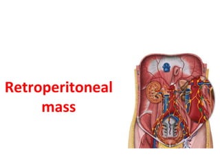
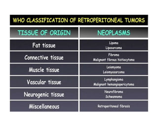
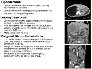
Retroperitoneal
mass
Learning Objectives
• Retroperitoneal anatomy
• Etiology of retroperitoneal mass
• Clinical features of retroperitoneal mass
• Investigations of retroperitoneal mas
• Management of retroperitoneal mass
Retroperitoneum
Boundaries
• Anteriorly : posterior parietal peritoneum
• Posteriorly : Vetebral column, iliopsoas ,
quadratus lumborum muscle and tendinous
part of transverse abdominis
• Superiorly : Diaphragm
• Inferiorly : Levator Ani and Pelvic
diaphragm
Retroperitoneal contents
SAD PUCKER
Suprarenal glands
Aorta and Inferior vena cava
Duodenum (except 1st part)
Pancreas (except tail)
Ureters
Colon (Ascending and Descending only)
Kidneys
Esophagus (Anterior and Left covered)
Rectum (Upper part)
Generally Retroperitoneal masses can
be classified into:
• Solid neoplastic masses
• Solid non neoplastic masses
• Cystic neoplastic masses
• Cystic non neoplastic masses
Solid Neoplastic Masses
Solid neoplasms in the retroperitoneum can be
broadly divided into four groups:
• mesodermal neoplasms
• neurogenic tumors
• germ cell, sex cord, and stromal tumors
• lymphoid and hematologic neoplasms.
Mesodermal Neoplasms
• Most of the retroperitoneal neoplasms are of mesodermal
origin, with liposarcomas, leiomyosarcomas, and malignant
fibrous histiocytomas making up more than 80% of these
tumors.
• Retroperitoneal sarcomas are commonly seen in the 5th
and 6th decades of life.
• These tumors are large at the time of clinical presentation
and often involve adjacent structures.
• The recurrence rates are high, and metastases to liver, lung,
bones, and brain may be seen .
Liposarcoma
• Liposarcoma is the most common (33%) primary
retroperitoneal sarcoma.
• Liposarcoma is usually large (average diameter, >20
cm) and is a slow growing tumor.
Leiomyosarcoma
• Leiomyosarcoma is the second most common (28%)
primary retroperitoneal sarcoma.
• from retroperitoneal smooth muscle tissue, blood
vessels, or wolffian duct remnants and can grow to a
large size (>10 cm).
• More common in women.
Malignant Fibrous Histiocytoma
• It is the third most common retroperitoneal sarcoma
(19%) and overall is the most common soft-tissue
sarcoma in the body.
• Malignant fibrous histiocytoma arises from primitive
mesenchymal elements, and 15% of these tumors
occur in the retroperitoneum.
• This tumor is more common in males (3:1),
particularly in the 50-60year age group.
• Neurogenic tumors constitute 10%-20% of primary
retroperitoneal tumors.
• Compared with the mesenchymal tumors, neurogenic
tumors occur in a younger age group and are more likely
to be benign and have a better prognosis.
• Neurogenic tumors can originate from the
• 1. nerve sheath (schwannoma, neurofibroma,
neurofibromatosis, malignant nerve sheath tumors),
• 2. ganglionic cells (ganglioneuroma,
ganglioneuroblastoma, neuroblastoma) .
• 3. paraganglionic cells (paraganglioma).
Neurogenic Tumors
Schwannoma
• Schwannoma, is a benign tumor that
arises from the perineural sheath of
Schwann cells.
• Schwannoma accounts for 6% of
retroperitoneal neoplasms and is
more common than neurofibroma.
• commonly seen along the
paravertebral sympathetic ganglia
• asymptomatic
• females (2:1)
• 20-50-year age group
Ganglioneuroma
• It is a rare benign tumor that
arises from the sympathetic
ganglia.
• The retroperitoneum (32%-52%
of cases) and mediastinum
(39%43% of cases) are the most
common sites for
ganglioneuroma, followed by the
cervical region (8%-9% of cases).
• Tumors that arise from the
chromaffin cells of the adrenal
medulla are called
pheochromocytomas, and those that
arise in an extraadrenal location
(10%) are referred to as
paragangliomas.
• Paraganglioma is commonly seen in
the 3rd to 4th decades, with no sex
predilection.
• In the retroperitoneum, the most
common site for a paraganglioma is
the organs of Zuckerkandl, which are
located anterior to the aorta at the
level of the origin of the inferior
mesenteric artery
Paraganglioma
Teratoma
• It is a germ cell tumor that
originates from pluripotent germ
cells
• Less than 10% of teratomas are
found in the retroperitoneum.
• It is third most common tumor in
the retroperitoneum in children,
after neuroblastoma and Wilms
tumor.
• More common in females
Lymphoma
• It is the most common retroperitoneal
malignancy, accounting for 33% of all of
these cases.
• seen in the 40-70-year age group
• frequently manifests with extranodal
disease in the liver, spleen, or bowel,
often at an advanced stage.
• History of fever , myalgia , night sweats ,
weight loss . Para-aortic lymph nodes are
involved in 25% of the patients with
Hodgkin lymphoma and 55% of the
patients with non-Hodgkin lymphoma
Solid non-neoplastic masses
• Retroperitoneal fibrosis
(Ormonds disease)
- Retroperitoneal fibrosis (RPF) is a relatively rare
condition that is characterized by a chronic
inflammatory and fibrotic process in the
retroperitoneum mostly around vessels like aorta and iliac,
and ureter.
- Approximately 70% of cases of retroperitoneal
fibrosis are idiopathic. (May be auto immune)
- The true incidence is unknown but is estimated
to be 1 per 200,000 to 500,000 per year.
• Pseudotumoral lipomatosis
• Retroperitoneal Erdheim-chester disease
• Retroperitoneal extramedullary hematopoiesis
Cystic Non-neoplastic Masses
Retroperitoneal hematoma
• Caused by trauma, blood dyscrasia,
anticoagulation therapy, rupture of an
abdominal aortic aneurysm, or interventional
or surgical procedures.
Urinoma
• A well-defined cystic lesion is seen in the
retroperitoneum, more commonly in the
perirenal space.
• caused by urinary tract leakage as a direct
result of obstruction, trauma, or
postinstrumentation.
Pancreatic Pseudocyst
• Pancreatic pseudocyst is a collection of
pancreatic fluid that occurs secondary to acute
pancreatitis and chronic pancreatitis also
especially alcoholic pancreatitis.
Cystic Neoplastic Masses
Cystic Change in Solid Neoplasms
Cystic changes may develop in solid lesions
such as paragangliomas and neurilemomas
Cystic Teratoma
Retroperitoneal cystic teratomas are cystic
tumors composed of well-differentiated
derivations from at least two of the three
germ layers (ectoderm, mesoderm,
endoderm). Most patients are female, and
the tumor is commonly diagnosed in
newborns, who are usually asymptomatic . A
cystic teratoma is likely to be benign,
whereas a solid teratoma is likely to be
malignant.
Lymphangioma
Lymphangioma is a noncancerous fluid
filled cystic tumor that is caused by failure of
communication of retroperitoneal lymphatic
tissue with the main lymphatic vessels.
Presentation
1- Most common presentation is huge abdominal lump
with compressive symptoms.
2- Asymptomatic: diagnosis is accidental
3- Presentation is usually late: because
a) tumors are slow growing & painless.
b) tumors displaces the adjacent structures. Infitration occurs in
late stage.
General Symptoms of Retroperitoneal Mass or Tumor
• Anemia
• Fatigue
• Weakness
• Loss of Appetite
• Weight Loss
• Retroperitoneal Pain or Back Pain
• Malabsorption
Specific symptoms
Pain
• Flank Pain
• Abdominal Pain
• Back Pain
Compression of Aorta
• Hypertension
• Renal Insufficiency
• Intermittent Claudication
Hematuria
Nerve Lesions
Urinary Retention
Constipation/ obstipation
Investigation
1. Routine blood investigations: to know about
A) hemoglobin: anemia
B) blood and serum creatinine raised on compression of kidney and ureter
C) liver function test
D) Effect of paraneoplastic syndrome
• Hypoglycemia:- due to increased insulin like hormone
• Hypercalcemia:- due to increased ectopic PTH hormone
• Catecholamines:- paraganglioma
E) Tumor markers:-AFP(alfafetoprotein), beta-HCG- Will be raised in
primary germ cell tumor
2. Plane X ray abdomen:- signs of intestinal obstruction,
obliterated psoas shadow, calcification of tumor mass.
3) US abdomen : nature of mass(solid/cystic) and
relation to the adjacent structures.
4) CT abdomen and pelvis
5) CT/US guided or laparoscopic core biopsy
6) FNAC has got limited role as the representative tissue
may not be obtained .
7) IVU - Can show obstruction and displacement of
kidney and ureter, distortion of renal pelvis and bladder
compression.
*Confirmation of diagnosis is only by tissue biopsy.
Investigation con.
Treatment
• Surgery : wide surgical resection is gold standard
• Pre & post-operative : Chemotherapy, Radiotherapy
only for some tumors
Key facts
• Retroperitoneal masses are rare and classified according to
tissue of origion, solid or cystic, tumor or non tumor.
• Lymphoma is most common retroperitoneal tumor.
• Liposarcoma is most common retroperitoneal sarcoma.
• Retroperitoneal sarcoma has got worse prognosis among all
soft tissue tumors.
• Liver followed by peritoneum is most common site of
distant metasatsis of retroperitoneal tumor .
• Contrast inhanced CT is investigation of choice for the
retroperitoneal masses.
• Surgery is the gold standard treatment.
Retroperitoneal mass.pptx

Retroperitoneal mass.pptx

  • 1.
  • 2.
    Learning Objectives • Retroperitonealanatomy • Etiology of retroperitoneal mass • Clinical features of retroperitoneal mass • Investigations of retroperitoneal mas • Management of retroperitoneal mass
  • 3.
    Retroperitoneum Boundaries • Anteriorly :posterior parietal peritoneum • Posteriorly : Vetebral column, iliopsoas , quadratus lumborum muscle and tendinous part of transverse abdominis • Superiorly : Diaphragm • Inferiorly : Levator Ani and Pelvic diaphragm
  • 4.
    Retroperitoneal contents SAD PUCKER Suprarenalglands Aorta and Inferior vena cava Duodenum (except 1st part) Pancreas (except tail) Ureters Colon (Ascending and Descending only) Kidneys Esophagus (Anterior and Left covered) Rectum (Upper part)
  • 5.
    Generally Retroperitoneal massescan be classified into: • Solid neoplastic masses • Solid non neoplastic masses • Cystic neoplastic masses • Cystic non neoplastic masses
  • 7.
    Solid Neoplastic Masses Solidneoplasms in the retroperitoneum can be broadly divided into four groups: • mesodermal neoplasms • neurogenic tumors • germ cell, sex cord, and stromal tumors • lymphoid and hematologic neoplasms.
  • 8.
    Mesodermal Neoplasms • Mostof the retroperitoneal neoplasms are of mesodermal origin, with liposarcomas, leiomyosarcomas, and malignant fibrous histiocytomas making up more than 80% of these tumors. • Retroperitoneal sarcomas are commonly seen in the 5th and 6th decades of life. • These tumors are large at the time of clinical presentation and often involve adjacent structures. • The recurrence rates are high, and metastases to liver, lung, bones, and brain may be seen .
  • 9.
    Liposarcoma • Liposarcoma isthe most common (33%) primary retroperitoneal sarcoma. • Liposarcoma is usually large (average diameter, >20 cm) and is a slow growing tumor. Leiomyosarcoma • Leiomyosarcoma is the second most common (28%) primary retroperitoneal sarcoma. • from retroperitoneal smooth muscle tissue, blood vessels, or wolffian duct remnants and can grow to a large size (>10 cm). • More common in women. Malignant Fibrous Histiocytoma • It is the third most common retroperitoneal sarcoma (19%) and overall is the most common soft-tissue sarcoma in the body. • Malignant fibrous histiocytoma arises from primitive mesenchymal elements, and 15% of these tumors occur in the retroperitoneum. • This tumor is more common in males (3:1), particularly in the 50-60year age group.
  • 10.
    • Neurogenic tumorsconstitute 10%-20% of primary retroperitoneal tumors. • Compared with the mesenchymal tumors, neurogenic tumors occur in a younger age group and are more likely to be benign and have a better prognosis. • Neurogenic tumors can originate from the • 1. nerve sheath (schwannoma, neurofibroma, neurofibromatosis, malignant nerve sheath tumors), • 2. ganglionic cells (ganglioneuroma, ganglioneuroblastoma, neuroblastoma) . • 3. paraganglionic cells (paraganglioma). Neurogenic Tumors
  • 11.
    Schwannoma • Schwannoma, isa benign tumor that arises from the perineural sheath of Schwann cells. • Schwannoma accounts for 6% of retroperitoneal neoplasms and is more common than neurofibroma. • commonly seen along the paravertebral sympathetic ganglia • asymptomatic • females (2:1) • 20-50-year age group
  • 12.
    Ganglioneuroma • It isa rare benign tumor that arises from the sympathetic ganglia. • The retroperitoneum (32%-52% of cases) and mediastinum (39%43% of cases) are the most common sites for ganglioneuroma, followed by the cervical region (8%-9% of cases).
  • 13.
    • Tumors thatarise from the chromaffin cells of the adrenal medulla are called pheochromocytomas, and those that arise in an extraadrenal location (10%) are referred to as paragangliomas. • Paraganglioma is commonly seen in the 3rd to 4th decades, with no sex predilection. • In the retroperitoneum, the most common site for a paraganglioma is the organs of Zuckerkandl, which are located anterior to the aorta at the level of the origin of the inferior mesenteric artery Paraganglioma
  • 14.
    Teratoma • It isa germ cell tumor that originates from pluripotent germ cells • Less than 10% of teratomas are found in the retroperitoneum. • It is third most common tumor in the retroperitoneum in children, after neuroblastoma and Wilms tumor. • More common in females
  • 15.
    Lymphoma • It isthe most common retroperitoneal malignancy, accounting for 33% of all of these cases. • seen in the 40-70-year age group • frequently manifests with extranodal disease in the liver, spleen, or bowel, often at an advanced stage. • History of fever , myalgia , night sweats , weight loss . Para-aortic lymph nodes are involved in 25% of the patients with Hodgkin lymphoma and 55% of the patients with non-Hodgkin lymphoma
  • 16.
    Solid non-neoplastic masses •Retroperitoneal fibrosis (Ormonds disease) - Retroperitoneal fibrosis (RPF) is a relatively rare condition that is characterized by a chronic inflammatory and fibrotic process in the retroperitoneum mostly around vessels like aorta and iliac, and ureter. - Approximately 70% of cases of retroperitoneal fibrosis are idiopathic. (May be auto immune) - The true incidence is unknown but is estimated to be 1 per 200,000 to 500,000 per year. • Pseudotumoral lipomatosis • Retroperitoneal Erdheim-chester disease • Retroperitoneal extramedullary hematopoiesis
  • 17.
    Cystic Non-neoplastic Masses Retroperitonealhematoma • Caused by trauma, blood dyscrasia, anticoagulation therapy, rupture of an abdominal aortic aneurysm, or interventional or surgical procedures. Urinoma • A well-defined cystic lesion is seen in the retroperitoneum, more commonly in the perirenal space. • caused by urinary tract leakage as a direct result of obstruction, trauma, or postinstrumentation. Pancreatic Pseudocyst • Pancreatic pseudocyst is a collection of pancreatic fluid that occurs secondary to acute pancreatitis and chronic pancreatitis also especially alcoholic pancreatitis.
  • 18.
    Cystic Neoplastic Masses CysticChange in Solid Neoplasms Cystic changes may develop in solid lesions such as paragangliomas and neurilemomas Cystic Teratoma Retroperitoneal cystic teratomas are cystic tumors composed of well-differentiated derivations from at least two of the three germ layers (ectoderm, mesoderm, endoderm). Most patients are female, and the tumor is commonly diagnosed in newborns, who are usually asymptomatic . A cystic teratoma is likely to be benign, whereas a solid teratoma is likely to be malignant. Lymphangioma Lymphangioma is a noncancerous fluid filled cystic tumor that is caused by failure of communication of retroperitoneal lymphatic tissue with the main lymphatic vessels.
  • 19.
    Presentation 1- Most commonpresentation is huge abdominal lump with compressive symptoms. 2- Asymptomatic: diagnosis is accidental 3- Presentation is usually late: because a) tumors are slow growing & painless. b) tumors displaces the adjacent structures. Infitration occurs in late stage.
  • 20.
    General Symptoms ofRetroperitoneal Mass or Tumor • Anemia • Fatigue • Weakness • Loss of Appetite • Weight Loss • Retroperitoneal Pain or Back Pain • Malabsorption
  • 21.
    Specific symptoms Pain • FlankPain • Abdominal Pain • Back Pain Compression of Aorta • Hypertension • Renal Insufficiency • Intermittent Claudication Hematuria Nerve Lesions Urinary Retention Constipation/ obstipation
  • 22.
    Investigation 1. Routine bloodinvestigations: to know about A) hemoglobin: anemia B) blood and serum creatinine raised on compression of kidney and ureter C) liver function test D) Effect of paraneoplastic syndrome • Hypoglycemia:- due to increased insulin like hormone • Hypercalcemia:- due to increased ectopic PTH hormone • Catecholamines:- paraganglioma E) Tumor markers:-AFP(alfafetoprotein), beta-HCG- Will be raised in primary germ cell tumor 2. Plane X ray abdomen:- signs of intestinal obstruction, obliterated psoas shadow, calcification of tumor mass.
  • 23.
    3) US abdomen: nature of mass(solid/cystic) and relation to the adjacent structures. 4) CT abdomen and pelvis 5) CT/US guided or laparoscopic core biopsy 6) FNAC has got limited role as the representative tissue may not be obtained . 7) IVU - Can show obstruction and displacement of kidney and ureter, distortion of renal pelvis and bladder compression. *Confirmation of diagnosis is only by tissue biopsy. Investigation con.
  • 24.
    Treatment • Surgery :wide surgical resection is gold standard • Pre & post-operative : Chemotherapy, Radiotherapy only for some tumors
  • 25.
    Key facts • Retroperitonealmasses are rare and classified according to tissue of origion, solid or cystic, tumor or non tumor. • Lymphoma is most common retroperitoneal tumor. • Liposarcoma is most common retroperitoneal sarcoma. • Retroperitoneal sarcoma has got worse prognosis among all soft tissue tumors. • Liver followed by peritoneum is most common site of distant metasatsis of retroperitoneal tumor . • Contrast inhanced CT is investigation of choice for the retroperitoneal masses. • Surgery is the gold standard treatment.