This document discusses primitive retroperitoneal tumors (PRTs). It begins by defining PRTs and noting their relative rarity. It then describes the radiological signs used to identify and characterize PRTs. Specifically, it outlines signs seen on CT like calcifications, adipose tissue, and necrosis that provide clues about tumor type. For MR, it discusses signal patterns related to histological components like myxoid stroma, collagen fibers, fat, and enhancement patterns. The rest of the document details the imaging appearance and characteristics of specific PRT types originating from connective tissue, lipomatous tissue, smooth muscle tissue, and other tissues.
![pending on the age.The tumors are mostly malignant in
both adults and infants (62%–86%) [2, 7–11].
In this chapter, following a short discussion on the
radiological semiotics necessary to define the exact lo-
cation of the lesions being examined, the macro- and
microscopic aspects of the most common primitive ret-
roperitoneal tumors recently dealt with in the WHO
classification (2002) (Table 1) will be reviewed,focusing
more on the characteristics that influence the appear-
ance at imaging rather than the purely histological and
cytogenetic aspects [12].
Radiological Semiotics
of Primitive Retroperitoneal Tumors
In order to define a PRT as such, it is absolutely neces-
sary to exclude its origin from retroperitoneal organs
[13].Many radiological signs have been described to as-
sist in making a differential diagnosis. These include
Introduction
Primitive retroperitoneal tumors (PRT) include masses
that originate in the retroperitoneal space, indepen-
dently of the organs present therein, and are thus histo-
logically primitive. They derive from tissues contained
in the retroperitoneal space (adipose, muscular, vessel
and nerve tissue),from embryonic remnants or heterot-
opies coming from one or more embryonic layers (ecto-
derm, mesoderm and endoderm) or from totipotent
embryonic germs. They do not include any growing le-
sions belonging to the retroperitoneal organs (kidneys,
adrenal glands, excretory tract, pancreas and colon) or
secondary invasive organs such as systemic masses
(lymphomas) and metastases [1, 2]. PRTs are relatively
rare (0.01%–0.3% of all tumors [3–6] in adults,5% in in-
fants [3, 7]), and they are rather different in histogene-
sis, structure and evolutionary potential. The only com-
mon feature is the site of development.
The average age for the appearance of PRTs is around
50 years even if no age is exempt. The percentage distri-
bution of the various forms differs considerably de-
Chapter
Contents
Introduction . . . . . . . . . . . . . . . . . . . . . . . . . . . 619
Radiological Semiotics of Primitive Retroperitoneal Tumors 619
Tumors Originating from Connective Tissue
(Fibrous, Myofibroblastic and Fibrohistiocytic) . . . . . . 624
Lipomatous Tumors . . . . . . . . . . . . . . . . . . . . . . . 625
Tumors Originating from Smooth Muscle Tissue . . . . . . 629
Tumors Originating from Skeletal Muscle Tissue . . . . . . 632
Vascular and Perivascular Tumors . . . . . . . . . . . . . . . 632
Neural Tumors . . . . . . . . . . . . . . . . . . . . . . . . . . 634
Extraskeletal Bone Tumors . . . . . . . . . . . . . . . . . . . 639
Unclassified Tumors . . . . . . . . . . . . . . . . . . . . . . 639
Miscellaneous . . . . . . . . . . . . . . . . . . . . . . . . . . 641
References . . . . . . . . . . . . . . . . . . . . . . . . . . . . 642
5.5
Retroperitoneal Tumors
Giovanni Carbognin, Lucia Pinali, Carlo Procacci (†)
Table 1. Histologic classification of PRT (modified from [12])
Tissue of origin Tumor
Connective (fibrous, Fibroma, fibrosarcoma, malignant
myofibroblastic, and fibrous histiocytoma
fibrohistiocytic tumors)
Fat Lipoma, liposarcoma
Smooth muscle Leiomyoma, leiomyosarcoma
Skeletal muscle Rhabdomyosarcoma
Vascular (blood vessels, Lymphangioma, lymphangiosar-
lymph vessels) and coma, hemangioma, angiosarco-
perivascular tumors ma, hemangiopericytoma
Neurogenic Neurofibroma, neurofibrosarco-
ma, neurilemmoma, malignant
schwannoma, ganglioneuroma,
ganglioneuroblastoma, neuroblas-
toma, paraganglioma (inactive),
pheochromocytoma
Tumors of uncertain Peripheral primitive neuroecto-
differentiation dermal tumor (PPNT), Ewing’s
sarcoma, synovial sarcoma
Miscellaneous Castleman’s disease](https://image.slidesharecdn.com/619-643-221225054947-456eaa1b/85/619-643-pdf-1-320.jpg)
![pending on the age.The tumors are mostly malignant in
both adults and infants (62%–86%) [2, 7–11].
In this chapter, following a short discussion on the
radiological semiotics necessary to define the exact lo-
cation of the lesions being examined, the macro- and
microscopic aspects of the most common primitive ret-
roperitoneal tumors recently dealt with in the WHO
classification (2002) (Table 1) will be reviewed,focusing
more on the characteristics that influence the appear-
ance at imaging rather than the purely histological and
cytogenetic aspects [12].
Radiological Semiotics
of Primitive Retroperitoneal Tumors
In order to define a PRT as such, it is absolutely neces-
sary to exclude its origin from retroperitoneal organs
[13].Many radiological signs have been described to as-
sist in making a differential diagnosis. These include
Introduction
Primitive retroperitoneal tumors (PRT) include masses
that originate in the retroperitoneal space, indepen-
dently of the organs present therein, and are thus histo-
logically primitive. They derive from tissues contained
in the retroperitoneal space (adipose, muscular, vessel
and nerve tissue),from embryonic remnants or heterot-
opies coming from one or more embryonic layers (ecto-
derm, mesoderm and endoderm) or from totipotent
embryonic germs. They do not include any growing le-
sions belonging to the retroperitoneal organs (kidneys,
adrenal glands, excretory tract, pancreas and colon) or
secondary invasive organs such as systemic masses
(lymphomas) and metastases [1, 2]. PRTs are relatively
rare (0.01%–0.3% of all tumors [3–6] in adults,5% in in-
fants [3, 7]), and they are rather different in histogene-
sis, structure and evolutionary potential. The only com-
mon feature is the site of development.
The average age for the appearance of PRTs is around
50 years even if no age is exempt. The percentage distri-
bution of the various forms differs considerably de-
Chapter
Contents
Introduction . . . . . . . . . . . . . . . . . . . . . . . . . . . 619
Radiological Semiotics of Primitive Retroperitoneal Tumors 619
Tumors Originating from Connective Tissue
(Fibrous, Myofibroblastic and Fibrohistiocytic) . . . . . . 624
Lipomatous Tumors . . . . . . . . . . . . . . . . . . . . . . . 625
Tumors Originating from Smooth Muscle Tissue . . . . . . 629
Tumors Originating from Skeletal Muscle Tissue . . . . . . 632
Vascular and Perivascular Tumors . . . . . . . . . . . . . . . 632
Neural Tumors . . . . . . . . . . . . . . . . . . . . . . . . . . 634
Extraskeletal Bone Tumors . . . . . . . . . . . . . . . . . . . 639
Unclassified Tumors . . . . . . . . . . . . . . . . . . . . . . 639
Miscellaneous . . . . . . . . . . . . . . . . . . . . . . . . . . 641
References . . . . . . . . . . . . . . . . . . . . . . . . . . . . 642
5.5
Retroperitoneal Tumors
Giovanni Carbognin, Lucia Pinali, Carlo Procacci (†)
Table 1. Histologic classification of PRT (modified from [12])
Tissue of origin Tumor
Connective (fibrous, Fibroma, fibrosarcoma, malignant
myofibroblastic, and fibrous histiocytoma
fibrohistiocytic tumors)
Fat Lipoma, liposarcoma
Smooth muscle Leiomyoma, leiomyosarcoma
Skeletal muscle Rhabdomyosarcoma
Vascular (blood vessels, Lymphangioma, lymphangiosar-
lymph vessels) and coma, hemangioma, angiosarco-
perivascular tumors ma, hemangiopericytoma
Neurogenic Neurofibroma, neurofibrosarco-
ma, neurilemmoma, malignant
schwannoma, ganglioneuroma,
ganglioneuroblastoma, neuroblas-
toma, paraganglioma (inactive),
pheochromocytoma
Tumors of uncertain Peripheral primitive neuroecto-
differentiation dermal tumor (PPNT), Ewing’s
sarcoma, synovial sarcoma
Miscellaneous Castleman’s disease](https://image.slidesharecdn.com/619-643-221225054947-456eaa1b/75/619-643-pdf-1-2048.jpg)
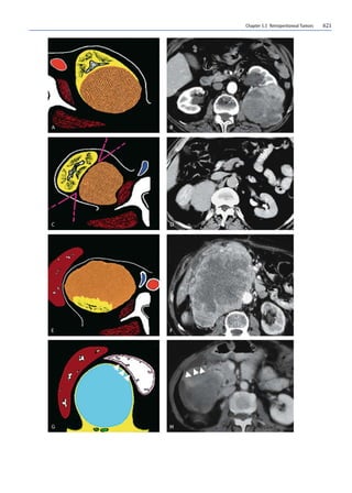
![620 Giovanni Carbognin, Lucia Pinali, Carlo Procacci (†)
valid signs common to every imaging method used and
other specific signs depending on the study technique.
The following are among the common signs:
쐌 The beak sign (positive when the mass causes the
edge of an adjacent organ to become beak shaped,
meaning that the mass arises from that organ)
(Fig. 1A–D).
쐌 The phantom organ sign (positive when a huge mass
arises from a small organ that then becomes unde-
tectable) (Fig. 1E, F).
쐌 The embedded organ sign (positive when part of a
hollow organ appears embedded in the tumor)
(Fig. 1G, H).
쐌 The prominent feeding artery sign (particularly useful
for hypervascular lesions supplied by arteries which
are prominent enough to be visualized at CT or MR).
Apart from these general signs, there are others that can
be specifically found with CT and MR.Specific diagnoses
can be suggested with CT when the following are present:
쐌 Calcifications (ganglioneuroma; malignant fibrohis-
tiocytoma)
쐌 Adipose tissue (homogeneous: lipoma (Fig. 2A); het-
erogeneous: liposarcoma (Fig. 2B)
쐌 Necrotic areas (tumors with high grade malignancy
such as leiomyosarcoma (Fig. 2C)
쐌 Hypervascularization (hemangioma, hemangioperi-
cytoma)
쐌 Areas of low homogeneous density (neurofibroma)
(Fig. 2D) [14]
MR,which is becoming the main method for examining
soft tissues,is not able to give specific information in all
cases,but the presence of some histological components
can be suggested by evaluating the signal characteristics
(intensity and enhancement) of the lesion. In the first
place, determination of the dominant histological com-
ponent can help narrow down the differential diagnosis
possibilities [15].
The myxoid stroma is hyperintense in T2- and hypo-
intense in T1-weighted images. After contrast medium
(CM) administration,the enhancement is slower.This is
common with ganglioneuroma (Fig. 3A–D), schwanno-
ma, neurofibroma, myxoid liposarcoma, malignant fi-
brohistiocytoma, ganglioneuroblastoma and malignant
tumor of the peripheral nerve sheaths.
The collagen fibers are hypointense in T1- and T2-
weighted images.After CM, the dense areas of the colla-
gen fibers enhance more slowly. Lesions that contain
collagen fiber include neurofibroma, ganglioneuroma
(Fig. 3A–D), leiomyosarcoma, malignant fibrohistiocy-
toma, malignant tumor of the peripheral nerve sheaths,
fibrosarcoma and retroperitoneal fibrosis.
Although better seen with CT,calcifications,especially
when large (ganglioneuroma,hemangioma,neuroblasto-
ma) appear as markedly hypointense areas with MR.
Fat is rather hyperintense in T1,moderately hyperin-
tense in T2 (fast or turbo spin-echo sequences) and hy-
pointense in fat-suppressed images. Lesions made up of
or often containing fat are lipoma, myelolipoma, an-
giomyolipoma and well-differentiated liposarcoma
(Fig. 3E).
Studying the signal behavior after CM administra-
tion also gives important information. Four enhance-
ment patterns have been described [15]:
쐌 No enhancement (benign lesions)
쐌 Early enhancement with rapid washout (benign le-
sions, Castleman’s disease)
쐌 Early enhancement with slow or no evident washout
(mostly malignant)
쐌 Delayed enhancement (benign masses (Fig. 3A–C)
and some malignant tumors with a myxoid compo-
nent such as myxoid liposarcoma, leiomyosarcoma)
There are other signs that are specifically appreciable
with MR [15]:
쐌 Target sign: central area with a low or intermediate
signal surrounded by a hyperintense ring in T2. His-
tologically it corresponds to fibrous tissue centrally
and myxoid tissue around the edge. It is frequently
seen in neurofibromas and schwannomas.
쐌 Bowl of fruit sign: low intensity mosaic,intermediate
and high signal in T2-weighted images due to a com-
bination of solid components, cystic degeneration,
hemorrhage, myxoid stroma and fibrous tissue. This
is often seen in malignant fibrohistiocytoma,synovi-
al sarcoma (Fig. 3F) and Ewing’s sarcoma.
쐌 Whorled appearance: a linear or curvilinear struc-
ture appearing hypointense in T2.It corresponds to a
band of Schwann cells and collagen fiber in the mass.
It is commonly seen in ganglioneuroma (Fig.3d) and
neurofibroma.
쐌 Flow void: this is often seen in hemangiopericytoma,
arteriovenous hemangioma and alveolar sarcoma of
the soft tissues.
쐌 Speckled enhancement: this can be found in T1-
weighted images after CM and corresponds to intratu-
moral structures similar to septa.It is more frequently
seen in leiomyosarcoma and rhabdomyosarcoma.
Fig. 1A–H. Origin of the mass. Positive beak sign: diagram (A) and
CT scan after CM administration (B).The appearance is supported
by parenchymal tokens that “envelop” the tumor. The lesion origi-
nates from the organ (renal mass).Negative beak sign: diagram (C)
and CT scan after contrast medium administration (D).The tumor
does not originate from the organ, which is also compressed. An
acute angle forms at the contact points between the resident organ
and the lesion as shown in (c) (retroperitoneal mass).E, F Phantom
organ sign: diagram (E) and CT scan after CM administration (F).
The tumor’s originating organ (right kidney) appears totally in-
corporated by the tumor and is no longer recognizable (F). Nega-
tive embedded organ sign: diagram (G) and CT scan after CM ad-
ministration (H).The wall of a hollow viscus is compressed extrin-
sically from the tumor creating a crescent shape (arrowheads)](https://image.slidesharecdn.com/619-643-221225054947-456eaa1b/85/619-643-pdf-2-320.jpg)

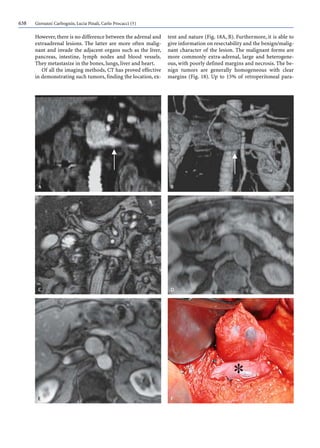
![624 Giovanni Carbognin, Lucia Pinali, Carlo Procacci (†)
Tumors Originating from Connective Tissue
(Fibrous, Myofibroblastic and Fibrohistiocytic)
The retroperitoneum is affected by border-line masses,
typically retroperitoneal fibromatosis and malignant
forms such as fibrosarcoma in adults.
Retroperitoneal fibromatosis is a group of benign fi-
brous tissue proliferations, with no sign of inflamma-
tion or tumor [14, 16]. The biological behavior is some-
where between benign fibrous lesions and fibrosarco-
ma: the proliferations tend to be locally aggressive and
can relapse locally but do not metastasize [17].They can
be primarily retroperitoneal or, more frequently, exten-
sions of mesenteric fibromatosis [17]. This disease can
occur at any age between 10 and 80 years (40 being the
average) and usually affects adults [17, 18]. However,
these lesions are rare: 10%–15% of cases are associated
with familial polyposis syndrome or Gardner syndrome
[19]. Macroscopically, the lesion appears as a solid mass
of variable size (1–30 cm) [18]. There is no real capsule
[19]. Microscopically, it is an infiltrating fibroprolifera-
tive process sustained by fibroblasts and myofibro-
blasts. Collagen with myxoid deposits can be found in
proximity to the fibroblasts. At US, fibromatosis shows
variable but generally low echotexture and smooth,
clear outlines [17, 20, 21].
At CT, it can have a homogeneous pattern and the
margins are usually well-defined.After CM administra-
tion, enhancement slows down because of the poor vas-
cularization. More particularly, the lesion appears gen-
erally isodense (47%) or hyperdense (41%) compared
to the muscle [18].
At MR the lesions appear hypointense compared to
the muscle in T1-weighted sequences while in T2-
weighted sequences the intensity is variable but gener-
ally low [19, 21].
Fibromatosis must be distinguished from idiopathic
retroperitoneal fibrosis (Ormond’s disease), which is a
rare pathological condition (1/100,000–200,000) char-
acterized by fibrous proliferation along the posterior
part of the retroperitoneum (Fig. 4) [22, 23].
The fibrosarcoma is a collagen-producing malignant
tumor deriving from cancerous fibroblast proliferation.
It can show variable degrees of cellularity,anaplasia and
mitotic index. It occurs more frequently in middle age,
typically develops in the more internal soft tissues and
is rarely located in the retroperitoneal [12] and medias-
tinal areas,with the exception of the so called inflamma-
tory subtype, more recently defined as inflammatory
myofibroblastic tumor. This is usually a large, well-de-
fined tumor within which necrotic focuses,which are as-
sociated with aggressive biological behavior, can often
be found (Fig. 5) [12, 24]. There are no further distinct
characteristics from the radiological aspect.
Up to now, the malignant fibrohistiocytoma (MFH)
has been the most common soft tissue tumor in adults,
also in view of the fact that, so far, the morphologic pat-
tern known as the pleomorphic MFH may be shared by
a wide variety of poorly differentiated malignant mass-
es [12].However,this lesion appears in the retroperiton-
eum only in about 16% of cases. It is clinically nonspe-
cific. Sometimes patients complain of indisposition, fe-
Fig. 4A–C. Retroperitoneal fibrosis (Ormond’s disease). CT exam-
ination in the pre- (A) and post- (B, C) contrastographic phases.
Solid tissue is visible with homogeneous density in the precontra-
stographic phase (A), surrounding the aorta outlined by lamellar
calcifications. In the dynamic contrast-enhanced phase (B, C) le-
sion enhancement is slow](https://image.slidesharecdn.com/619-643-221225054947-456eaa1b/85/619-643-pdf-6-320.jpg)
![Chapter 5.5 Retroperitoneal Tumors 625
ver, weight loss and abdominal pain. Hypoglycemia can
be present but is very rare [25].
Macroscopically, it appears as a lobulated, gelatinous
mass and is usually more than 5 cm in diameter. There
are five different subtypes,depending on the main com-
ponent, but the most useful to recognize are the myxoid
MFH and the pleomorphic MFH, as they have charac-
teristics that can assist diagnosis.
The myxoid MFH has an abundant amount of funda-
mental and mucopolysaccharide substance. It is biolog-
ically less aggressive and hardly ever leads to metastatic
spread. With the MR examination, the myxoid compo-
nent, hypointense in T1- and considerably hyperintense
in T2-weighted images, is particularly useful in assist-
ing diagnosis [26].
The pleomorphic variety generally affects areas that
have previously undergone radiotherapy. Macroscopi-
cally, it is a multilobulated, large mass (about 10 cm).
Microscopically, it shows a complex structure with mul-
tiple hemosiderin deposits and numerous gigantic cells
similar to osteoclasts [27]. The signal intensity at MR is
the same as that of the muscular tissue in both T1- and
T2-weighted sequences, and enhancement is poor after
administration of gadolinium. But the anamnestic find-
ings of previous radiotherapy in the area of a mass with
the above characteristics can lead to the correct diagno-
sis [28].
At imaging, the malignant fibrohistiocytoma is not
easily distinguished from other soft tissue sarcomas.
The combined use of the various methods, especially
MR, together with clinical evaluations, can be useful in
making a differential diagnosis, particularly in the case
of a large lesion that extensively infiltrates the adjacent
structures (Fig. 6).
Lipomatous Tumors
Adipose tissue tumors are common and represent
about half of soft tissue tumors in the surgical series
[29], and about 6% of all soft tissue tumors in children.
They can be divided into benign (lipoma and hiberno-
ma) and malignant (liposarcoma in its different vari-
ants) tumors.
The lipoma is the most common benign soft tissue
tumor.Nevertheless, its location in the retroperitoneum
is so rare that some authors do not take it into consider-
ation at all [12].It arises at different ages but is more fre-
quently found in Patients of between 40 and 60 years of
age while it is very rarely found in children. It involves
women more than men with a ratio of 1:2 and causes a
large increase in abdominal volume, with dislocation of
the contiguous abdominal structures [30,31].It is easily
removed and usually does not degenerate or relapse.
Histologically, it is a tumor of the white adipose tis-
sue with no lobulation composed of mature adipocytes
with a fibromembranous or fibrous capsule and tends to
expand without infiltrating the nearby organs. It can be
difficult to make a differential diagnosis with other adi-
pose tissue tumors such as the hibernoma and lipoblas-
toma and especially the well-differentiated liposarcoma
[21].
At imaging it appears with the typical characteristics
of a fatty, hypovascularized mass: homogeneously ra-
Fig. 5A, B. Fibrosarcoma. CT examination after CM administration
(A) and macroscopic section (B). At CM-enhanced CT (A) the le-
sion shows clear margins and inhomogeneous enhancement due
to the presence of necrotic spaces within it, confirmed by the re-
sected specimen (B)](https://image.slidesharecdn.com/619-643-221225054947-456eaa1b/85/619-643-pdf-7-320.jpg)
![626 Giovanni Carbognin, Lucia Pinali, Carlo Procacci (†)
diolucent in conventional radiograms, hyperechogenic
at US,and with low intensity at CT (Figs.2A,7),where it
displays well-defined outlines and the occasional intra-
lesional fibrous septa, which appear with greater den-
sity as internal linear stria.There is no enhancement af-
ter contrast medium administration (Fig. 7). At MR the
lipoma is hyperintense on T1- and hypointense on T2-
weighted images.The T1 sequence with selected fat sup-
pression leads to a definite diagnosis, especially when
the lesion shows a homogenous aspect [26].
Hibernomas are extremely rare tumors of the brown
adipose tissue. Only two of the 100 cases that have been
described in the literature were found in the retroperi-
toneal region. They appear at any age with an incidence
peak in the 30s. Clinically, it is a slow-growing, benign
tumor that can reach considerable dimensions. Macro-
scopically, it appears as a mass that is well circum-
scribed by a reddish-brown capsule, which may be due
to the high degree of vascularization. Imaging findings
are generally nonspecific and a differential diagnosis
between the other adipose soft tissue tumors is difficult
to make. A biopsy is therefore necessary for classifying
the mass. At CT, the density of the hibernoma can ap-
pear somewhere between that of the adipose tissue and
the muscle, with clear enhancement in the arterial con-
trast phase because of the rich vascularization.At MR, a
signal hyperintensity in both T1- and T2-weighted im-
ages has been described [32].
Fig. 6A–D. Giant cell malignant fibrous histiocytoma.At US exam-
ination, the paracoronal scan on the left flank (A) shows a solid
and quite homogeneous mass with a multilobular appearance in
strict contiguity with the spine. At MR examination, in the SE T2-
weighted scan (B), the lesion appears inhomogeneously hyperin-
tense; its medial portion infiltrates the paravertebral muscles and
the spine,invading the canal (arrow).In the T1-weighted scans be-
fore (C) and after (D) CM, the marked vascularization of the mass
is confirmed infiltrating the L2 body and cranially displacing the
left kidney](https://image.slidesharecdn.com/619-643-221225054947-456eaa1b/85/619-643-pdf-8-320.jpg)
![Chapter 5.5 Retroperitoneal Tumors 627
The liposarcoma is a mesenchymal tumor with an
uncertain pathogenesis that takes on an adipose diffe-
rentiation pattern. It represents about 16%–18% of all
sarcomas of the soft tissue in adults and is the second
most common tumor after malignant fibrohistiocyto-
ma.Although it is the most frequent retroperitoneal tu-
mor,only 14% of cases can be found at this level,since it
prefers to locate around the kidneys [33]. It is slightly
more common in men than in women [34]. It affects
people of all ages but occurs more frequently in the 50s
and 60s.
Clinical symptoms are the same as those of other ret-
roperitoneal tumors, i.e., nonspecific and ambiguous,
and usually in the advanced stage: vague and diffuse ab-
dominal pain, weight loss, abdominal growth with pal-
pable mass, gastroenteric or genitourinary symptoms.
The clinical outcome depends on the size,site and histo-
logical variant. Only 50% of tumors at the moment of
diagnosis can be completely removed, so an early diag-
nosis with imaging is therefore of great importance.The
best treatment is still surgery since neither radiothera-
py nor chemotherapy have proved to have any influence
on the prognosis so far [33].
The anatomopathologic aspect is variable, with
widespread overlapping between the benign and malig-
nant characteristics with the possibility of identifying
five different subtypes: the well-differentiated (Fig. 8),
myxoid, pleomorphic, undifferentiated and round cell
forms [34–37].
Macroscopically, the liposarcoma appears well-
circumscribed, encapsulated, typically multilobulated
(Fig. 8B); fibrous septa can be seen internally [38]. Mi-
croscopically, the well-differentiated form is made up of
lipoblasts, irregularly shaped cells with hyperchromatic
nuclei and adipocytes (Fig.8C),which are often situated
along the fibrous septa. The myxoid variant has three
main tissue components: lipoblasts in proliferation, a
delicate plexiform capillary pattern and a mucopolysac-
charide myxoid matrix. The extracellular mucin often
forms large lakes (lake-like aspect). Pleomorphic lipo-
sarcomas are more aggressive and tend to metastasize
early. There is a high degree of cellular pleomorphism,
including giant cells, with areas of necrosis and hemor-
rhage. If there are no characteristic lipoblasts, the ana-
tomopathologic differential diagnosis with malignant
fibrohistiocytoma can be difficult [35]. The round cell
liposarcoma is extremely aggressive from a clinical and
a biological standpoint [35]. The undifferentiated form
derives from a dedifferentiation of a well-differentiated
liposarcoma, a previous retroperitoneal tumor that was
treated with chemo- or radiotherapy.It can also recur as
a slow-developing, spontaneous process. This variant is
highly malignant. Its cellular population is mixed and
heterogeneous and reflects the origin of the mesenchy-
mal tumor from which it has dedifferentiated.This pop-
ulation not only includes multinucleated cells and large
pleomorphic cells with abundant cytoplasm, but also
nests of smaller cellular elements with a high nucle-
us/cytoplasm ratio (Fig. 9) [39].
Imaging,traditionally CT and MR,plays a key role in
the diagnosis of liposarcomas, so much so that today,
MR is considered as the gold standard in their study.
Ultrasound is usually used more to define the extent of
the tumor than to diagnose its nature. It is, however,
very useful during follow-up as it can recognize any
cancerous recurrence early. Recurrence arises in 90% of
cases within 10 years of surgical intervention. Since ple-
omorphic liposarcomas recur more often than the well-
Fig. 7A, B. Lipoma,same case as Fig.2a.CT examination in the ear-
ly (A) and late (B) phases after CM.At enhanced CT,an encapsulat-
ed lesion can be seen,considerably hypointense in all the phases of
enhancement,which displaces,without infiltrating,the body of the
pancreatic gland and the superior mesenteric artery (A)](https://image.slidesharecdn.com/619-643-221225054947-456eaa1b/85/619-643-pdf-9-320.jpg)
![628 Giovanni Carbognin, Lucia Pinali, Carlo Procacci (†)
differentiated forms, it is good to follow these patients
up with frequent ultrasound examinations. The major-
ity of well-differentiated liposarcomas have high echog-
enicity but they are sometimes very large and have a
complex internal structure in which the internal echoes
are difficult to recognize. Only well-differentiated lipo-
sarcomas have a particularly suggestive echogenic pat-
tern with a wavy aspect stemming from hyperechoic
horizontal or concentric lines, depending on whether a
linear or curved probe is used. These correspond ana-
tomopathologically to the fibrous septa inside the lipo-
sarcoma [37].The absence of cystic degeneration can be
indicative of the liposarcomatous nature of a retroperi-
toneal lesion even if it is a rather nonspecific sign.Color
Doppler examination can often highlight compressed
vessels but it does not give any further information in
terms of diagnosis [37].
Fig. 8A–C. Lipoma-like liposarcoma. In the T2-weighted coronal
scan, a voluminous expansive mass occupies the right flank dis-
placing the intestinal loops and the bladder to the left (arrow in A).
At the subsequent intervention, the lesion was resected (B) and
proved to have quite clear, multilobular margins. Histologically,
the mass appears to be made up of adipoblasts with hyperchromic
nuclei and adipocytes (C)
Fig. 9A, B. Dedifferentiated liposarcoma. The contrast-enhanced
CT scan (A) shows an inhomogeneously dense mass in the front
left pararenal space. The irregular aspect is due to the presence of
necrotic areas. In the low-magnified histologic section (B), the
dedifferentiated component is appreciable, heralded by a hyper-
cellular fibrous area](https://image.slidesharecdn.com/619-643-221225054947-456eaa1b/85/619-643-pdf-10-320.jpg)
![Chapter 5.5 Retroperitoneal Tumors 629
At CT, the liposarcoma appears as a large mass,
which displaces, compresses and distorts the outlines of
the contiguous organs. It has a negative attenuation co-
efficient and can have large internal fibrous septa. The
well-differentiated forms are hypodense (Fig. 2B), while
the pleomorphic forms have a rather inhomogeneous
appearance (Fig. 9A).
At MR the mass shows up with a typically thick pe-
ripheral ring and linear septa with internal nodules.The
adjacent structures are preserved. The signal intensity
of the well-differentiated liposarcoma is identical to
that of subcutaneous adipose tissue in all the sequences
(Fig. 3E). It is largely composed of fat and thin or thick
fibrous septa, as well as intralesional nodules. The septa
are hypointense in T1-weighted images. After CM ad-
ministration,there is no noticeable enhancement,only a
weak signal increase compared to the unenhanced
phase, which is better seen in the fat-suppressed se-
quences [35, 38]. The myxoid variant is isodense com-
pared to the muscle in MR sequences.It can appear cys-
tic in the unenhanced phase (Fig.10),but has a solid as-
pect after CM administration. Furthermore, after CM
administration, the pattern of the lesion is heterogene-
ous with areas that do not enhance,which histologically
correspond to mucin accumulation, and others that en-
hance more, corresponding to areas of greater cellular
concentration.The pleomorphic subtype has a poor ad-
ipose content and appears inhomogeneous in T1 and T2
sequences, often with areas of hemorrhage and necro-
sis. It is hypointense compared to the adipose tissue in
T1 and hyperintense in T2 with an internal structure
that is quite heterogeneous [35, 38].
Tumors Originating from Smooth Muscle Tissue
These tumors originate from smooth muscle, particu-
larly that of the retroperitoneal vessel walls.
The leiomyoma is a lesion that is only occasionally
found in the retroperitoneum. It is a small mass (less
than 5 cm), usually located in the female pelvis. Micro-
scopically, it is well differentiated and neither presents
mitosis or pleomorphism. At imaging it is difficult to
distinguish from the malignant counterpart when small
[25].
The leiomyosarcoma represents about 15% of soft
tissue sarcomas in adults and 2% in children.It more of-
ten arises in the gastroenteric and genitourinary tracts
and in the retroperitoneum where 30% of cases occur
and where its frequency is second only to liposarcoma.
Quite often retroperitoneal tumors originate from the
wall of the inferior vena cava, under the kidneys or at
the mid-upper third of the hepatic segments [40]. The
pelvic location is rather rare and differential diagnosis
with other pelvic masses is a problem, especially with
gynecological, uterine or adnexal masses [41].
Occurrence is usually in adults aged between 50 and
70 years. Females are more commonly affected, and,
perhaps due to the influence of estrogens, locations are
more frequently retroperitoneal and at the vena cava
[40].
At diagnosis, the leiomyosarcoma is already usually
in the advanced stage and considerably large, with a di-
ameter of 5–35 cm. Therefore the clinical signs and
symptoms resolve little and are nonspecific. Weight
loss, pain and the appearance of an abdominal mass are
Fig. 10A, B. Myxoid liposarcoma.T2- (A) and T1- (B) weighted parasagittal scans.The mixed appearance of the lesion with cystic compo-
nent, T2 hyperintense and T1 hypointense (asterisk), can be seen, located cranially](https://image.slidesharecdn.com/619-643-221225054947-456eaa1b/85/619-643-pdf-11-320.jpg)
![630 Giovanni Carbognin, Lucia Pinali, Carlo Procacci (†)
often reported, and the clinical result is sometimes of
intestinal obstruction. Among the complications is ob-
struction of the vena cava with the development of col-
lateral circulation.
Macroscopically, it is a well-circumscribed tumor
that tends to grow in size rather than invade the adja-
cent structures,with the exception of the vessels,partic-
ularly the inferior vena cava [42]. The lesion may also
spread by means of local or hematogenous dissemina-
tion. Microscopically, it is composed of spindle cells
with eosinophilic cytoplasm and a spindle aspect,or the
nucleus may be cigar-shaped. It contains many hyali-
nized and necrotic areas,hemorrhage and areas of stro-
ma with myxoid tissue, fibrosis or inflammation. Three
different variants can be distinguished: the totally ex-
travascular form (extraluminal), the intravascular form
(intraluminal) and the more common mixed variant,
both intra- and extraluminal [41, 42].
Intravascular leiomyosarcomas are subdivided on
the basis of their origin: intimal or tunica media leiom-
yosarcoma. The former has limited dimensions and can
mimic a parietal thrombus or a dissected aneurysm at
imaging.
The leiomyosarcoma usually appears at imaging as a
voluminous tumor with a nonadipose, partially necrot-
ic central area,that takes up the CM unevenly both at CT
and MR. The periphery of the tumor has clear outlines
and is clearly distinguishable from the adjacent struc-
tures. The tumor rarely extends to the intraspinal area
[40], but a voluminous retroperitoneal mass can origi-
nate from vessels or invade them, and often, proximity
to the adrenal glands and the displacement of the vena
cava can lead to the mistaken suspicion of the supraren-
al origin of the tumor [43]. US shows a solid content
(Figs. 11A, 12A, B) or cystic lesion and is the method of
choice for carrying out a needle biopsy, which is useful](https://image.slidesharecdn.com/619-643-221225054947-456eaa1b/85/619-643-pdf-12-320.jpg)
![Chapter 5.5 Retroperitoneal Tumors 631
for diagnosis. Both angiography (Fig. 11B) and color
Doppler sonography can be used to examine the vascu-
lar flow. CT is a sensitive method for diagnosis and fol-
low-up,as it highlights the relationship of the mass with
the adjacent structures and the involvement of the ret-
roperitoneal vessels (Fig. 12). The leiomyosarcoma ap-
pears as a solid,lobular retroperitoneal mass with cystic
areas caused by necrosis. Calcifications are rarely found
within it and seldom is it completely cystic [44]. MR is
preferable to the other methods for classifying the tu-
mor,as it can highlight the different signal intensities of
the individual tissues and carry out a dynamic contra-
stographic study [40]. Retroperitoneal leiomyosarco-
mas are hypointense with intermediate intensity in T1-
and hyperintense with intermediate intensity in T2-
weighted images. Enhancement of the lesion after CM
administration mainly involves the muscular or fibrous
content of the lesion but it is usually delayed (thus the
differential diagnosis with other hypervascularized ret-
roperitoneal tumors such as the hemangiopericytoma,
the rhabdomyosarcoma and the extraskeletal Ewing’s
sarcoma is easier) [41] (Fig. 11C, D).
Fig. 12A–D. Leiomyosarcoma with extra- and intraluminal pattern
(type 3).At US,axial (A) and parasagittal (B) scans on the left flank
show a solid mass with heterogeneous echotexture occupying the
lumen of the vena cava and extending outside of it. CT examina-
tion in the unenhanced phase (C) highlights a clear-marginated,
hypodense lesion that replaces the vessel lumen. There are two hy-
podense hepatic metastases (arrows). Surgery (D) demonstrates
the lesions origin from the vessel wall
Fig. 11A–D. Leiomyosarcoma with extra- and intraluminal pattern
(type 3).At US examination, the parasagittal scan on the left flank
(A), shows a solid and quite homogeneous mass, which partially
develops inside the vena cava. The T1-weighted axial (B) and sag-
ittal (C) scans confirm the presence of a rounded mass with exten-
sions both external and internal of the vena cava. The angio-MR
(2D time-of-flight,(D) highlights a rich network of collateral circu-
lation resulting from the caval obstruction](https://image.slidesharecdn.com/619-643-221225054947-456eaa1b/85/619-643-pdf-13-320.jpg)
![632 Giovanni Carbognin, Lucia Pinali, Carlo Procacci (†)
Tumors Originating
from Skeletal Muscle Tissue
The rhabdomyosarcoma is the most common soft tissue
tumor in children and adolescents, accounting for
roughly 60% of sarcomas.It is however,relatively rare in
adults. The PRT rhabdomyosarcoma accounts for about
5% of all rhabdomyosarcomas and has the worst prog-
nosis [45]. There are three variants: the embryonic and
alveolar, which are more common in children, and the
pleomorphic, which is more common in adults. Macro-
scopically, this tumor appears well circumscribed, usu-
ally large (5–15 cm) [12, 25], lobular and often sur-
rounded by a fibrous pseudocapsule. On dissection, it
appears grayish white with a compact appearance and
variable amounts of hemorrhage. Sometimes necrotic
focuses can be found.
Definite diagnosis is immunohistochemical.Diagno-
sis is nonspecific at imaging, especially when the lesion
is smaller than 2.5 cm.At US it appears as a well-circum-
scribed, solid, hypoechogenic lesion. At CT, the lesion’s
density is identical to that of the skeletal muscle tissue.
Sometimes, low-density foci are recognizable, sugges-
tive of necrosis. At MR, the lesion appears hypo- to iso-
dense compared to the muscle tissue in T1- and irregu-
larly hyperintense in T2-weighted images, depending
on the presence or absence of necrotic foci [26].As with
the other PRTs, the treatment of choice is resection.
Nevertheless,the tumor is also highly sensitive to chem-
otherapy [45].
Vascular and Perivascular Tumors
The lymphangioma is a rare, benign, vascular tumor
that typically extends into spaces between preexisting
structures and surrounds vessels without compressing
their lumina (Fig. 13) [2, 46]. Histologically, it is a well-
circumscribed mass made up of vascular spaces of var-
iable size. The stroma is composed of collagen fiber and
fibrous tissue and its walls can contain focal accumula-
tion of lymphoid tissue.
The lymphangioma is classified into three categories
depending on the size of the vascular spaces: capillary
(or simple), cavernous and cystic. The last two catego-
ries are the only ones found in the retroperitoneal area
in less than 5% of cases [47].
The tumor shows no particular preference for any
specific age or sex. Its location in the retroperitoneum
more often affects older children or adults.
The mass grows in a benign manner and can some-
times reach such sizes as to cause compression on the
adjacent structures (Fig. 13). It can provoke a range of
symptoms that go from abdominal pain and enlarge-
ment, fever, fatigue, weight loss and hematuria to acute
abdomen.Other clinical manifestations can be linked to
complications arising in the mass such as infection,
hemorrhage, torsion or rupture. Lastly, it can be found
by chance in asymptomatic patients during examina-
tions for other clinical reasons.
Fig. 13A–C. Lymphangioma. US (A) shows a voluminous hyper-
echoic mass with well-defined margins. The mass appears consid-
erably hyperintense in the T2-weighted (B) axial scan and notably
hypodense with multicyclic margins at the CT examination after
CM administration (C). The lesion, without mass effect, stretches
along the mesenteric root, where it circumscribes the mesenteric
vessels. These findings should give rise to the suspicion of lym-
phangioma](https://image.slidesharecdn.com/619-643-221225054947-456eaa1b/85/619-643-pdf-14-320.jpg)
![Chapter 5.5 Retroperitoneal Tumors 633
Diagnosis is quite easy with imaging. Plain film of
the abdomen can be useful in recognizing complica-
tions such as dislocation of intestinal loops or obstruc-
tion. US and CT are very sensitive and relatively specif-
ic in evaluating the cystic variant of this mass.US exam-
inations demonstrate the internal structure of the lesion
with its typically well-defined margins.If it is of the cys-
tic type, it can have a uni- or multilocular aspect and at
the subsequent US controls, it shows progressive
growth, wall thickening and septa, and the fluid content
becomes increasingly echoic (Fig. 13A) [46].
CT confirms the US findings and gives further infor-
mation on the tumor’s size, composition, extent and its
relationship with the surrounding anatomic structures.
Sometimes at CT, the lymphangioma has a high lipidic
content, mimicking a lipoma (Figs. 13c, 14) [48]. The
cystic lymphangioma appears as a well-circumscribed
cystic mass and has a homogeneous aspect with liquid-
type toning (Fig. 14). Septa, all of similar thickness, are
often found [47].
MR, thanks to its ability to directly acquire multipla-
nar images,delineates the relationship of the mass more
precisely, especially with the skeletal system, also dem-
onstrating the particular signal characteristics, which
are considerably higher in the T2-weighted sequences
(Fig. 13B) [47]. After CM administration, there is mini-
mal contrast enhancement [2].
The differential diagnosis is made mainly with cystic
or apparently cystic forms in the retroperitoneum area
such as cysts and pseudocysts, hematomas, abscesses
and lymphocele; with tumor-like lesions, such as lym-
phangioleiomyoma, which differentiates histologically
from lymphangioma due to the presence of smooth
muscle elements; with liposarcoma,leiomyosarcoma,fi-
brosarcoma and teratoma, which may have a cystic-like
aspect. It should be remembered that sometimes, some
retroperitoneal metastatic lymph nodes may have a cys-
tic aspect [47].
From a therapeutic point of view, the lymphangioma
is surgically removed and has an excellent prognosis
with total resection [46]. Otherwise it tends to recur
[47].
The angiosarcoma is a rare tumor of mesodermic or-
igin originating from atypical endothelial cells. The
most common sites found are the skin,soft tissues,liver,
spleen and upper air tracts. Location in the retroperito-
neal area is rather infrequent [49].It occurs in advanced
age.Clinical manifestations,mainly abdominal pain,are
linked to organ compression or displacement. In some
cases urinary and/or hematuria symptoms may appear.
The tumor’s histologic aspect can vary from that of a
well-differentiated angiosarcomatous pattern to that of
a poorly differentiated solid pattern.The mass compris-
es small or medium-sized blood vessels that widely in-
filtrate the fibroadipose tissue and the bordering irreg-
ular vascular spaces. It also has a capsule.
US imaging is useful for identifying the lesion but
not for classifying it. In fact, it shows a heterogeneous
hypoechogenic mass. CT confirms the presence, loca-
tion, size, internal consistency and extent of the tumor.
Furthermore, it evaluates the presence of distant metas-
tases. Nevertheless, it cannot establish the tumor’s ori-
gin.The CT findings are of a solid mass with a heteroge-
neous content, well-defined margins and irregular en-
hancement after intravenous CM administration [49].
MR sequences show an isointense mass compared to
the muscular tissue in T1-weighted images, with a high
signal intensity in T2-weighted images. The tumor en-
hances inhomogeneously in fat-suppressed T1-weight-
ed sequences after intravenous administration of gado-
linium-DTPA.This evaluation confirms the vascular na-
ture of the mass. The MR angiographic (MRA) exam-
ination is also very informative regarding this aspect, as
it assesses the true vascularity of the mass [49]. The sig-
nal characteristics of the lesion are however,identical to
those of other PRTs.
The angiosarcoma enters into differential diagnosis
with tumor and nontumoral pathologies. The former
are represented by lymphoma,hypervascular liposarco-
ma, leiomyosarcoma, metastases of renal cell carcino-
ma, choriocarcinoma, endothelioma, hemangiopericy-
toma and Kaposi’s sarcoma.The following can be found
within the nontumoral pathologies: retroperitoneal fi-
brosis, pancreatitis, aortic aneurysm, and retroperito-
neal fluid accumulations (blood, abscess, lymphocele,
urinoma).
Fig. 14. Cystic lymphangioma. CT examination in the contrast-en-
hanced phase. A mass with lobulated morphology, notably hypo-
dense after CM administration can be seen lying both in front and
behind the body and tail of the pancreas. The appearance can
mimic a lipoma (see Fig. 7)](https://image.slidesharecdn.com/619-643-221225054947-456eaa1b/85/619-643-pdf-15-320.jpg)
![634 Giovanni Carbognin, Lucia Pinali, Carlo Procacci (†)
Neural Tumors
Retroperitoneal neural tumors also originate from the
central nervous system or are associated with it. They
are therefore not included in the WHO classification of
soft tissue. However, bearing in mind the site and the
imaging aspects, as well as the problems in differential
diagnosis that these tumors create, they are included in
this work. Two types of tumors are considered: those
originating from the nerves and those originating from
support (Schwann) cells. The first category includes be-
nign masses (ganglioneuroma) and malignant ones
(neuroblastoma and ganglioneuroblastoma). The sec-
ond group also includes benign forms (neurofibroma
and schwannoma), infrequent in the retroperitoneum,
and malignant forms (malignant schwannoma). Tu-
mors originating in the nerves derive from primordial
neural cells that migrate toward the sympathetic gangli-
ons and the suprarenal gland medullary.
The ganglioneuroma is a benign mass of the periph-
eral and central nervous system. It develops from ma-
ture ganglion cells, and is the most mature form among
the autonomous nervous system masses. It occurs in
children and young adults (the majority of cases occur
before the age of 20), with a slight prevalence for fe-
males. The main site of origin is the posterior mediasti-
num followed by the retroperitoneal space. This type of
tumor is benign, its growth is slow but expansive, with
progressive mass effect and involvement of the adjacent
structures.It can extend to the spine,but this is not very
common [50].
The ganglioneuroma can originate from the trans-
formation of a neuroblastoma or a ganglioneuroblasto-
ma through a maturing process that can be spontaneous
or induced by radio-chemotherapeutic treatment. In
fact, tumors originating from nerves are expressions of
three developmental stages of the cancerous process it-
self. Neuroblastomas are made up of immature neuro-
blasts, while ganglioneuroblastomas are composed of a
mixture of neuroblasts and mature ganglion cells. Last-
ly, ganglioneuromas, as stated above, are composed of
mature ganglion cells and Schwann cells.
The ganglioneuroma appears as a rounded or oval
mass with clear margins, and is usually well-circum-
scribed by a capsule. Its diameter is generally around
10 cm but in some cases can reach 50 cm. The mass has
an abundant myxoid matrix, which raises suspicions of
its diagnosis at imaging, especially MR [2, 26, 50], and it
frequently contains calcifications.As it is generally hor-
monally inactive,the mass often remains asymptomatic
for a long time due to its slow growth and can be detect-
ed as a palpable abdominal mass and/or with the non-
specific complaint of abdominal pain, encopresis and
diarrhea, depending on its location and size [51, 52].
US imaging shows a solid,homogeneous mass whose
echotexture is similar or inferior to that of hepatic or
splenic parenchymas (Fig. 15A) [53]. At plain CT it has
low density and is moderately enhanced after intrave-
nous CM administration (Fig. 15B). This finding is due
to the histopathologic abundance of myxoid matrix and
the relatively low ganglion cell component that make up
the mass. CT therefore leads to a differential diagnosis
in terms of neuroblastomas and pheochromocytomas,
which usually demonstrate a high concentration of CM.
Moreover, CT imaging is more sensitive in identifying
any calcifications that US and MR did not recognize
(Fig. 15b). This finding can lead to the differentiation
between a ganglioneuroma and its malignant form, the
neuroblastoma. This is the most important differential
diagnosis within the area of retroperitoneal tumors in
children and young adults [50].
In T1-weighted MR images, the mass has a homoge-
neously low signal intensity (the same as muscle tissue)
(Fig.3B) unless the mass contains hemorrhagic areas or
an adipose component,which then give an intermediate
signal intensity in T1. In T2-weighted images, the mass
has a high signal intensity (like fluids),in relation to the
myxoid component (Fig. 3A). After injecting CM, the
T1-weighted images gradually show a homogenous
marked enhancement of the mass (Fig. 3B–D). When
compared, the histological findings and the imaging as-
pect correlate well (Fig. 15). Endocrinologically active
masses are rare and are found almost exclusively in chil-
dren. The literature reports only two adult cases. The
childhood forms describe a high urinary excretion of
catecholamine metabolites (vanilmandelic acid and ho-
movanillic acid) and high plasmatic levels of adrenalin
and dopamine [51].
In the functioning masses found in adults, there was
a moderate increase in plasmatic and urinary levels of
norepinephrine and dopamine. Since the literature re-
ports cases of malignant transformation of ganglioneu-
romas, it is necessary to make an early diagnosis, com-
pletely remove the mass surgically and then carry out a
long postoperative follow-up [52].The prognosis is gen-
erally good, as the tumor does not usually recur after
surgical resection.
The neuroblastoma is a rare tumor in adults, while it
is one of the most common malignant pathologies in
children, holding third place after leukemias and brain
tumors [54,55].The most common locations are the ad-
renals (48%), the retroperitoneum (32%), the kidneys
(10%), and it can also be found in the mediastinum, the
cervical and sacral region and, since the tumor origi-
nates along the sympathetic ganglion chain,the Zucker-
kandl organ and the adrenal medullary.
Histologically, the neuroblastoma is composed of
small rounded cells with hyperchromatic nuclei and
poor cytoplasm, positive for neuron-specific enolase
(NSE) and synaptophysin.These elements make up var-
ious types of totipotent cells of the neural crest in differ-
ent stages of development. The neuroblastoma is highly](https://image.slidesharecdn.com/619-643-221225054947-456eaa1b/85/619-643-pdf-16-320.jpg)
![Chapter 5.5 Retroperitoneal Tumors 635
malignant and in most cases, there are already bone,
lymph node,hepatic and skin metastases at the moment
of diagnosis.
The tumor’s clinical manifestation has nonspecific
symptoms such as fever, general indisposition and pain
caused by the growth of the mass. In rare cases, the tu-
mor has created an obstructive jaundice [54].
The US findings for this tumor are of a hypoechoic
and normally large mass. This examination can be car-
ried out as a preliminary study of the mass for evaluat-
ing any involvement of the biliary system by recogniz-
ing dilation of the intrahepatic biliary tracts and the
gallbladder [54].
At CT,the mass appears large (about 6–8 cm in diam-
eter), lobular and covered by a thin capsule. It is com-
mon to find intratumoral calcifications, which can
sometimes also be seen at ultrasound. Intravenous CM
administration produces a low-density nonspecific pat-
tern.T1-weighted MR images show a mass with low sig-
nal intensity, which in T2-weighted images presents
Fig. 15A–E. Ganglioneuroma. At US examination the parasagittal
scan on the right flank (A) shows a solid and mainly hypoechogen-
ic mass adhering tightly to the upper pole of the right kidney. At
CT examination after CM administration (B), the lesion appears
hypovascularized and homogenous, with large calcifications on
the ventral side. Another lesion is visible at the left upper renal
pole (arrow).MR examination,T2-weighted axial scan (C) and gra-
dient recalled echo (GRE) T1-weighted coronal scan after CM ad-
ministration (D).Both lesions have a high signal intensity in T2.In
the contrast-enhanced phase (D),the mass shows intense enhance-
ment comparable to the renal parenchyma. The surgical specimen
(E) demonstrates the whitish lesion that was resected along with
the right upper renal pole](https://image.slidesharecdn.com/619-643-221225054947-456eaa1b/85/619-643-pdf-17-320.jpg)
![636 Giovanni Carbognin, Lucia Pinali, Carlo Procacci (†)
transition toward high signal intensity. From the above
description, is it evident that US, CT and MR are not
able to make a definite differential diagnosis between a
neuroblastoma and other malignant tumors.
Therefore other evaluations are necessary to reach a
definitive diagnosis such as an immunohistochemical
analysis for neuron-specific enolase (NSE) and synap-
tophysin and the dosage of a specific neuroblastoma
marker, i.e., vanilmandelic acid (VMA) in the urine.
Neuroblastoma cells can give rise to the production
of catecholamine, but usually only an increase in their
inactive urinary metabolite level is found [54].
The plexiform neurofibroma appears as an intricate
network of cancerous fronds with a serpiginous appear-
ance along a nerve branch or its branches [56, 57]. Mi-
croscopically, it is composed of a chaotic series of
Schwann cells dispersed in an extracellular matrix
made of mucopolysaccharides and collagen.
Plexiform retroperitoneal neurofibromas are de-
scribed in adolescents or young adult patients. Typical-
ly they are bilateral and symmetrical masses in the par-
aspinal or presacral area. With their elongated and cy-
lindrical aspect, they extend into the neural foramen.
At CT, the mass appears with a low homogeneous
toning after intravenous contrast medium administra-
tion. It sometimes mimics a lymphangioma (Fig. 16).
The finding can be attributed to the intratumoral pres-
ence of adipose tissue,cystic degeneration and the pres-
ence of myxoid matrix.Less frequently,these lesions can
contain calcifications and/or demonstrate, after CM ad-
ministration, a serpiginous or peripheral enhancement.
The tumor does not usually recur after radical surgical
resection [56].
The benign Schwannoma is also known as neurino-
ma or neurilemmoma. This is a solitary, slow-growing
benign tumor that develops from Schwann cells. It can
occur at any age but its incidence rate peaks between the
30s and 60s. In a few cases it can locate at the retroperi-
toneal level [58], representing approximately 1%–5% of
all retroperitoneal masses [59]. There is no sex prefer-
ence. Macroscopically, it appears as a rounded or oval
mass in the paravertebral site or along the route of a
nerve and is well encapsulated. It is grayish in color at
dissection with irregular, degenerative, cystic and he-
morrhagic aspects. At immunohistochemical examina-
tion, the benign schwannoma is positive for S-100, the
neural protein that is found inside Schwann cells. It is
sometimes possible to identify the originating nerve ad-
hering closely to the tumoral capsule. It is not usually
bigger than 5 cm in diameter, although it can be larger
in the retroperitoneal location. Schwannomas are sel-
dom multiple and in this case appear in subjects affect-
ed by von Recklinghausen’s disease [57]. It rarely tends
to become malignant, again seen mostly in cases of von
Recklinghausen’s disease [60]. The clinical aspect of the
benign schwannoma is often represented by vague ab-
dominal or low dorsal pain.In these cases,because of its
location,the tumor can cause urethral obstruction.This
mass enters into the differential diagnosis with patho-
logic processes that involve the structures adjacent to its
site of origin: kidneys, suprarenal glands (tumoral pro-
cesses),pancreas (cystic tumor),liver (tumor of the cau-
date lobe), psoas muscle (inflammation, hematoma, ab-
scess, tumor) [59, 61].
When the formation is in its early stages,CT imaging
shows a solid, homogeneous mass with a capsule. In the
more advanced stage, a cystic-type aspect more com-
monly appears because of hemorrhage or necrosis.This
is unlike other retroperitoneal tumors that usually show
no cystic-type change, thus making the differential di-
Fig. 16A, B. Multicentric neurofibroma mimicking a lymphangio-
ma. CT examination after CM administration (A, B): axial scans at
the level of the celiac trunk.An expansile hypodense mass,without
mass effect, is appreciable surrounding the celiac branching and
extending along the hepatoduodenal ligament](https://image.slidesharecdn.com/619-643-221225054947-456eaa1b/85/619-643-pdf-18-320.jpg)
![Chapter 5.5 Retroperitoneal Tumors 637
agnosis easier. CT images reproduce the heterogeneous
character of a benign Schwannoma in the advanced
stage less faithfully than MR sequences. In T1-weighted
MR images, the lesion is usually hypointense or isoin-
tense, while in the T2-weighted sequences, it appears
hyperintense (Fig. 17A). After injection of gadolinium,
inhomogeneous areas inside the tumor enhance. This
allows for a more definite characterization of the struc-
tural components of the tumor (Fig. 17B–D) [60]. MR
imaging also helps to establish the tumor’s relationship
with the surrounding structures more precisely, thus
leading to an effective resection (Fig. 17). Resection is
the only efficient treatment [58].
The paraganglioma is formed by neural crest cells
[62, 63]. In the retroperitoneum, about 20% of such tu-
mors are located in the extra-adrenals area with the or-
igin prevalently at the level of the Zuckerkandl organ.
The paraganglioma can be associated with MEN IIA,
bronchial or gastrointestinal carcinoid, von Hippel-Lin-
dau syndrome and neurofibromatosis.The literature re-
ports a predominance in males. The average age is
about 40 years.They are usually functioning tumors,es-
pecially the benign lesions of suprarenal origin [64]. In
the majority of cases (60%), they secrete epinephrine
and norepinephrine.
Macroscopically, the paraganglioma appears as a
brownish,partially encapsulated mass.Its natural histo-
ry is variable. Retroperitoneal paragangliomas are po-
tentially metastatic (20%–40%) [63]. Of adrenal origi-
nating tumors, 10%–15% are malignant and the per-
centage is even higher for aorticosympathetic paragan-
gliomas. The histological aspect does not distinguish
the benign forms from the malignant ones,as both have
cellular pleomorphism and mitotic figures [64].
In general, the benign or functioning tumors are
smaller than the malignant or nonfunctioning ones.
Fig. 17A–D. Benign Schwannoma extending into the left gluteal re-
gion through the major ischiatic foramen, MR examination. In the
T2-weighted axial scan (A), the lesion appears quite hyperintense
and well defined.A hypointense nodule is visible at the medial side
of the lesion (asterisk).In the unenhanced GRE T1-weighted coro-
nal scan (B),the lesion appears hypodense; at the medial side there
is a dubious, slightly more intense nodular image which, after CM
administration, shows intense enhancement in the axial (C) and
coronal (D) scans. The piriform muscle is displaced cranially](https://image.slidesharecdn.com/619-643-221225054947-456eaa1b/85/619-643-pdf-19-320.jpg)
![Chapter 5.5 Retroperitoneal Tumors 639
gangliomas contain punctate calcifications. Enhance-
ment after CM administration is variable [64], but it is
nevertheless usually high. The tumor’s hypervascular-
ization is associated with the frequent presence of he-
morrhagic necrosis inside the lesion, which can be seen
as fluid–fluid levels. This aspect can be recognized both
with CT and MR imaging (Fig. 18C–E) [2, 64].
Extraskeletal Bone Tumors
Extraskeletal osteosarcomas account for 1%–2% of all
soft tissue sarcomas and approximately 4% of all osteo-
sarcomas [65–67]. When they are located in the retro-
peritoneum (17% of cases) they appear as a growing
mass, are often large with calcified areas within and can
cause swelling, and vague, nonspecific abdominal pain
may or may not be present [65, 66, 68, 69].
Macroscopically,the extraskeletal sarcoma varies de-
pending on the relative quantity of osteoid, bone and
other components. It is made up of partially ossified
solid areas and cystic areas where necrotic-hemorrhag-
ic phenomena prevail. It is pseudoencapsulated or infil-
trating and varies in size from 5 to 15 cm.
Histologically, it is common to find osteoid, chon-
droid and bony substance with a different representa-
tion of the osteoblastic, fibroblastic and chondroblastic
elements. It has been observed that, in the presence of
giant multinucleated cells of the osteoclast type, poor
sinusoidal vascularization or intratumoral hemorrhage
is frequently found [65, 70].
Conventional X-rays and CT both commonly high-
light masses that contain a large quantity of osteoid
and/or calcified material.Moreover,CT can evaluate the
presence of low density areas, expressions of suspected
necrosis or hemorrhage, and also whether or not the
mass is well-outlined [68].At bone scintigraphy, intense
captation is demonstrated, as for lively osteoblastic ac-
tivity [65, 68]. At angiography, the tumor is frequently
highly vascularized [71].
MR can determine the size and the characteristics of
the tumor very precisely. It can reveal cystic areas, he-
morrhagic and necrotic zones and solid components,
which correspond to the resected specimen, and it can
also demonstrate the margins that, if clear, correspond
to the pseudocapsule when examined pathologically.
However,hemorrhage and necrosis can be found in oth-
er soft tissue sarcomas [68].
This tumor enters into the differential diagnosis with
several benign and malignant processes that can pro-
duce bone or osteoid. The most important benign le-
sions are calcified posttraumatic hematoma and ossi-
fied myositis. Among the malignant forms are malig-
nant fibrohistiocytoma with osteocartilaginous meta-
plasia, synovial sarcoma with calcifications, epithelioid
sarcoma, liposarcoma and many other mesenchymal
and epithelial tumors that can have bone or osteoid for-
mations within them [65, 67, 68, 71]. Nevertheless, the
absence of osteoid or calcified material, which could be
visible radiologically within a soft tissue mass, should
not stop the radiologist from suspecting extraskeletal
osteosarcoma,because there are reports in the literature
of some cases where such findings were not found ra-
diologically. In these cases, the differential diagnosis is
directed toward other sarcomas that do not contain
bony and osteoid tissue such as myxoid liposarcoma,
synovial sarcoma, malignant fibrohistiocytoma and ex-
traskeletal myxoid chondrosarcoma [68].
The prognosis of extraskeletal osteosarcoma is rath-
er severe. Almost all patients die following recurrence
and/or metastases (pulmonary, hepatic, lymph node,
bone) within 2–3 years of the operation, but relapse is,
in most cases, very quick (a few months after surgery).
The size of the tumor (<5 vs ≥5 cm) is a very important
prognostic factor since retroperitoneal tumors or tu-
mors that arise inside the abdomen tend to be very large
and total resection of the mass is very difficult [65, 67].
Unclassified Tumors
Unclassified retroperitoneal tumors are masses whose
origin and histologic differentiation is uncertain or un-
defined.
The peripheral primitive neuroectodermal tumor
(PPNT) and Ewing’s extraskeletal sarcoma are rare ma-
lignant tumors of poorly understood histogenesis,
whose cells have a varied morphology and irregular nu-
clei.It is thought that they represent the same type of tu-
mor but with a different level of neuroendocrine diffe-
rentiation (the PPNT represents the well-differentiated
form and Ewing’s sarcoma the undifferentiated).In 90%
of cases,they occur before the age of 30 and have a pref-
erence for males. The clinical symptoms at the begin-
ning are pain, swelling, fever and weight loss. The most
typical sites of origin are the lower limbs,the paraverte-
Fig. 18A–F. Paraganglioma. CT examination after CM administra-
tion: MPR reconstruction in the coronal plane (A) and maximum-
intensity-projection (MIP) reconstruction (B). A hypervascular,
rounded and clearly defined lesion is recognizable displacing the
left renal vein downward (arrow). MR examination, fast imaging
with steady-state precision (true FISP) coronal (C) sequence, un-
enhanced axial GRE T1 (D) and axial GRE T1 after CM administra-
tion (E). The lesion appears inhomogeneously hyperintense and
well defined in (C). It is intensely bright in the contrast-enhanced
phase (E). At surgery (F) it is easily separable from the left renal
vein (asterisk)](https://image.slidesharecdn.com/619-643-221225054947-456eaa1b/85/619-643-pdf-21-320.jpg)
![640 Giovanni Carbognin, Lucia Pinali, Carlo Procacci (†)
bral regions, the thoracic wall and the retroperitoneum.
Ewing’s extraskeletal sarcoma accounts for 4% of all
sarcomas in childhood [72].
Macroscopically, the PPNT and Ewing’s sarcoma ap-
pear lobular and well circumscribed. They sometimes
have irregular margins and a rather inhomogeneous
content due to the presence of necrotic-hemorrhagic
and cystic areas. They can reach considerable dimen-
sions of up to 40 cm in diameter. Microscopically, they
are made up of small, rounded cells with a round nucle-
us and are rich in periodic acid Schiff (PAS)-positive
glycogen cytoplasm, organized in lamina [73]. Progno-
sis is severe since the occurrence of metastases and
postsurgical relapse is high.
CT often shows an infiltrating paravertebral mass
with internal calcifications [72]. MR, thanks to multi-
planar acquisitions and the high level of tissue contrast
between cancerous and adipose tissue, is the best imag-
ing method for evaluating the spatial extent of the tu-
mor (Fig. 19) [73].
The primitive retroperitoneal synovial sarcoma is a
very rare and extremely malignant tumor with a high
rate of relapse and mortality [74]. It is of mesenchymal
origin and mainly affects children and young adults,
with females being more at risk. The usual site of origin
is periarticular,especially at the level of the lower limbs.
The lesions are generally large, deeply located and grow
rapidly. Clinical and objective findings are nonspecific
[75]. Macroscopically, only the smaller masses have a
pseudocapsule; the others are characterized by poorly
defined and infiltrating margins. The internal content
appears inhomogeneous with necrotic areas and irregu-
lar calcifications (present in 15%–20% of cases). When
it arises at the retroperitoneal level, it usually does not
present characteristics at imaging that are able to safely
distinguish it from other mesenchymal tumors [74]. At
US, the lesion has a mixed appearance (Fig. 20A).At CT
(Fig.20B,C),the lesion appears well bordered,solid and
enhancement is irregular (Fig. 20C). MR can help with
diagnosis. In fact, at MR the synovial sarcoma appears
as a very large mass with an intermediate signal inten-
sity in both T1- and T2-weighted sequences, with a het-
erogeneous signal pattern above all in T2, directly cor-
related with the considerable size of the tumor (bowl of
fruit sign, Fig. 3F). After intravenously injecting gado-
Fig. 19A–C. Ewing’s sarcoma. In the US scan in the axial plane (A),
a voluminous, mainly hyperechoic mass can be seen with a solid,
irregular content. There are multiple hypodense areas within (ne-
crotic). In the CT scan after CM administration (B), a voluminous
hyperdense mass is visible, within which multiple hypodense are-
as are confirmed, corresponding to the hypointense areas visual-
ized in the SE T1-weighted coronal MR scans after gadolinium ad-
ministration (C)](https://image.slidesharecdn.com/619-643-221225054947-456eaa1b/85/619-643-pdf-22-320.jpg)
![Chapter 5.5 Retroperitoneal Tumors 641
linium, the synovial sarcoma shows an inhomogeneous
enhancement (Fig. 20D). Moreover, the presence of he-
morrhagic areas, of lesions with fluid content and hyp-
er-, hypo- or iso-intense areas compared to the adipose
tissue (the so-called triple-signal) in the T2 sequences,
can suggest this diagnosis. MR is also able to highlight
the sarcoma’s invasion of soft tissues and contiguous
bone in 21%–28% of cases [75].
Miscellaneous
Even if it does not strictly enter into the classification of
PRTs, Castleman’s disease is taken into consideration by
virtue of its frequency and the characteristics that usu-
ally lead to its diagnosis. It is a rare lymphoproliferative
syndrome that generally appears as a benign isolated
mediastinal mass [76]. Histologically, it is characterized
by a large vascular proliferation surrounding the nor-
mal lymphoid follicles. Two histologic patterns are dis-
tinguishable: hyaline vascular (85%–90%) and plasma
cellular (10%–15%). The former is characterized by ab-
normal lymphoid follicles,numerous vessels and exten-
sive fibrous septa.The latter has mature polyclonal plas-
ma cells and few vessels.
Castleman’s disease is ubiquitous, nevertheless it
predominates at the lymph node stations. The local
form is more frequent. It is a solitary mass, generally
asymptomatic and nonfiltrating and causes compres-
sion on the surrounding organs. The most common site
is the mediastinum (60%–70%). Abdominal forms are
rare (10%–17%),the majority being in the retroperiton-
eum. Different hypotheses have been made on its path-
ogenesis: a cancerous anomaly in cellular differentia-
tion, an immunologic disorder, a variety of hamartoma.
It can be associated with paraneoplastic manifestations
such as pemphigus,a recently defined autoimmune syn-
drome.
Fig. 20A–D. Synovial sarcoma. The US scan in the axial plane (A)
shows a voluminous mass with irregular echogenic content due to
the presence of minute transonic spaces within it (necrotic).At the
CT examination before CM administration (B), the lesion appears
irregularly isodense compared to the muscle. Its margins are rath-
er clear.After CM administration (C), the lesion has an inhomoge-
neous enhancement, as demonstrated in the sagittal FS GRE T1-
weighted scan (D)](https://image.slidesharecdn.com/619-643-221225054947-456eaa1b/85/619-643-pdf-23-320.jpg)
![642 Giovanni Carbognin, Lucia Pinali, Carlo Procacci (†)
The histologic diagnosis of the lesion, essentially
based on the cellular architecture, is quite difficult, and
a biopsy can provide poor or sometimes erroneous in-
formation. At CT, the lesion shows clear margins and
high vascularization, which is responsible for the high
degree of enhancement [77].
MR evaluation of Castleman’s disease has recently
been described [76]. In T1-weighted sequences, the sig-
nal is isointense or slightly hyperintense compared to
the muscle tissue and hypointense compared to the he-
patic parenchyma.In T2-weighted sequences,the signal
is variable but usually shows a considerable homogene-
ous hyperintensity.After administration of gadolinium,
there is pronounced enhancement in the arterial phase
that persists in the later sequences.
Calcifications, fibrous septa or vessels with linear as-
pects of hypointensity are sometimes recognizable
within the mass. The presence of an extensive fibrous
component produces a particularly heterogeneous sig-
nal, like the enhancement after CM administration.
The disease can be cured with total surgical resection
as relapse is unusual.However,in some cases this can be
hindered by the hypervascularity of the mass.
References
1. Serio G,Danieli D,Procacci C,Caudana R,Mangiante G,Nifo-
si F,Benetti R,Mongelli D (1983) Retroperitoneal tumors.Tac-
tics, technics and surgical results. Chir Ital 35:3–37
2. Nishino M,Hayakawa K,Minami M,Yamamoto A,Ueda H,Ta-
kasu K (2003) Primary retroperitoneal neoplasms: CT and
MR imaging findings with anatomic and pathologic diagnos-
tic clues. Radiographics 23:45–57
3. D’Amico DF, Frego M (1994) I tumori retroperitoneali. In:
Collana monografica della Società Italiana di Chirurgia (ed),.
Collana monografica della Società Italiana di Chirurgia.Vol 2.
Rome, pp 20–34
4. Ebbe M (1993) A 6 kg retroperitoneal liposarcoma in a 42-
year-old woman. Scand J Urol Nephrol 27:409–412
5. Storm FK,SondakV,Economou JS (1987) Sarcomas of the ret-
roperitoneum. In: Eilber FR (ed) The soft tissue sarcomas.
Grune & Stratton, , Orlando, pp 239–247
6. Tramontano R,Ponzio S,Fraccalini M,Fossati G (1998) Retro-
peritoneal tumors. Observations on 8 cases. Minerva Chir 53:
539–541
7. Engelken JD, Ros P (1997) Retroperitoneal MR imaging. MRI
Clin North Am 5:165–178
8. Heslin MJ, Smith JK (1999) Imaging of soft tissue sarcomas.
Surg Oncol Clin North Am 8:91–107
9. Bautista N, Su W, O’Connell TX (2000) Retroperitoneal soft-
tissue sarcomas: prognosis and treatment of primary and re-
current disease.Am Surg 66:832–836
10. Jaques DP, Coit DG, Brennan MF (1989) Soft tissue sarcomas
of the retroperitoneum. In: Shiu MH, Brennan MF (eds) Sur-
gical management of soft tissue sarcoma. Lea & Febiger, Phil-
adelphhia, pp 12:157–169
11. Van Dalen T, Coebergh JWW, Casparie MK, Gimbrere CHF,
Hoekstra HJ, Van Geel BN, Van Coevorden F, Hennipman A
(2001) Soft tissue sarcoma: the predominant primary malig-
nancy in the retroperitoneum. Sarcoma 5:5–8
12. Fletcher CDM, Unni KK, Mertens F (2002) Tumours of soft
tissue and bone.WHO Classification of Tumours.IARC Press,
Lyon
13. Pistolesi GF, Procacci C, Caudana R, Bergamo Andreis IA,
Manera V, Recla M, Grasso C, Florio C (1984) CT criteria of
the differential diagnosis in primary retroperitoneal masses.
Eur J Radiol 4:127–138
14. Lane RH,Stephens DH,Reiman HM (1989) Primary retroper-
itoneal neoplasms: CT findings in 90 cases with clinical and
pathologic correlation.AJR Am J Roentgenol 152:83–89
15. Nishimura H, Zhang Y, Ohkuma K, Uchida M, Hayabuchi N,
Sun S (2001) MR imaging of soft-tissue masses of the extrap-
eritoneal spaces. Radiographics 21:1141–1154
16. Sumi Y, Shindoh N, Komura S, Minowa O, Ozaki Y, Kyogoku S,
Katayama H (2000) Paratesticular aggressive fibromatosis: CT
findings.Abdom Imaging 25:210–212
17. Al Jadaan SA, Al Rabeeah A (1999) Mesenteric fibromatosis:
case report and literature review.J Pediatr Surg 34:1130–1132
18. Einstein DM, Tagliabue JR, Desai RK (1991) Abdominal de-
smoids: CT findings in 25 patients.AJR Am J Roentgenol 157:
275–279
19. Casillas J,Sais GJ,Greve JL,Iparraguirre MC,Morillo G (1991)
Imaging of intra- and extra-abdominal desmoid tumors. Ra-
diographics 11:959–968
20. Mantello MT, Haller JO, Marquis JR (1989) Sonography of ab-
dominal desmoid tumors in adolescents.J Ultrasound Med 8:
467–470
21. Ayuso JR, Garcia-Criado A, Caralt TM, Ayuso C, Torras A, Ri-
balta T (1999) Atypical retroperitoneal fibrosis: MRI findings.
Eur Radiol 9:937–939
22. Braun J, Schuldes H, Berkefeld J, Zanella F, Jonas D, Usadel
KH,Badenhoop K (2001) Panhypopituitarism associated with
severe retroperitoneal fibrosis. Clin Endocrinol (Oxf) 54:
273–276
23. Vivas I, Nicolas AI, Velazquez P, Elduayen B, Fernandez-Villa
T, Martinez-Cuesta A (2000) Retroperitoneal fibrosis: typical
and atypical manifestations. Br J Radiol 73:214–222
24. Jaques DP, Coit DG, Hajdu SI, Brennan MF (1990) Manage-
ment of primary and recurrent soft-tissue sarcoma of the ret-
roperitoneum.Ann Surg 212:51–59
25. Kempson RL, Fletcher CDM, Evans HL, Hendrickson MR,
Sibley RK (2001) Atlas of tumor pathology.Tumors of the soft
tissues.Armed Forced Institute of Pathology,Washington DC
26. Nishimura H, Zhang Y, Ohkuma K, Uchida M, Hayabuchi N,
Sun S (2001) MR imaging of soft-tissue masses of the extrap-
eritoneal spaces. Radiographics 21:1141–1154
27. August C, Holzhausen HJ, Zornig C, Harms D, Schroder S
(1994) Plexiform fibrohistiocytic tumor. Histology, immuno-
histology and ultrastructure. Pathologe 15:49–53
28. Habermann CR, Nicolas V, Steiner P (1995) Magnetic reso-
nance tomography diagnosis of plexiform fibrohistiocytic tu-
mor.Aktuelle Radiol 5:243–245
29. Kransdorf MJ,Brancroft LW,Peterson JJ,Murphey MD,Foster
WC,Temple HT (2002) Imaging of fatty tumors: distinction of
lipoma and well-differentiated liposarcoma. Radiology 224:
99–104
30. Hicks J, Dilley A, Patel D, Barrish J, Zhu SH, Brandt M (2001)
Lipoblastoma and lipoblastomatosis in infancy and child-
hood: histopathologic, ultrastructural and cytogenetic fea-
tures. Ultrastruct Pathol 25:321–333
31. Yalcin B,Õztûrk H,Kismet E,Deveci S,Centinkur0un S,Özcan
O (2001) Giant retroperitoneal lipoma in a child. Pediatr Ra-
diol 31:304
32. Sansom HE, Blunt DM, Moskovic EC (1999) Large retroperi-
toneal hibernoma-CT findings with pathologic correlation.
Clin Radiol 54:625–627
33. Niedergethmann M, Hildenbrand R, Winkler M (2001) Mon-
strous, retroperitoneal liposarcoma – a case report. Swiss
Surg 7:32–35
34. Israel GM, Bosniak MA, Slywotzky CM, Rosen RJ (2002) CT
differentiation of large exophytic renal angiomyolipomas and
perirenal liposarcomas.AJR Am J Roentgenol 179:769–773
35. Arkun R,Memis A,Akalin T,Emin Ustun E,Sabah D,Kandilo-
glu G (1997) Liposarcoma of soft tissue: MRI finding with
pathologic correlation. Skeletal Radiol 26:167–172
36. Barile A,Zugaro L,Catalucci A,Caulo M,Di Cesare E,Splendi-
ani A, Gallucci M, Masciocchi C (2002) Soft tissue liposarco-](https://image.slidesharecdn.com/619-643-221225054947-456eaa1b/85/619-643-pdf-24-320.jpg)
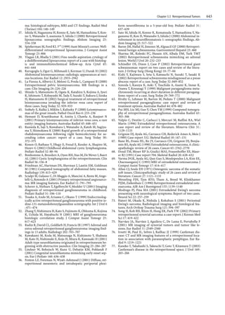


![CONGENITAL DIAPHRAGMATIC HERNIA [Recovered].pptx](https://cdn.slidesharecdn.com/ss_thumbnails/congenitaldiaphragmaticherniarecovered-240421180720-5c4f960f-thumbnail.jpg?width=640&height=640&fit=bounds)


