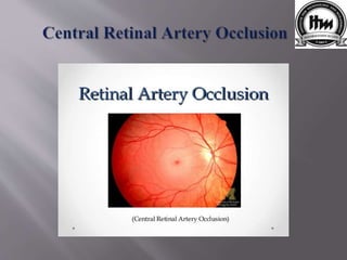
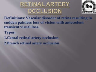
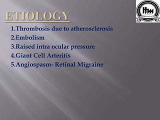
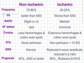
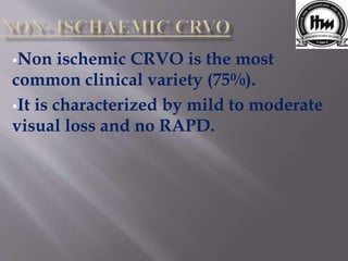
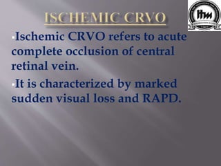
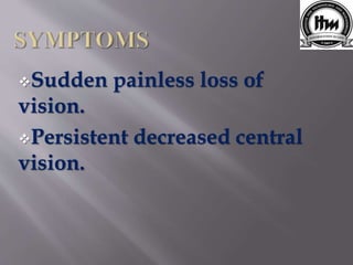
Harshali Vijay Mestri
2nd Year
B-optometry
ITM-IHS
#Central Retinal Artery Occlusion{CRAO}#
It occurs due to obstruction at the level of Lamina
cribrosa.
Clinically Retinal Artery Occlusion may present as
Central Retinal Artery occlusion (60%).
It is more common in males than females.
It is usually unilateral but rarely may be bilateral(1
to 2%cases).
Definitions: Vascular disorder of retina resulting in
sudden painless loss of vision with antecedent
transient visual loss.
Types:
1.Cenral retinal artery occlusion
2.Branch retinal artery occlusion
1.Thrombosis due to atherosclerosis
2.Embolism
3.Raised intra ocular pressure
4.Giant Cell Arteritis
5.Angiospasm- Retinal Migraine
Patients complains of sudden
painless loss of vision occurring
over seconds.
Patients may give history of
transient visual loss in (amaurosis
fugax) the past
Visual Acuity severely reduced between
counting finger and light perception.
Direct pupillary light reflex- absent.
Fundus Examination- whitish appearance of
retina. Thin retinal arteries, segmented blood
column in retinal vessels.
Cherry red spot at macula , pale appearance of
optic disc
Intraocular pressure lowered immediately by
anterior chamber paracentasis or I.V.
Acetazolamide.
Inhaled oxygen and carbon dioxide mixture to
improve oxygen delivery to retina.
Thrombolytic therapy.
Definition : It’s a common vascular disorder
characterized by retinal vein occlusion resulting in
edema and hemorrhages on retina in the affected
region with potential blinding complications.
Types :
1. Central retinal vein occlusion
2. Branch retinal vein occlusion
CRVO is an end vein, its occlusion leads to
complete defective of vision in the morning.
These typically affect elderly patients in sixth or
seventh decade of life.
It may be Non-ischemic CRVO (venous stasis
retinopathy) or Ischemic CRVO (haemorrhagic
retinopathy).
Age of age above 30 years.
Systemic diseases like
hyperlipidemia, diabetes, chronic
renal failure.
Chronic open angle glaucoma.
Non ischemic CRVO is the most
common clinical variety (75%).
It is characterized by mild to moderate
visual loss and no RAPD.
Ischemic CRVO refers to acute
complete occlusion of central
retinal vein.
It is characterized by marked
sudden visual loss and RAPD.
Sudden painless loss of
vision.
Persistent decreased central
vision.
Visual acuity- severe visual loss, up to 20/200.
Intra ocular pressure raised
Fundus Examination- dilated tortuous veins,
retinal and macular edema, flame shape
hemorrhages and cotton wool spots .
FLUORESCEIN ANGIOGRAPHY
ECG
BLOOD CP
BLOOD GLUCOSE LEVEL.
COMPLICATION
CHRONIC MACULAR EDEMA
RETINAL NEOVASCULARIZATION
NEOVASCULAR GLAUCOMA
CONVERSION FROM NON-ISCHEMIC
TO ISCHEMIC TYPE.
CRAO AND CRVO

CRAO AND CRVO

  • 1.
    Harshali Vijay Mestri 2ndYear B-optometry ITM-IHS
  • 2.
    #Central Retinal ArteryOcclusion{CRAO}# It occurs due to obstruction at the level of Lamina cribrosa. Clinically Retinal Artery Occlusion may present as Central Retinal Artery occlusion (60%). It is more common in males than females. It is usually unilateral but rarely may be bilateral(1 to 2%cases).
  • 4.
    Definitions: Vascular disorderof retina resulting in sudden painless loss of vision with antecedent transient visual loss. Types: 1.Cenral retinal artery occlusion 2.Branch retinal artery occlusion
  • 5.
    1.Thrombosis due toatherosclerosis 2.Embolism 3.Raised intra ocular pressure 4.Giant Cell Arteritis 5.Angiospasm- Retinal Migraine
  • 6.
    Patients complains ofsudden painless loss of vision occurring over seconds. Patients may give history of transient visual loss in (amaurosis fugax) the past
  • 7.
    Visual Acuity severelyreduced between counting finger and light perception. Direct pupillary light reflex- absent. Fundus Examination- whitish appearance of retina. Thin retinal arteries, segmented blood column in retinal vessels. Cherry red spot at macula , pale appearance of optic disc
  • 8.
    Intraocular pressure loweredimmediately by anterior chamber paracentasis or I.V. Acetazolamide. Inhaled oxygen and carbon dioxide mixture to improve oxygen delivery to retina. Thrombolytic therapy.
  • 9.
    Definition : It’sa common vascular disorder characterized by retinal vein occlusion resulting in edema and hemorrhages on retina in the affected region with potential blinding complications. Types : 1. Central retinal vein occlusion 2. Branch retinal vein occlusion
  • 11.
    CRVO is anend vein, its occlusion leads to complete defective of vision in the morning. These typically affect elderly patients in sixth or seventh decade of life. It may be Non-ischemic CRVO (venous stasis retinopathy) or Ischemic CRVO (haemorrhagic retinopathy).
  • 13.
    Age of ageabove 30 years. Systemic diseases like hyperlipidemia, diabetes, chronic renal failure. Chronic open angle glaucoma.
  • 14.
    Non ischemic CRVOis the most common clinical variety (75%). It is characterized by mild to moderate visual loss and no RAPD.
  • 15.
    Ischemic CRVO refersto acute complete occlusion of central retinal vein. It is characterized by marked sudden visual loss and RAPD.
  • 16.
    Sudden painless lossof vision. Persistent decreased central vision.
  • 17.
    Visual acuity- severevisual loss, up to 20/200. Intra ocular pressure raised Fundus Examination- dilated tortuous veins, retinal and macular edema, flame shape hemorrhages and cotton wool spots .
  • 18.
    FLUORESCEIN ANGIOGRAPHY ECG BLOOD CP BLOODGLUCOSE LEVEL. COMPLICATION CHRONIC MACULAR EDEMA RETINAL NEOVASCULARIZATION NEOVASCULAR GLAUCOMA CONVERSION FROM NON-ISCHEMIC TO ISCHEMIC TYPE.