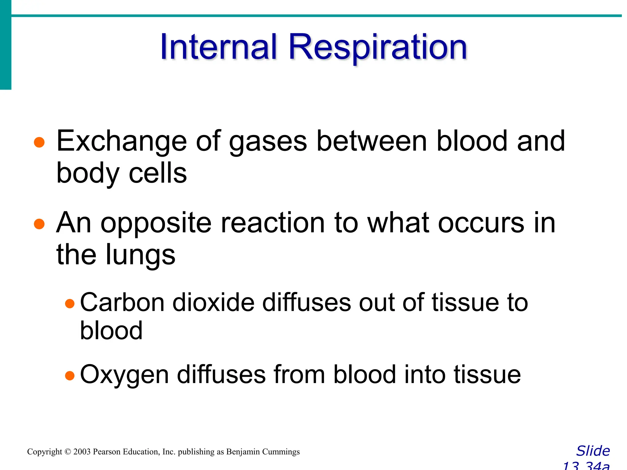
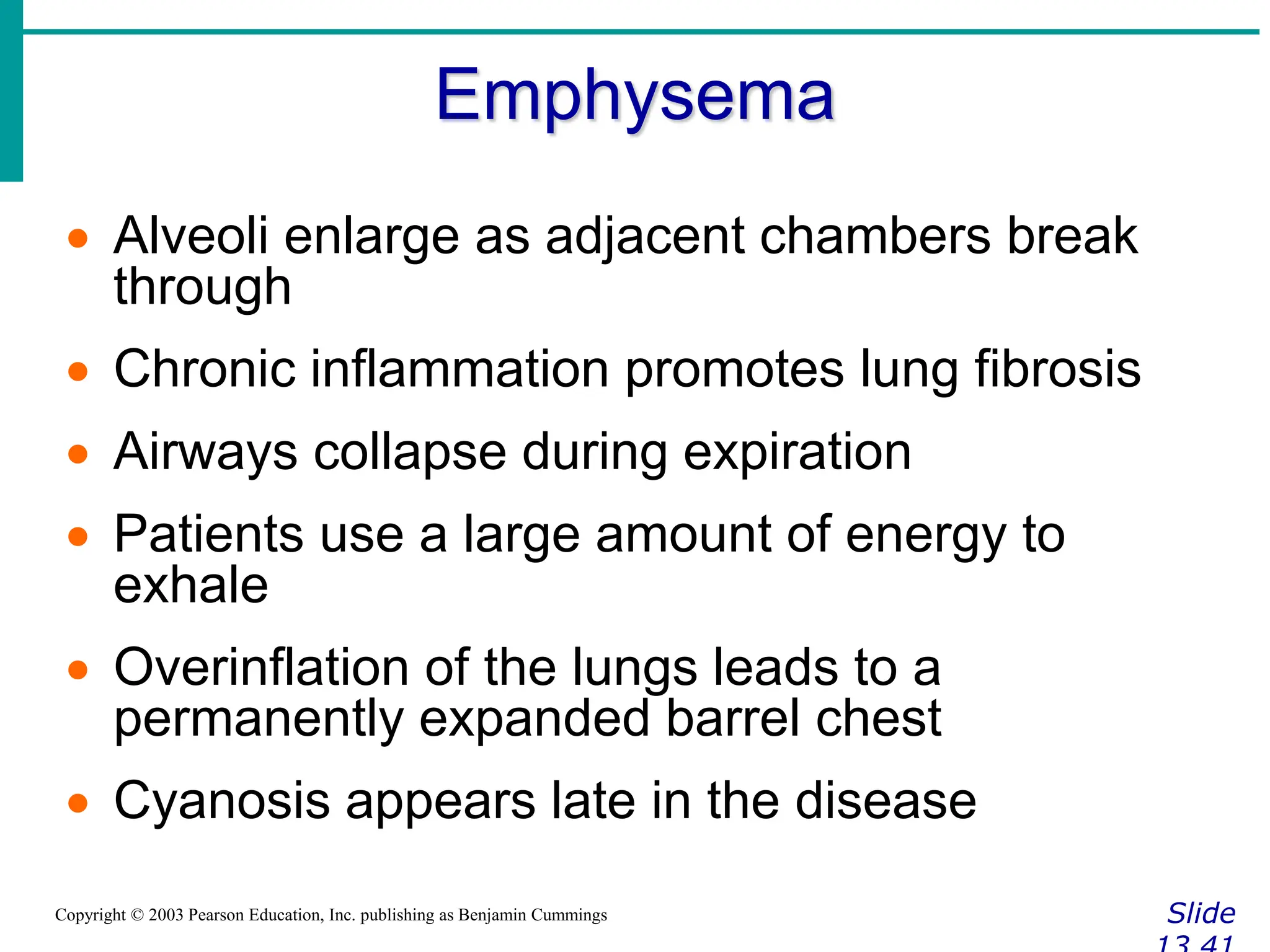
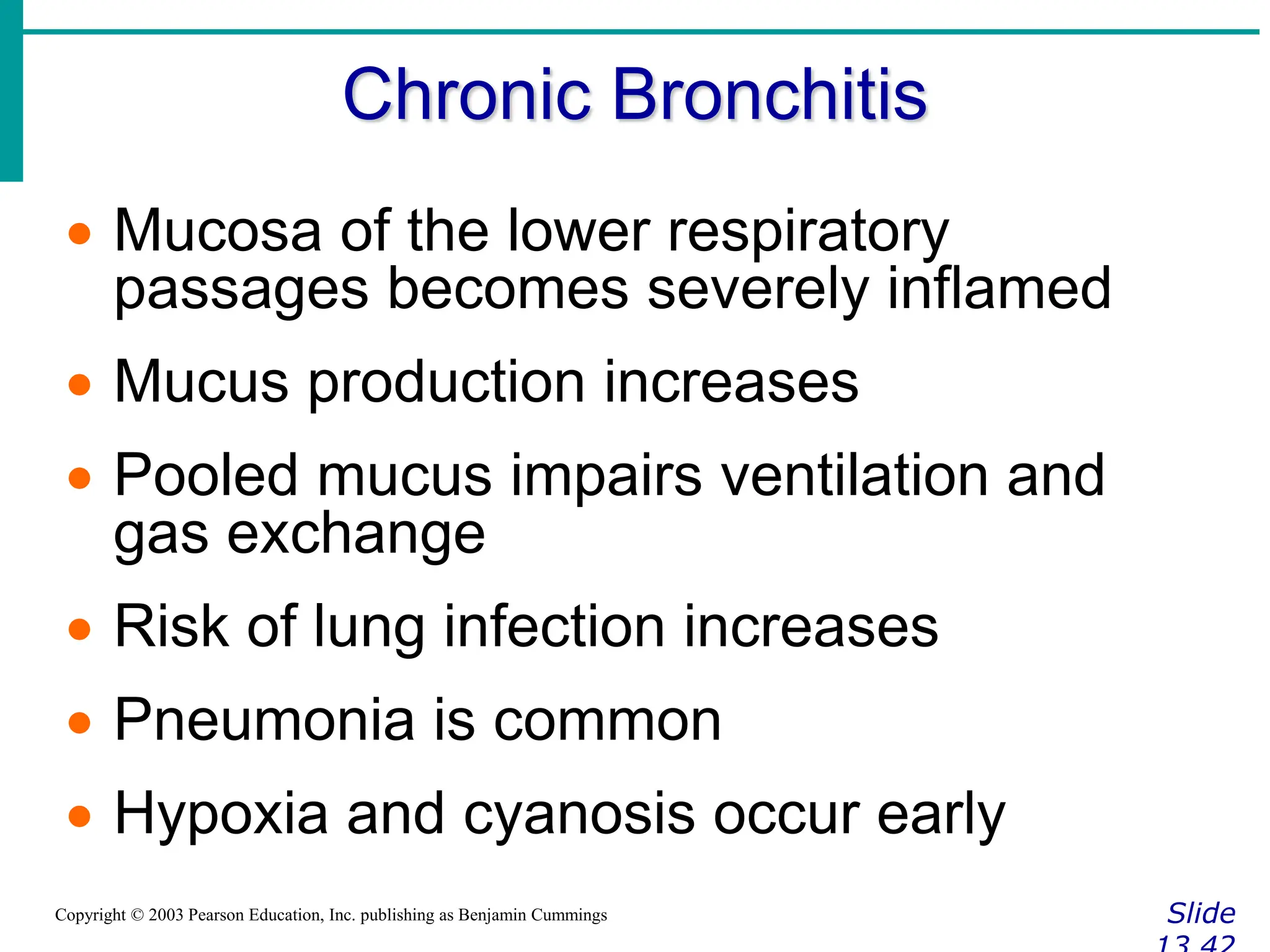
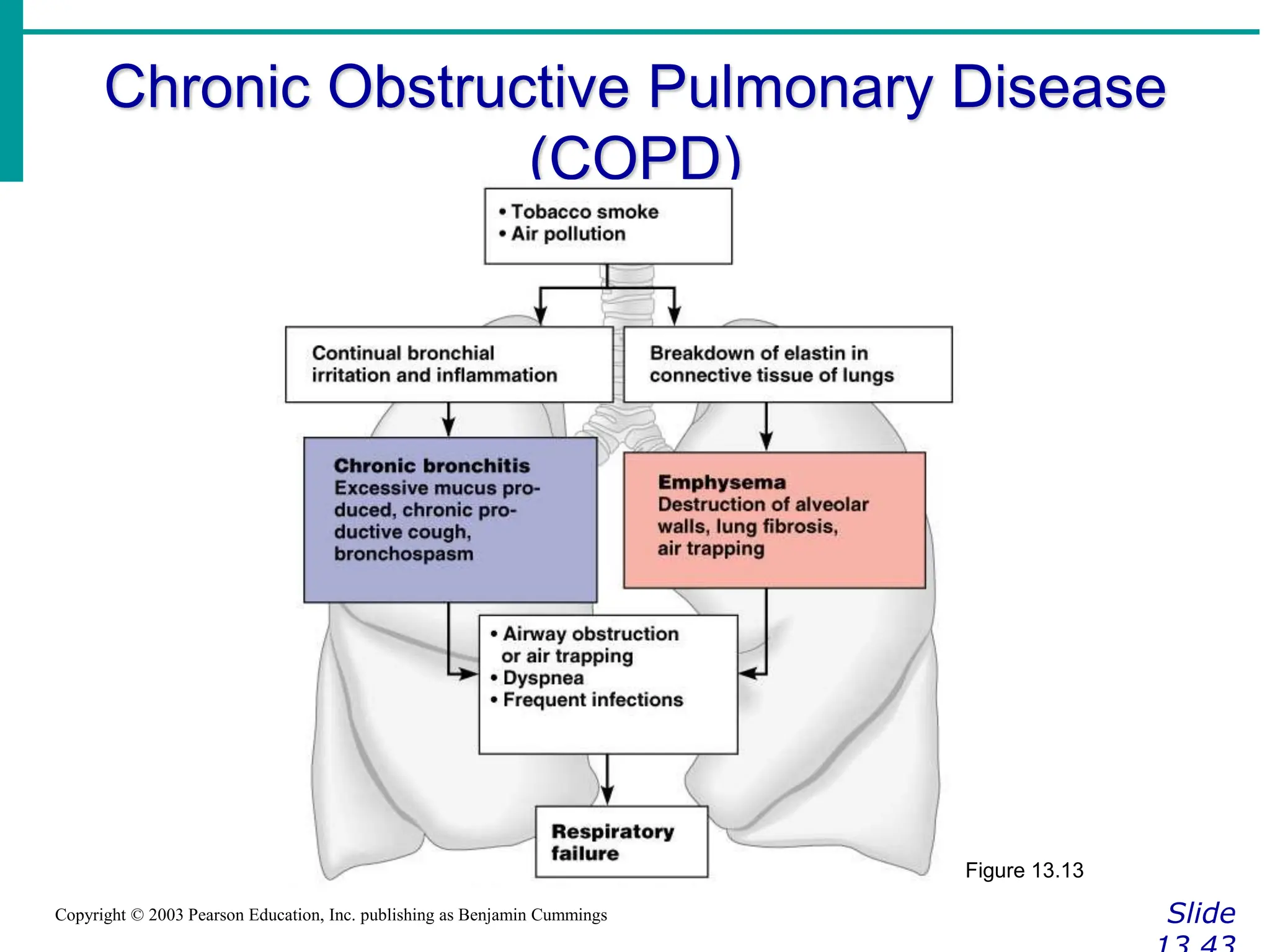
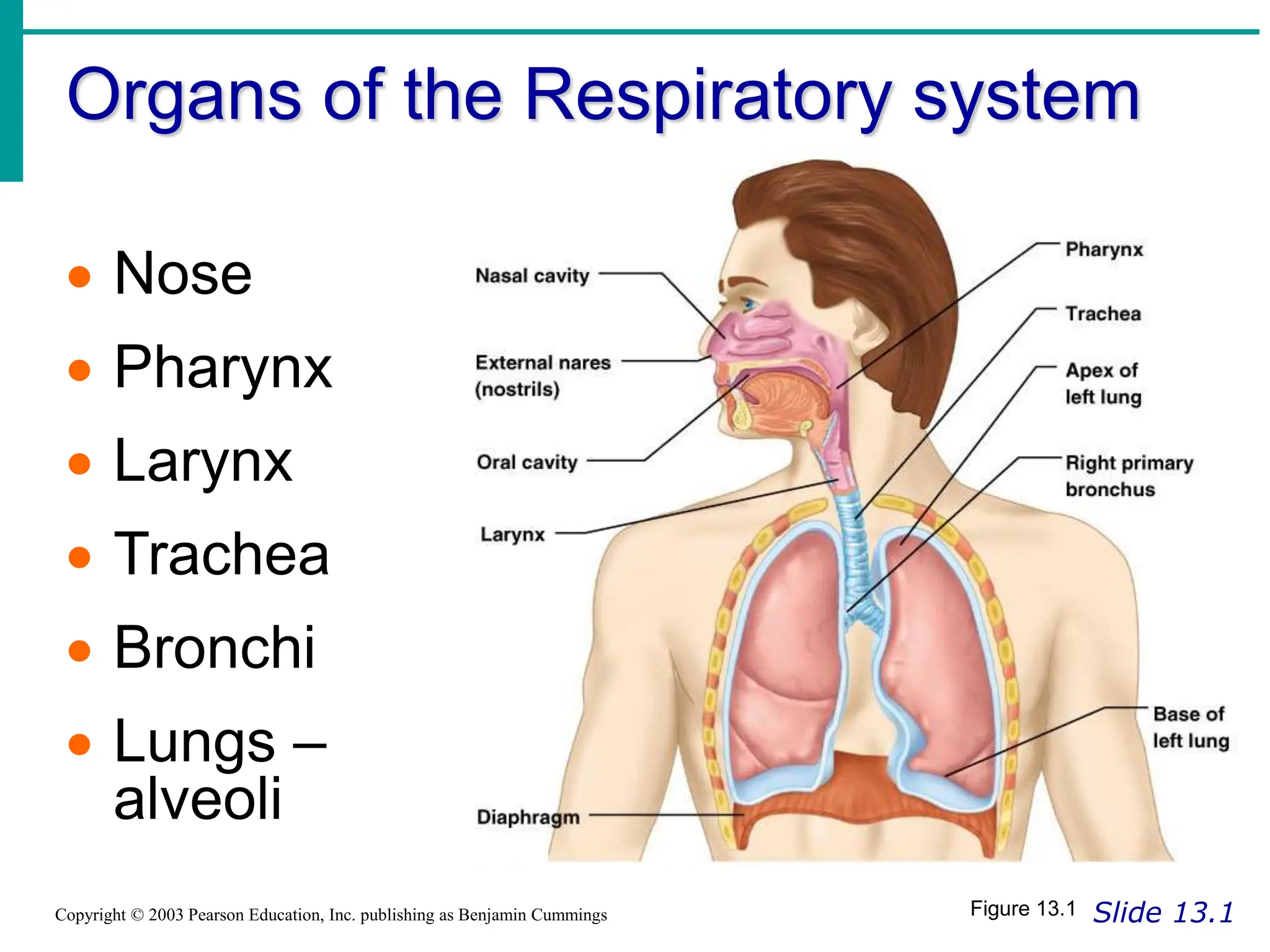
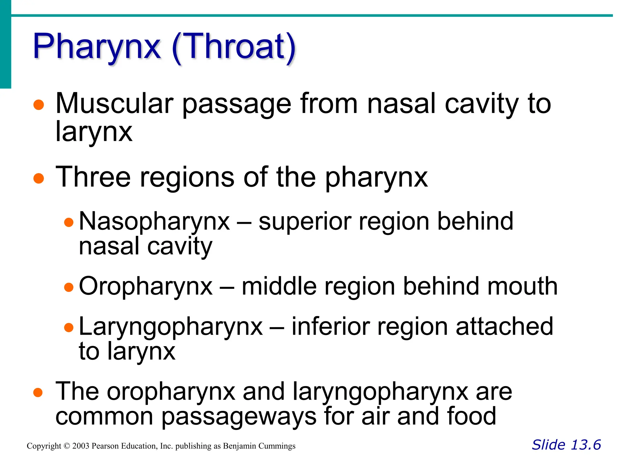
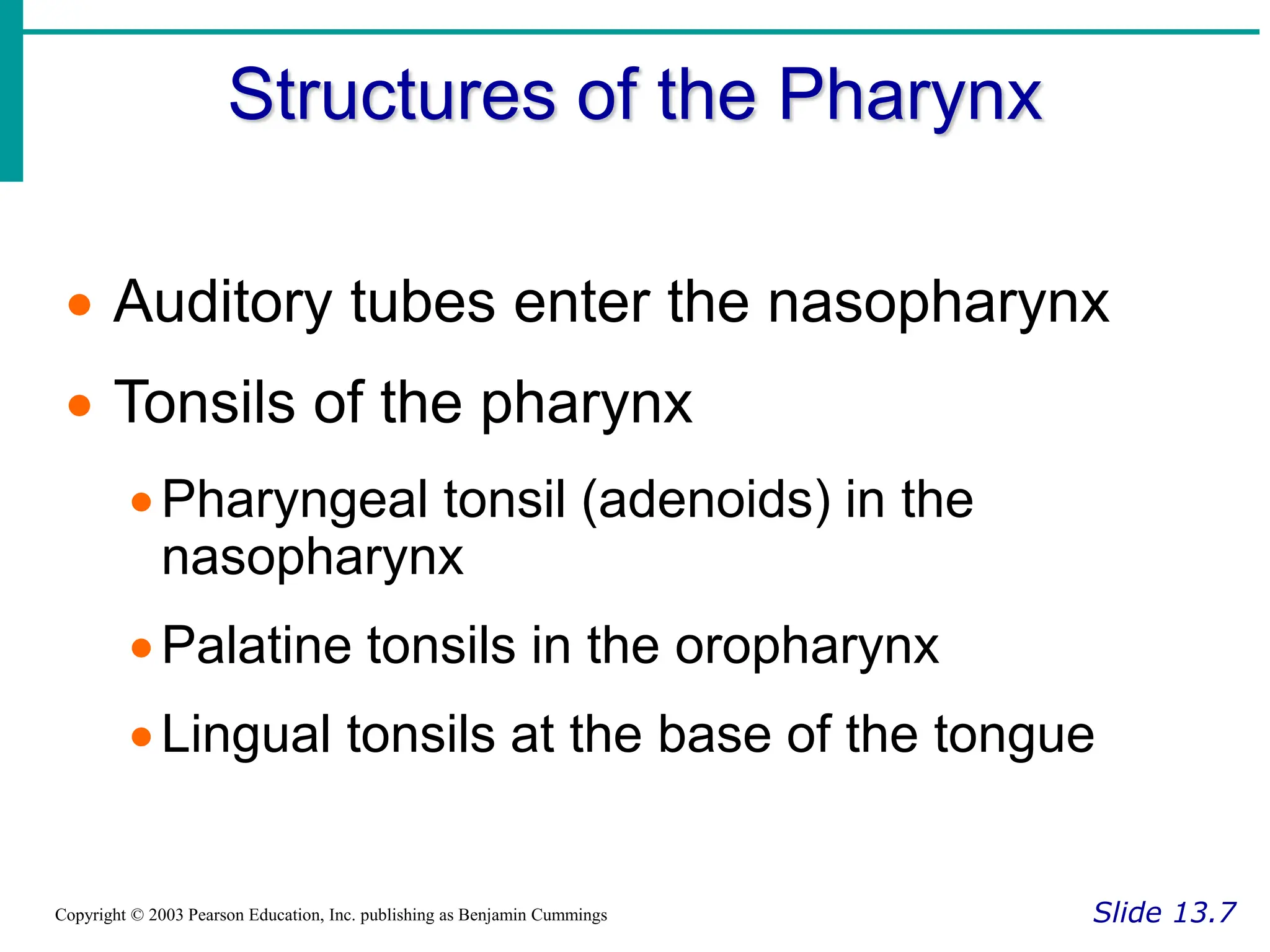
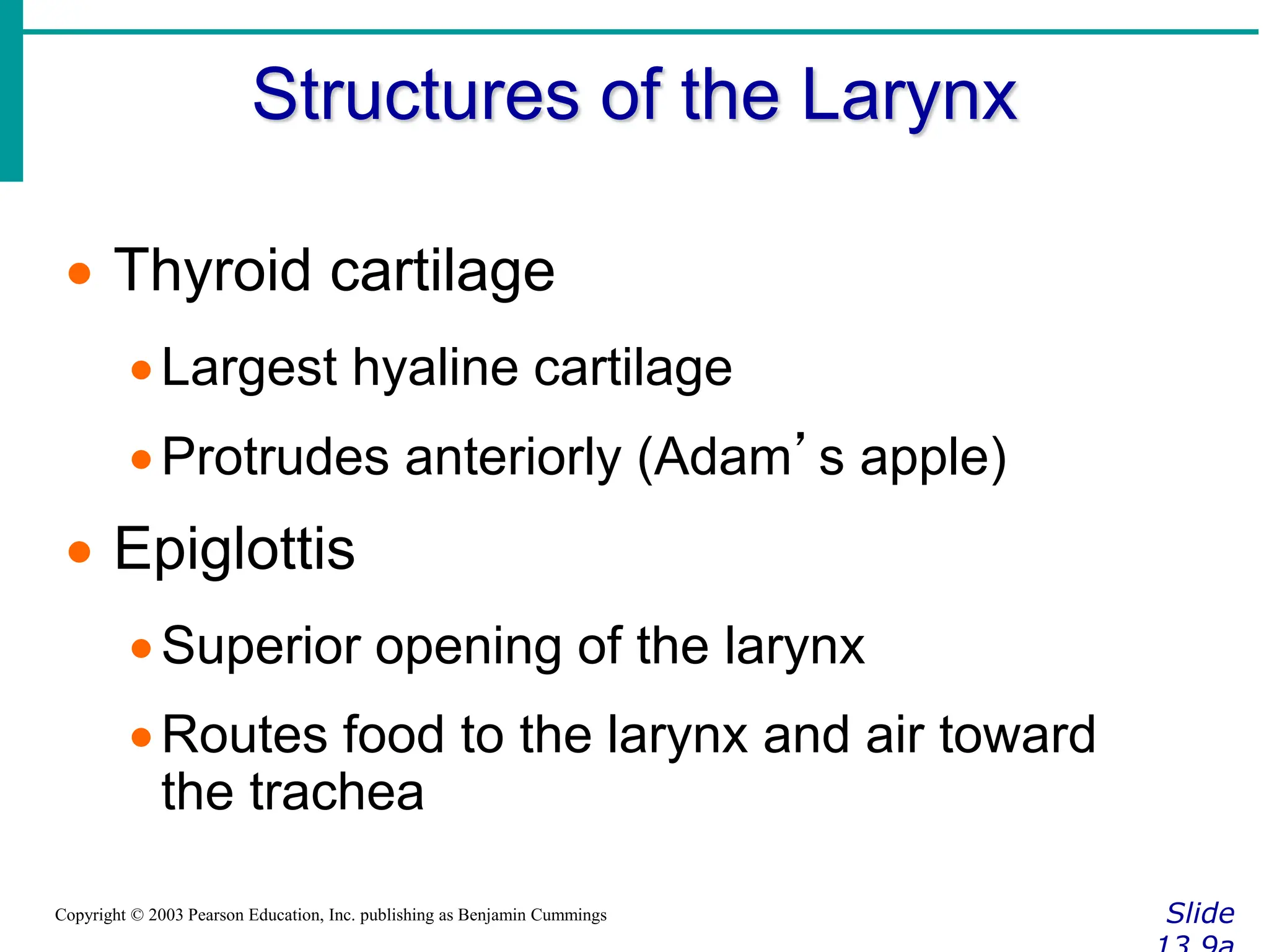
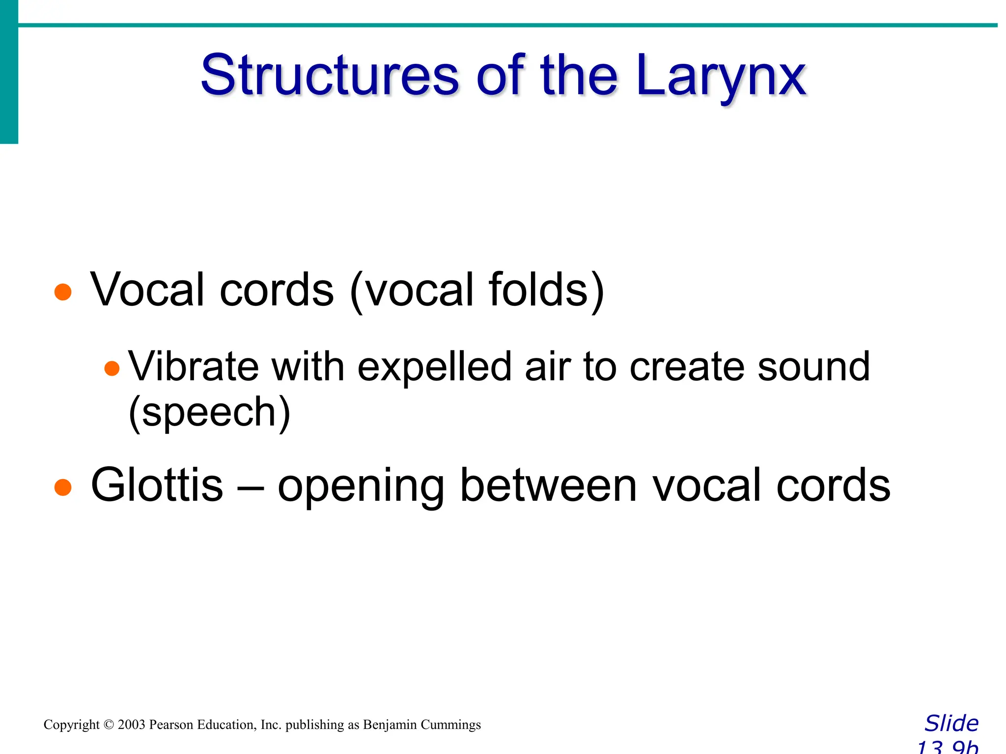
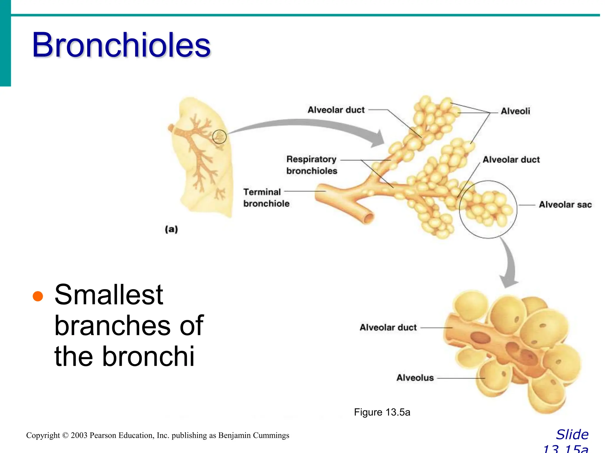
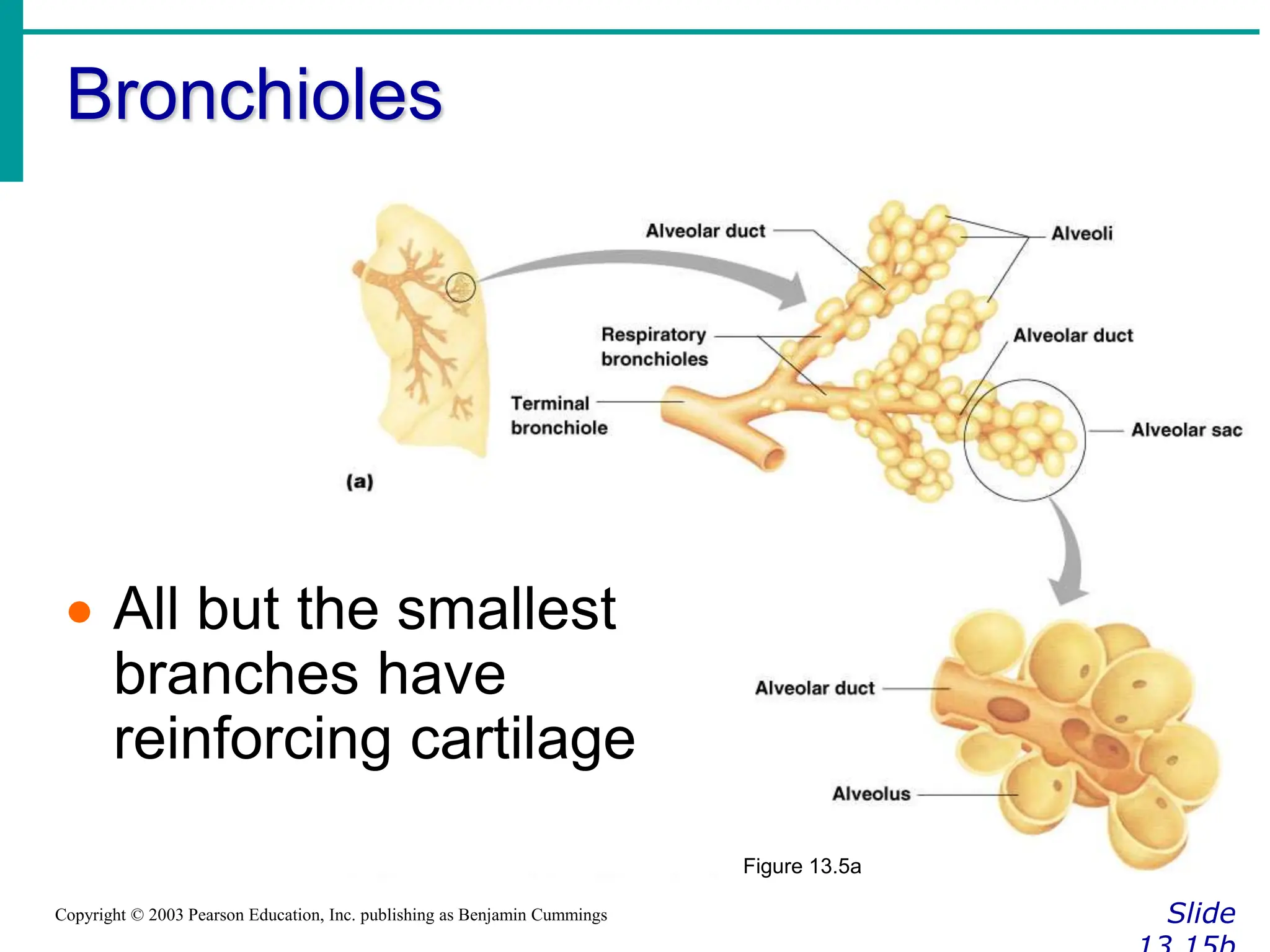
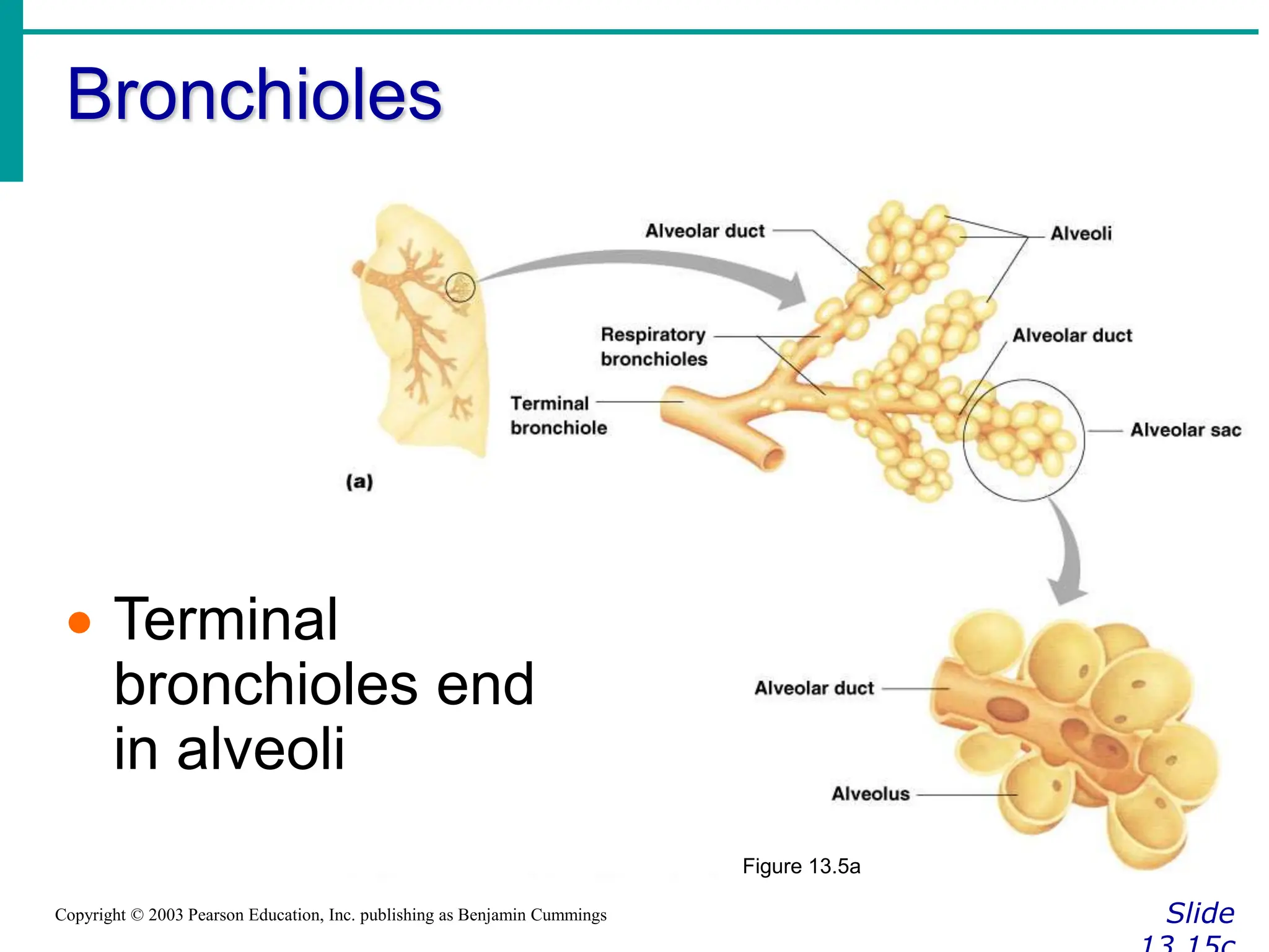
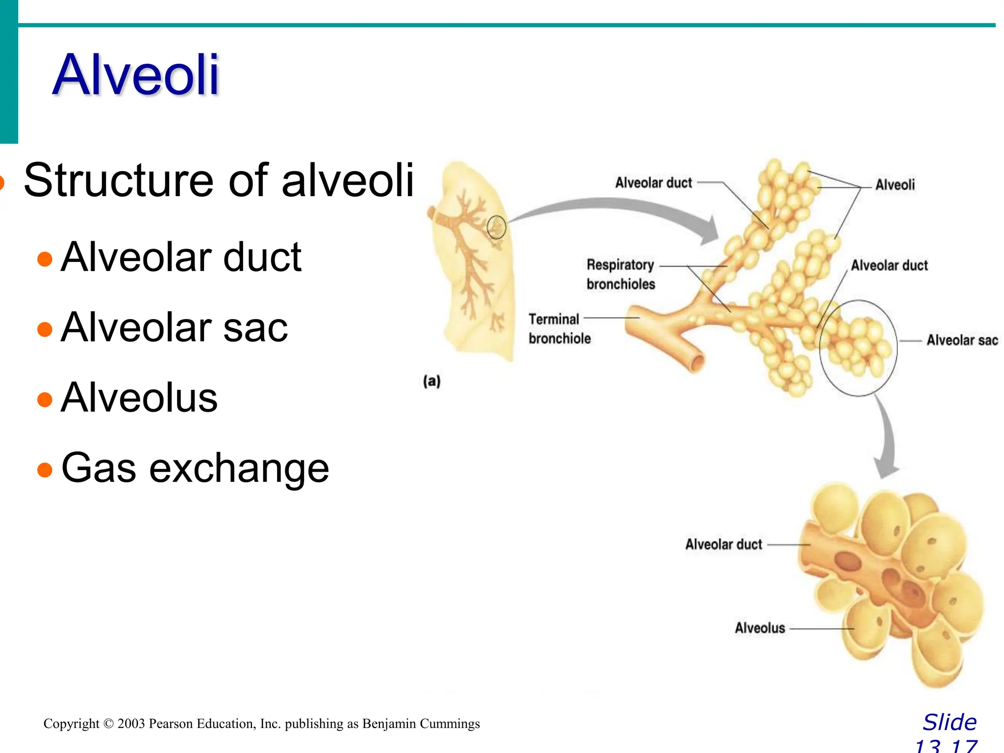
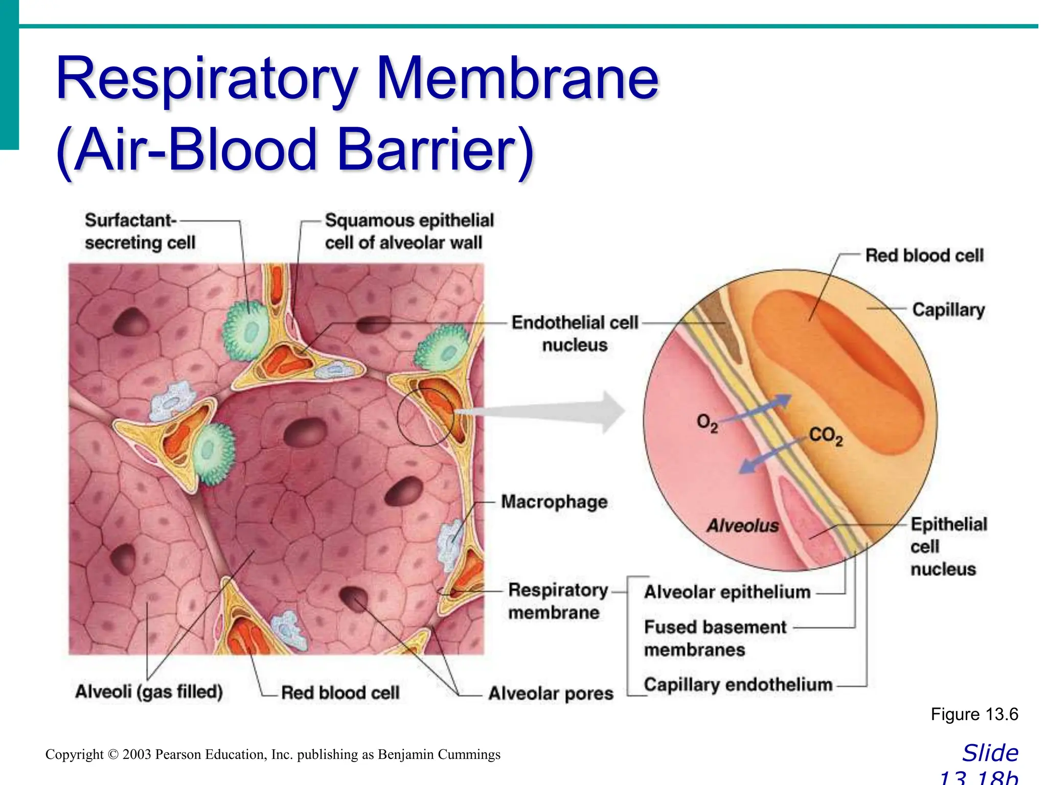
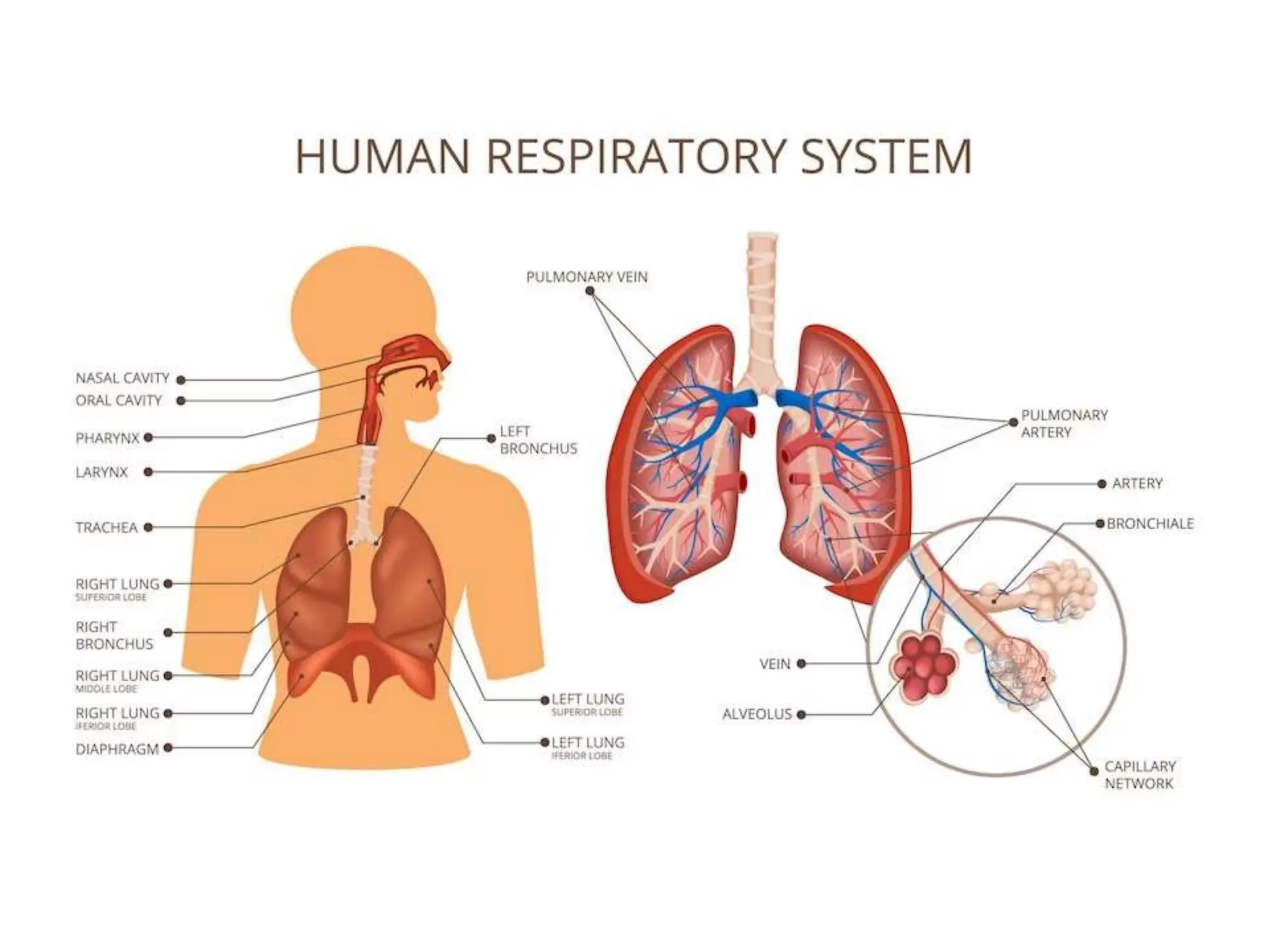
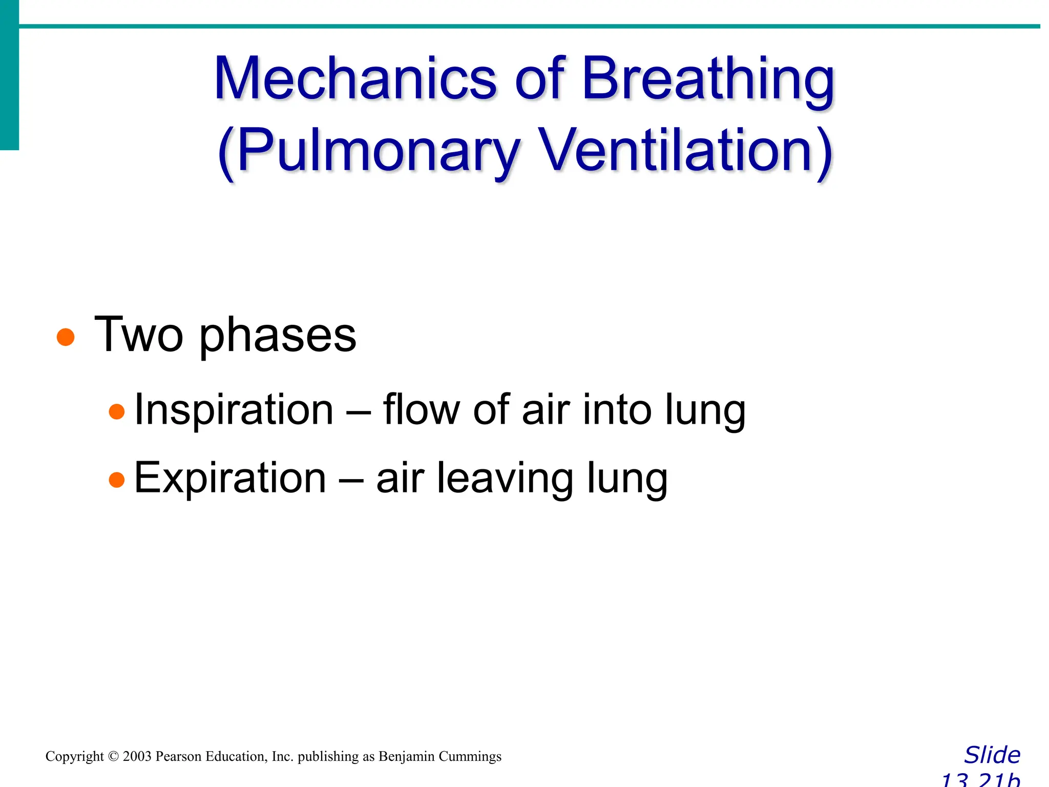
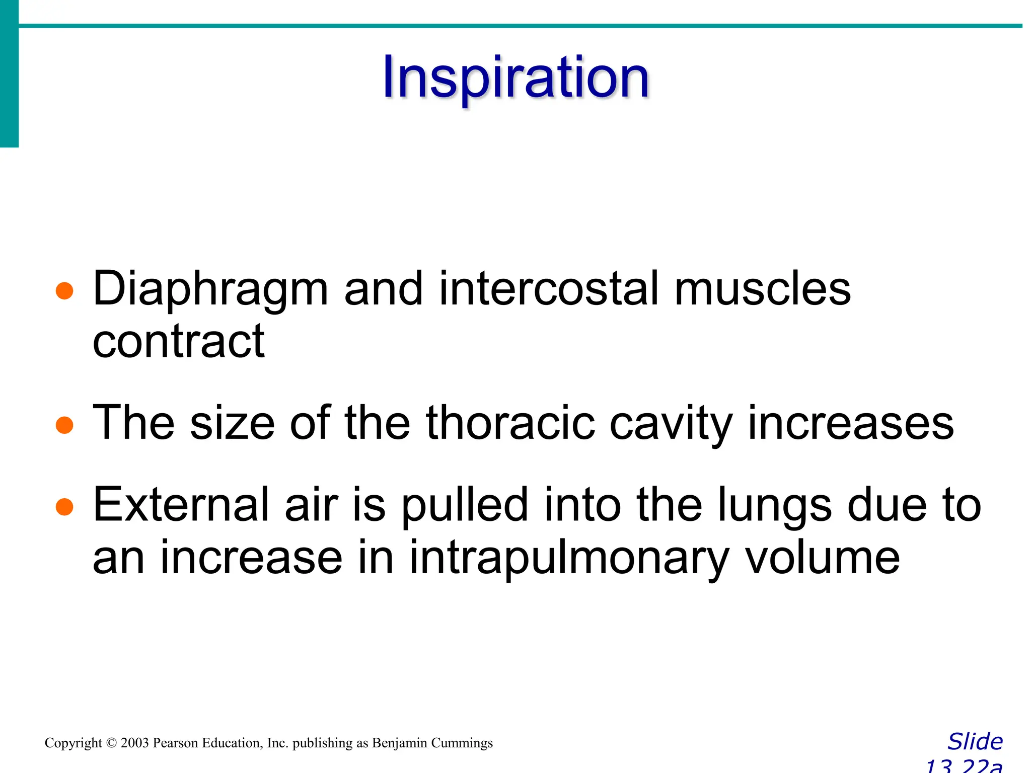
The document provides an overview of the structure and function of the respiratory system. It describes the key organs involved, including the nose, pharynx, larynx, trachea, bronchi, lungs and alveoli. It explains that the respiratory system facilitates gas exchange between the blood and air, allowing oxygen to diffuse into the blood and carbon dioxide to diffuse out. It also summarizes several respiratory diseases like emphysema, chronic bronchitis, lung cancer and asthma. Developmental aspects and the effects of aging on the respiratory system are also briefly discussed.


![Gas Transport in the Blood
Slide
Copyright © 2003 Pearson Education, Inc. publishing as Benjamin Cummings
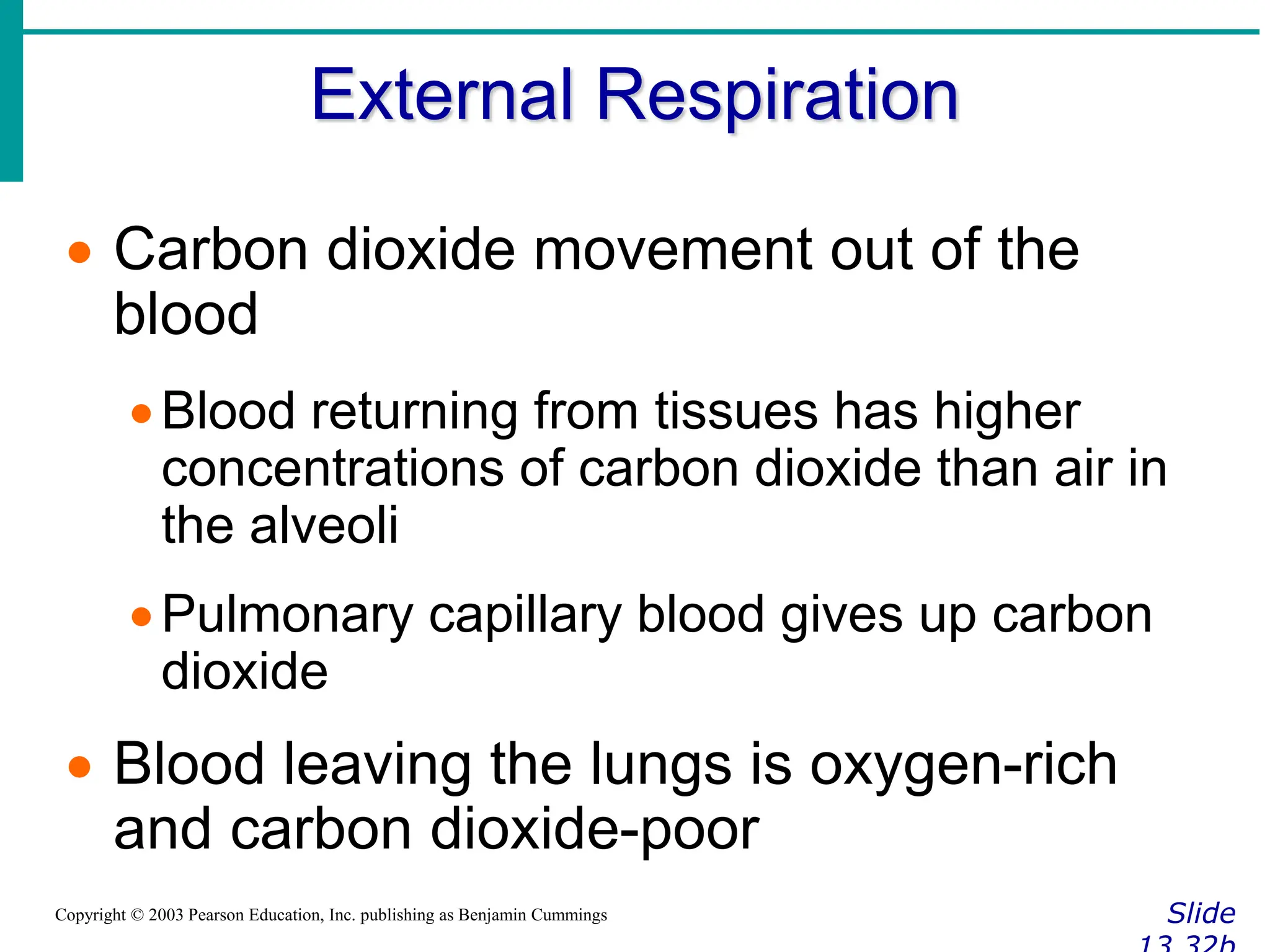
Oxygen transport in the blood
Inside red blood cells attached to
hemoglobin (oxyhemoglobin [HbO2])
A small amount is carried dissolved in the
plasma](https://image.slidesharecdn.com/respiratoryppt-240403130058-c32a393e/75/respiratoryppt-ppt-slide-presentation-for-grade-9-33-2048.jpg)