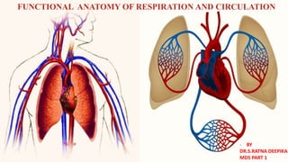
The document provides an overview of the functional anatomy of the respiratory and circulatory systems. It describes the major structures of the respiratory system including the nasal cavities, pharynx, larynx, trachea, bronchi, lungs, and associated blood vessels and nerves. It discusses the divisions of the upper and lower respiratory tract. It also describes the structural features and functions of key components like the lungs, trachea, bronchi and associated blood supply.
Anatomy of Tracheobronchial Tree and Bronchopulmonary Segments with summary o...Jega Subramaniam
Edited version of my Presentation in College.
Hope its useful for you rather than sleeping in my desktop.
Sorry if there is any mistakes.
Thanks and god bless.
Introduction
Functions
Development
Structure
Nasal cavity
Nasal septum
Lateral wall
Applied anatomy and pathology –
- danger area of nose
- nose bleeding
- foreign body in nose
- developmental nasal deformities
- nasal polyps
- mouth breathing
- rhinitis
Dentist in pune.(BDS. MDS) - Dr. Amit T. Suryawanshi. Nose & Paranasal sinuses.All Good Things
Dentist in pune. (BDS. MDS) - Dr. Amit T. Suryawanshi. Seminar- Nose & Paranasal sinuses.
Email ID- amitsuryawanshi999@gmail.com
Contact -Ph no.-9405622455
Subscribe our channel on youtube - Copy and paste this URL. https://www.youtube.com/channel/UC_gylEXTrjmEbbOTSXjuZ4Q/videos?view_as=public
Follow us on slideshare
Join live classes, download study aids, sell your documents, join or host your own classes online, get tutoring, tutor students, take practices tests and more at Examville.com
Duplicating and refractory materials used in removable partial/endodontic co...Indian dental academy
The Indian Dental Academy is the Leader in continuing dental education , training dentists in all aspects of dentistry and
offering a wide range of dental certified courses in different formats.for more details please visit
www.indiandentalacademy.com
Title: Sense of Smell
Presenter: Dr. Faiza, Assistant Professor of Physiology
Qualifications:
MBBS (Best Graduate, AIMC Lahore)
FCPS Physiology
ICMT, CHPE, DHPE (STMU)
MPH (GC University, Faisalabad)
MBA (Virtual University of Pakistan)
Learning Objectives:
Describe the primary categories of smells and the concept of odor blindness.
Explain the structure and location of the olfactory membrane and mucosa, including the types and roles of cells involved in olfaction.
Describe the pathway and mechanisms of olfactory signal transmission from the olfactory receptors to the brain.
Illustrate the biochemical cascade triggered by odorant binding to olfactory receptors, including the role of G-proteins and second messengers in generating an action potential.
Identify different types of olfactory disorders such as anosmia, hyposmia, hyperosmia, and dysosmia, including their potential causes.
Key Topics:
Olfactory Genes:
3% of the human genome accounts for olfactory genes.
400 genes for odorant receptors.
Olfactory Membrane:
Located in the superior part of the nasal cavity.
Medially: Folds downward along the superior septum.
Laterally: Folds over the superior turbinate and upper surface of the middle turbinate.
Total surface area: 5-10 square centimeters.
Olfactory Mucosa:
Olfactory Cells: Bipolar nerve cells derived from the CNS (100 million), with 4-25 olfactory cilia per cell.
Sustentacular Cells: Produce mucus and maintain ionic and molecular environment.
Basal Cells: Replace worn-out olfactory cells with an average lifespan of 1-2 months.
Bowman’s Gland: Secretes mucus.
Stimulation of Olfactory Cells:
Odorant dissolves in mucus and attaches to receptors on olfactory cilia.
Involves a cascade effect through G-proteins and second messengers, leading to depolarization and action potential generation in the olfactory nerve.
Quality of a Good Odorant:
Small (3-20 Carbon atoms), volatile, water-soluble, and lipid-soluble.
Facilitated by odorant-binding proteins in mucus.
Membrane Potential and Action Potential:
Resting membrane potential: -55mV.
Action potential frequency in the olfactory nerve increases with odorant strength.
Adaptation Towards the Sense of Smell:
Rapid adaptation within the first second, with further slow adaptation.
Psychological adaptation greater than receptor adaptation, involving feedback inhibition from the central nervous system.
Primary Sensations of Smell:
Camphoraceous, Musky, Floral, Pepperminty, Ethereal, Pungent, Putrid.
Odor Detection Threshold:
Examples: Hydrogen sulfide (0.0005 ppm), Methyl-mercaptan (0.002 ppm).
Some toxic substances are odorless at lethal concentrations.
Characteristics of Smell:
Odor blindness for single substances due to lack of appropriate receptor protein.
Behavioral and emotional influences of smell.
Transmission of Olfactory Signals:
From olfactory cells to glomeruli in the olfactory bulb, involving lateral inhibition.
Primitive, less old, and new olfactory systems with different path
Explore natural remedies for syphilis treatment in Singapore. Discover alternative therapies, herbal remedies, and lifestyle changes that may complement conventional treatments. Learn about holistic approaches to managing syphilis symptoms and supporting overall health.
micro teaching on communication m.sc nursing.pdfAnurag Sharma
Microteaching is a unique model of practice teaching. It is a viable instrument for the. desired change in the teaching behavior or the behavior potential which, in specified types of real. classroom situations, tends to facilitate the achievement of specified types of objectives.
Ethanol (CH3CH2OH), or beverage alcohol, is a two-carbon alcohol
that is rapidly distributed in the body and brain. Ethanol alters many
neurochemical systems and has rewarding and addictive properties. It
is the oldest recreational drug and likely contributes to more morbidity,
mortality, and public health costs than all illicit drugs combined. The
5th edition of the Diagnostic and Statistical Manual of Mental Disorders
(DSM-5) integrates alcohol abuse and alcohol dependence into a single
disorder called alcohol use disorder (AUD), with mild, moderate,
and severe subclassifications (American Psychiatric Association, 2013).
In the DSM-5, all types of substance abuse and dependence have been
combined into a single substance use disorder (SUD) on a continuum
from mild to severe. A diagnosis of AUD requires that at least two of
the 11 DSM-5 behaviors be present within a 12-month period (mild
AUD: 2–3 criteria; moderate AUD: 4–5 criteria; severe AUD: 6–11 criteria).
The four main behavioral effects of AUD are impaired control over
drinking, negative social consequences, risky use, and altered physiological
effects (tolerance, withdrawal). This chapter presents an overview
of the prevalence and harmful consequences of AUD in the U.S.,
the systemic nature of the disease, neurocircuitry and stages of AUD,
comorbidities, fetal alcohol spectrum disorders, genetic risk factors, and
pharmacotherapies for AUD.
Prix Galien International 2024 Forum ProgramLevi Shapiro
June 20, 2024, Prix Galien International and Jerusalem Ethics Forum in ROME. Detailed agenda including panels:
- ADVANCES IN CARDIOLOGY: A NEW PARADIGM IS COMING
- WOMEN’S HEALTH: FERTILITY PRESERVATION
- WHAT’S NEW IN THE TREATMENT OF INFECTIOUS,
ONCOLOGICAL AND INFLAMMATORY SKIN DISEASES?
- ARTIFICIAL INTELLIGENCE AND ETHICS
- GENE THERAPY
- BEYOND BORDERS: GLOBAL INITIATIVES FOR DEMOCRATIZING LIFE SCIENCE TECHNOLOGIES AND PROMOTING ACCESS TO HEALTHCARE
- ETHICAL CHALLENGES IN LIFE SCIENCES
- Prix Galien International Awards Ceremony
Recomendações da OMS sobre cuidados maternos e neonatais para uma experiência pós-natal positiva.
Em consonância com os ODS – Objetivos do Desenvolvimento Sustentável e a Estratégia Global para a Saúde das Mulheres, Crianças e Adolescentes, e aplicando uma abordagem baseada nos direitos humanos, os esforços de cuidados pós-natais devem expandir-se para além da cobertura e da simples sobrevivência, de modo a incluir cuidados de qualidade.
Estas diretrizes visam melhorar a qualidade dos cuidados pós-natais essenciais e de rotina prestados às mulheres e aos recém-nascidos, com o objetivo final de melhorar a saúde e o bem-estar materno e neonatal.
Uma “experiência pós-natal positiva” é um resultado importante para todas as mulheres que dão à luz e para os seus recém-nascidos, estabelecendo as bases para a melhoria da saúde e do bem-estar a curto e longo prazo. Uma experiência pós-natal positiva é definida como aquela em que as mulheres, pessoas que gestam, os recém-nascidos, os casais, os pais, os cuidadores e as famílias recebem informação consistente, garantia e apoio de profissionais de saúde motivados; e onde um sistema de saúde flexível e com recursos reconheça as necessidades das mulheres e dos bebês e respeite o seu contexto cultural.
Estas diretrizes consolidadas apresentam algumas recomendações novas e já bem fundamentadas sobre cuidados pós-natais de rotina para mulheres e neonatos que recebem cuidados no pós-parto em unidades de saúde ou na comunidade, independentemente dos recursos disponíveis.
É fornecido um conjunto abrangente de recomendações para cuidados durante o período puerperal, com ênfase nos cuidados essenciais que todas as mulheres e recém-nascidos devem receber, e com a devida atenção à qualidade dos cuidados; isto é, a entrega e a experiência do cuidado recebido. Estas diretrizes atualizam e ampliam as recomendações da OMS de 2014 sobre cuidados pós-natais da mãe e do recém-nascido e complementam as atuais diretrizes da OMS sobre a gestão de complicações pós-natais.
O estabelecimento da amamentação e o manejo das principais intercorrências é contemplada.
Recomendamos muito.
Vamos discutir essas recomendações no nosso curso de pós-graduação em Aleitamento no Instituto Ciclos.
Esta publicação só está disponível em inglês até o momento.
Prof. Marcus Renato de Carvalho
www.agostodourado.com
HOT NEW PRODUCT! BIG SALES FAST SHIPPING NOW FROM CHINA!! EU KU DB BK substit...GL Anaacs
Contact us if you are interested:
Email / Skype : kefaya1771@gmail.com
Threema: PXHY5PDH
New BATCH Ku !!! MUCH IN DEMAND FAST SALE EVERY BATCH HAPPY GOOD EFFECT BIG BATCH !
Contact me on Threema or skype to start big business!!
Hot-sale products:
NEW HOT EUTYLONE WHITE CRYSTAL!!
5cl-adba precursor (semi finished )
5cl-adba raw materials
ADBB precursor (semi finished )
ADBB raw materials
APVP powder
5fadb/4f-adb
Jwh018 / Jwh210
Eutylone crystal
Protonitazene (hydrochloride) CAS: 119276-01-6
Flubrotizolam CAS: 57801-95-3
Metonitazene CAS: 14680-51-4
Payment terms: Western Union,MoneyGram,Bitcoin or USDT.
Deliver Time: Usually 7-15days
Shipping method: FedEx, TNT, DHL,UPS etc.Our deliveries are 100% safe, fast, reliable and discreet.
Samples will be sent for your evaluation!If you are interested in, please contact me, let's talk details.
We specializes in exporting high quality Research chemical, medical intermediate, Pharmaceutical chemicals and so on. Products are exported to USA, Canada, France, Korea, Japan,Russia, Southeast Asia and other countries.
Lung Cancer: Artificial Intelligence, Synergetics, Complex System Analysis, S...Oleg Kshivets
RESULTS: Overall life span (LS) was 2252.1±1742.5 days and cumulative 5-year survival (5YS) reached 73.2%, 10 years – 64.8%, 20 years – 42.5%. 513 LCP lived more than 5 years (LS=3124.6±1525.6 days), 148 LCP – more than 10 years (LS=5054.4±1504.1 days).199 LCP died because of LC (LS=562.7±374.5 days). 5YS of LCP after bi/lobectomies was significantly superior in comparison with LCP after pneumonectomies (78.1% vs.63.7%, P=0.00001 by log-rank test). AT significantly improved 5YS (66.3% vs. 34.8%) (P=0.00000 by log-rank test) only for LCP with N1-2. Cox modeling displayed that 5YS of LCP significantly depended on: phase transition (PT) early-invasive LC in terms of synergetics, PT N0—N12, cell ratio factors (ratio between cancer cells- CC and blood cells subpopulations), G1-3, histology, glucose, AT, blood cell circuit, prothrombin index, heparin tolerance, recalcification time (P=0.000-0.038). Neural networks, genetic algorithm selection and bootstrap simulation revealed relationships between 5YS and PT early-invasive LC (rank=1), PT N0—N12 (rank=2), thrombocytes/CC (3), erythrocytes/CC (4), eosinophils/CC (5), healthy cells/CC (6), lymphocytes/CC (7), segmented neutrophils/CC (8), stick neutrophils/CC (9), monocytes/CC (10); leucocytes/CC (11). Correct prediction of 5YS was 100% by neural networks computing (area under ROC curve=1.0; error=0.0).
CONCLUSIONS: 5YS of LCP after radical procedures significantly depended on: 1) PT early-invasive cancer; 2) PT N0--N12; 3) cell ratio factors; 4) blood cell circuit; 5) biochemical factors; 6) hemostasis system; 7) AT; 8) LC characteristics; 9) LC cell dynamics; 10) surgery type: lobectomy/pneumonectomy; 11) anthropometric data. Optimal diagnosis and treatment strategies for LC are: 1) screening and early detection of LC; 2) availability of experienced thoracic surgeons because of complexity of radical procedures; 3) aggressive en block surgery and adequate lymph node dissection for completeness; 4) precise prediction; 5) adjuvant chemoimmunoradiotherapy for LCP with unfavorable prognosis.
TEST BANK for Operations Management, 14th Edition by William J. Stevenson, Ve...kevinkariuki227
TEST BANK for Operations Management, 14th Edition by William J. Stevenson, Verified Chapters 1 - 19, Complete Newest Version.pdf
TEST BANK for Operations Management, 14th Edition by William J. Stevenson, Verified Chapters 1 - 19, Complete Newest Version.pdf
The prostate is an exocrine gland of the male mammalian reproductive system
It is a walnut-sized gland that forms part of the male reproductive system and is located in front of the rectum and just below the urinary bladder
Function is to store and secrete a clear, slightly alkaline fluid that constitutes 10-30% of the volume of the seminal fluid that along with the spermatozoa, constitutes semen
A healthy human prostate measures (4cm-vertical, by 3cm-horizontal, 2cm ant-post ).
It surrounds the urethra just below the urinary bladder. It has anterior, median, posterior and two lateral lobes
It’s work is regulated by androgens which are responsible for male sex characteristics
Generalised disease of the prostate due to hormonal derangement which leads to non malignant enlargement of the gland (increase in the number of epithelial cells and stromal tissue)to cause compression of the urethra leading to symptoms (LUTS
Pulmonary Thromboembolism - etilogy, types, medical- Surgical and nursing man...VarunMahajani
Disruption of blood supply to lung alveoli due to blockage of one or more pulmonary blood vessels is called as Pulmonary thromboembolism. In this presentation we will discuss its causes, types and its management in depth.
Knee anatomy and clinical tests 2024.pdfvimalpl1234
This includes all relevant anatomy and clinical tests compiled from standard textbooks, Campbell,netter etc..It is comprehensive and best suited for orthopaedicians and orthopaedic residents.
These simplified slides by Dr. Sidra Arshad present an overview of the non-respiratory functions of the respiratory tract.
Learning objectives:
1. Enlist the non-respiratory functions of the respiratory tract
2. Briefly explain how these functions are carried out
3. Discuss the significance of dead space
4. Differentiate between minute ventilation and alveolar ventilation
5. Describe the cough and sneeze reflexes
Study Resources:
1. Chapter 39, Guyton and Hall Textbook of Medical Physiology, 14th edition
2. Chapter 34, Ganong’s Review of Medical Physiology, 26th edition
3. Chapter 17, Human Physiology by Lauralee Sherwood, 9th edition
4. Non-respiratory functions of the lungs https://academic.oup.com/bjaed/article/13/3/98/278874
3. INTRODUCTION
• Respiratory system consists of
respiratory surfaces of lungs , and the air
passages provided by the nose, pharynx,
larynx and respiratory tree.
• Associated with the nasal cavities are
series of bony sacs, paranasal sinuses,
which are uncertain and included here
with respiratory tract
4. • Besides its primary respiratory role in gaseous exchange and
ventilation, a number of accessory activities are performed by
respiratory system as a whole
- Production of sound (phonation) by larynx and its
related structures.
- Odorant sampling by olfactory senses in the nasal
chambers.
- Mechanical stabilisation of thorax during
mechanical exertion.
- Biochemical functions such as conversion of
Angiotensin I to Angiotensin II
5. STRUCTURAL FEATURES OF RESPIRATORY SYSTEM
Respiratory surface:
• Very large as much as 200m2 .
• Forms very thin, moist barrier between air and blood capillaries
around perimeters of many millions of blind-ending sacs(alveoli),
constituting much of muscle mass.
• Because of their delicate structure these respiratory surfaces are
very vulnerable to mechanical damage from inhaled particles, and
also to infectious organisms.
6. Conducting passages:
• Protecting surface area from
dehydration, heat loss and abrasive
particles, conducting passages form
a series of moist, warm adhesive
channels between alveoli and the
pharynx.
• In addition, to the mucus secreted
on to these surfaces, these tubes are
lined by cilia which beat towards
the pharynx, so continually
removing most inhaled particles
from respiratory system(the
monocilliary clearance current).
7. • DURING INSPIRATION:
When volume of thorax increases
Intra thoracic pressure decreases
Air flows through the respiratory
passages
In to enlarging alveoli
• In expiration reverse process occurs.
9. UPPER RESPIRATORY TRACT
• It can be defined as those parts of the air passages which lie above the
inlet of larynx.
It includes
‒ Nasal cavities
‒ Nasopharynx
‒ Oropharynx
10.
11. LOWER RESPIRATORY TRACT
‒ Larynx
‒ Trachea
‒ Bronchi
‒ Rest of respiratory tree and
respiratory surfaces of
lungs
12.
13.
14.
15.
16.
17.
18. EXTERNAL NOSE
• Externally the nose is pyramidal in shape.
Upper angle or roof being
continuous with forehead.
And free tip forming the apex.
• Inferiorly are two ellipsoidal apertures,
the external nares or nostrils, separated
by nasal septum.
• The upper part of the nose is kept patent
by the nasal bones and the frontal
process by the maxillae below this
cartilage form the walls.
19. • The lateral surfaces end below in the rounded
alae nasi.
Cartilages:
• SEPTAL
• MAJOR ALAR
• LATERAL NASAL
20. BLOOD SUPPLYAND NERVE SUPPLY
Arteries:
Alar and septal branches of facial artery –
Alae and Lower septum,
Dorsal nasal branch of ophthalmic artery
and infraorbital branch of maxillary artery
–Lateral aspects and Dorsum of nose.
Veins:
Facial and Ophthalmic vein.
Nerves:
Buccal branches of facial nerve.
Ophthalmic nerve through infratrochlear
and external nasal branches of nasociliary
nerve.
Nasal branch of maxillary nerve.
21. NASAL CAVITY
• The nasal cavity is divided sagittally in to
right and left halves by the nasal septum.
• These two halves open on the face through the
nares and are continuous posteriorly with the
nasopharynx through the posterior nasal
apertures.
• The nasal cavity has a floor, roof, and a lateral
and medial wall.
• It is divisible in to three regions
-Nasal vestibule
-Respiratory region
-Olfactory area
• The respiratory regions constitutes majority of
the nasal cavity.
22. VESSELS AND NERVES OF THE NASAL CAVITY
ARTERIES:
The anterior and posterior
ethmoidal branches-Ethmoidal and
frontal sinus and nasal roof.
Sphenopalatine branch of maxillary
artery-Mucosa of conchae, meatus
and septum.
The septal branch of the superior
labial ramus of the facial artery
supplies the septum in the region of
the vestibule; anastomoses with
sphenopalatine artery; common site
of bleeding from nose(Epistaxis).
23. Veins:
These form a rich submucosal
cavernous plexus which is
especially dense in the lower
part of the septum and in the
middle and inferior conchae;
arteriovenous anastomoses also
occur(Harper 1947).
Venous drainage in to
- Sphenopalatine vein
- Facial vein
- Opthalmic vein
24. Innervation:
Anterior ethmoidal
branch of nasociliary
nerve.
Infraorbital nerve.
Anterior superior
alveolar nerve.
Lateral posterior superior
nasal and medial
posterior superior nasal
nerves.
Branches from nerve to
pterygoid canal.
25. PARANASAL SINUSES
Paranasal sinuses include bilaterally
paired -Frontal
-Ethmoidal
-Sphenoidal
-Maxillary
All sinuses open into the lateral
wall of nasal cavity.
Most of the sinuses are rudimentary
or absent at birth; they enlarge
appreciably during the eruption of
permanent teeth and after puberty,
markedly altering the size and shape
of the face.
26. LARYNX
The larynx, which is an air passage, a
sphincteric device and an organ of
phonation, extends from the tongue to
the trachea.
It projects ventrally between the great
vessels of neck and is covered
anteriorly by skin, fasciae and the hyoid
depressor muscles.
Above-opens into laryngopharynx and
forms its anterior wall.
Below-opens into trachea.
It lies opposite to third to sixth cervical
vertebrae.
28. LARYNGEAL VESSELS AND NERVES
Arteries:
Branches of superior and inferior
thyroid arteries.
Veins:
Superior thyroid vein opening into
internal jugular.
Inferior thyroid draining into left
brachiocephalic.
Nerve supply
External and internal branches of
superior laryngeal nerve.
Recurrent laryngeal nerve.
Sympathetic nerves.
29. TRACHEA
Trachea or windpipe is the
patent tube for the passage of
air from the lungs.
It is a wide tube lying more or
less in the midline, in the lower
part of the neck and in the
superior mediastinum.
Its upper end is continuous with
the lower end of the larynx.
At its lower end the trachea
ends by dividing into the right
and left principal bronchi.
30. Arterial Supply:
Inferior thyroid artery
Venous drainage:
Into the left
brachiocephalic vein
Nervous supply:
Parasympathetic: Nerves
through vagi and recurrent
laryngeal nerves.
Sympathetic: Fibres from
the middle cervical
ganglion reach it along the
inferior thyroid arteries.
31. BRONCHIAL TREE
The trachea divides at the level of
lower border of the fourth thoracic
vertebrae into two primary
principal bronchi, one for each
lung.
The right principal bronchus is
2.5cm long.
The left principal bronchus is 5cm,
longer, narrower and more oblique
than the right bronchus.
Angulation of the principal bronchi
with the tracheal bifurcation
-Right bronchus: 250
-Left bronchus: 450
32. Each principal bronchus enters the lungs through the hilium, and
divides into secondary lobar bronchi, one for each lobe of the lungs.
There are three lobar bronchi on the right side, and only two on the
left side.
Each lobar bronchus divides into tertiary or segmental bronchi, one
for each bronchopulmonary segment(10 on right and 10 on left).
The segmental bronchi divide repeatedly to form very small branches
called terminal bronchioles
33. Still smaller branches are called respiratory bronchioles.
Each respiratory bronchiole aerates a small part of lung
known as pulmonary unit.
The respiratory bronchioles ends in microscopic passages
which are termed as
Alveolar ducts Atria Air sacules Alveoli
34.
35. • From functional point of view, therefore the whole trachea-
bronchial tree can be divided into two major zones:
1. Conducting zone:
-This includes the portion of air passage where no exchange of
gases is possible, called dead space.
-This extends from nose and mouth upto the terminal bronchioles.
-The total capacity of this zone is approximately 150ml.
2. Respiratory zone:
-This includes portion of air passage where gaseous exchange takes
place.
-This is made up of respiratory bronchioles, alveolar ducts and the
alveoli.
-Its volume is approx. 4 litres.
36. LUNGS
The lungs occupy the major portion
of the thoracic cavity.
Each lung is conical in shape
1. An apex at the upper end
2. A base resting on the diaphragm
3. Three borders, i.e. anterior, posterior
and inferior
4. Two surfaces, i.e. costal and medial
The medial surface is divided into
-Vertebral
-Mediastinal
37. Apex:
Is blunt and lies above the level
of the anterior end of the first
rib
It reaches nearly 2.5cms above
the medial one-third of the
clavicle, just medial to the
supraclavicular fossa.
It is covered by the cervical
pleura and the suprapleural
membrane
It is grooved by the subclavian
artery on the medial side and
anteriorly.
38. Base:
It is semilunar and concave.
It rests on the diaphragm which separates
the right lung from the right lobe of the
liver and the left lung from the left lobe of
the liver, the fundus of the stomach, and
the spleen.
Anterior border:
Thin, shorter than the posterior border.
On the right side, it is vertical and
corresponds to the anterior or
costomediastinal line of pleural reflection.
The anterior border of the left lung shows
a wide cardiac notch below the level of
the fourth costal cartilage.
39. Posterior border:
Thick and ill defined.
It corresponds to the medial margins of
the heads of the ribs.
It extends from the level of the seventh
cervical spine to the tenth thoracic
spine.
Inferior border:
Separates the base from the costal and
the medial surfaces.
Costal surface:
Large and convex.
It is in contact with the costal pleura
and the overlying thoracic wall.
40. Medial surface:
It is divided into a posterior or
vertebral part, and an anterior or
mediastinal part.
The vertebral part is related to
- Vertebral bodies
- Intervertebral discs
- Posterior intercostal vessels
- Splanchnic nerves
The mediastinal part is related to
the mediastinal septum, and shows
a cardiac impression, the hilium
and a number of impressions that
differ on the two sides.
41. BLOOD SUPPLYAND NERVE SUPPLY
Arterial supply:
On the right side, there is one bronchial
artery which arises from the third right
posterior intercostal artery.
On the left side, there are two bronchial
arteries both of which arise from the
descending thoracic aorta, the upper
opposite fifth thoracic vertebra and the
lower just below the left bronchus.
Deoxygenated blood is brought to the
lungs by the two pulmonary arteries and
oxygenated blood is returned to the heart
by the four pulmonary veins.
42. There are precapillary anastomoses
between the bronchial and pulmonary
arteries. These connections enlarge
when any one of them is obstructed in
disease.
Venous drainage:
There are two bronchial veins on each
side.
The right bronchial vein drains into the
Azygos vein.
The left bronchial veins drain into the
hemiazygous vein.
The greater part of the venous blood
from the lungs drained by the
pulmonary veins.
43. Nerve supply:
Parasympathetic nerves are
derived from the vagus.
The sympathetic nerves are
derived from second to fifth
sympathetic ganglia.
Auscultation of lung:
Upper lobe is auscultated
above 4th rib on both sides.
Lower lobes are best heard on
the back.
Middle lobe is auscultated
between 4th and 6th ribs on
right side.
44. THE RESPIRATORY MEMBRANE
The air in the alveoli is
separated from the blood in the
pulmonary capillaries by a wall
called alveolar-capillary
membrane or respiratory
membrane.
It has a thickness in the range
of 0.3 to 1 micro meter.
Due to its thinness, the gaseous
exchange between the alveoli
and the blood capillaries is
completed within fraction of
second.
45. PLEURA
The lungs are enveloped by pleura
which has two layers
1. Parietal
2. Visceral
Parietal pleura: It is adherent to
parieties i.e. inner side of the
chest wall and the thoracic side of
the diaphragm. Therefore, when
these structures move, the parietal
pleura has to move.
Visceral pleura: It is adherent to
the underlying viscus i.e. the
lungs itself. Therefore when the
lungs move, it has to follow the
viscus.
46. In between the two layers there is a
potential space, called pleural cavity.
This space is filled with a very small
amount (approx. 2ml) of serous
lubricating fluid, called pleural fluid.
The fluid is adhesive and non-
expansile and keeps the two pleurae
together, therefore, when one moves,
others follow.
That is why the lungs slide easily on
the chest wall but resist by being
pulled away from it.
47. Functions of pulmonary circulation:
1. Reservoir for left ventricle- If LV output becomes transiently
greater than systemic venous return, LV output can be maintained
for a few strokes by drawing out blood stored in pulmonary
circulation.
2. Fluid exchange and drug absorption:
I. Low pulmonary hydrostatic pressure tends to pull fluids from
alveoli into pulmonary capillaries and keeps the alveolar surface
free from liquids.
II. Drugs that rapidly pass through the alveolar-capillary barrier by
diffusion, rapidly enter the systemic circulation.
Therefore, these are administered by inhalation e.g.
-Anaesthetic gases
-Aerosol and other bronchodilators
48. Metabolic and endocrine functions of the lungs:
1. Substances synthesized and used in the lungs: surfactant.
2. Substances synthesized or stored and released into the blood: Prostaglandins
and histamine.
3. Substances removed from the blood: many vasoactive substances are
inactivated, altered or removed from the blood as they pass through the lungs.
For example,
-prostaglandins
-Bradykinin
-Adenine derivatives
-Seratonin
-Nor-epinephrine
-Acetyl-choline
4. Substances activated in the lungs e.g.
Angiotensin I Angiotensin II
Decapeptide Octapeptide
49. TRANSPORT OF GASES
Oxygen transport
Distribution of oxygen in the body:
pO2 (mmHg) O2 content
Inspired air 158 21 ml%
Expired air 116 16 ml%
Alveolar air 100-104 13-14 ml%
Arterial blood 98-100 19 ml%
Venous blood 40 14 ml%
For each 100 ml of inspired air – 5 ml of O2 is extracted by the
blood.
For each 100 ml of arterial blood – 5 ml of O2 is extracted by the
tissues.
50. Significance of alveolar pO2 :
pO2 difference across the alveolar capillary membrane
determines the diffusion of O2
It is practically kept constant, because air is continuously
going to the blood and coming from the inspired air.
It determines the pO2 and O2 content of the arterial blood.
Normal arterial pO2 is 98-100 mmHg.
Venous blood pO2 at rest is 40 mmHg. It varies according to
degree of body activity, since active tissue will utilize more O2
and pO2in venous blood decreases.
51. Carriage of oxygen in blood:
O2 is carried in 2 forms:
-Dissolved form
-In combination with haemoglobin
Dissolved form:
• Amount of O2 is 0.3 ml per 100 ml of blood per 100 mmHg of pO2
• Amount of dissolved O2 increases in linearity with arterial pO2 i.e. greater the
arterial pO2, more the amount in dissolve form.
Combined with haemoglobin:
• Each haemoglobin molecule has four heme groups which have an iron in
ferrous form.
• Fe2+ combines with 1 mole(2 atoms) of O2.
Therefore, 4 moles(8 atoms) of O2 combine with one mole of haemoglobin.
52. The reaction is rapid requiring
<0.01 sec.
The deoxygenation of Hb4O8 is
also very rapid.
The O2 carrying power of
haemoglobin is given by Oxygen
Haemoglobin Dissociation Curve
i.e. the curve relating percentage
O2 saturation of the haemoglobin
to the pO2 .
It has a characteristic sigmoid
shape.
53. Bohr Effect:
All the factors which shift the O2- haemoglobin dissociation curve to
right, decrease the affinity of haemoglobin for O2, therefore CO2 enters
the blood from tissues and helps unloading of O2 . This phenomenon is
called Bohr Effect i.e. decrease in O2 affinity of haemoglobin when pH
of blood falls.
54. CARRIAGE OF OXYGEN IN BODY:
A. In the tissues:
pO2 O2 content
Arterial blood 100 mmHg 19 ml % i. 0.3 ml% in
dissolved form
ii. 18.7 ml% bound to
haemoglobin
Venous blood 40 mmHg 14 ml% i. 0.12% in dissolved
form
ii. 13.88 ml% bound
to haemoglobin
B. In the Lungs:
Venous blood pO2 is 40 mmHg and alveolar air pO2 is 100 mmHg.
Thus, because of pressure gradient O2 rapidly diffuses from alveoli through
the thin pulmonary and capillary endothelium into plasma, therefore, arterial
blood finally leaves the lungs almost fully saturated with O2 at pO2 100
mmHg with O2 content of 19 ml% .
55. CARBON DIOXIDE TRANSPORT
Tissue activity produces CO2 which enters the blood
due to :
1. Difference in pCO2 between arterial blood and tissues.
Arterial blood pCO2 is 40 mmHg and tissues pCO2 is
46 mmHg.
2. CO2 has high diffusion coefficient, 20 times more
than O2 ; therefore, even this small pressure gradient
of 6 mmHg is sufficient for CO2 transport.
3. Decrease in O2 content, shifts “CO2 dissociation”
curve to left, causing further loading of CO2 from the
tissues to the blood.
56.
57. HALDANE EFFECT:
When the haemoglobin
is oxygenated, the CO2
dissociation curve
shifts to right , i.e. the
blood begins to lose
some CO2 as it
becomes oxygenated.
This is called Haldane
effect.
58. HAMBURGER PHENOMENON:
Also known as Chloride shift.
It refers to the exchange of bicarbonate(HCO3-) and chloride (Cl-)
across the membrane of the red blood cells.
59. LUNG VOLUMES AND CAPACITIES
Tidal Volume: It is the volume of air breathed in or out of the
lungs during quiet respiration. Normal:500 ml.
Inspiratory Reserve Volume: It is the maximal volume of air
which can be inspired after completing a normal tidal
inspiration. Normal:2000-3200 ml.
Expiratory Reserve Volume: It is the maximal volume of air
which can be expired after a normal tidal expiration.
Normal:750-1000ml
Residual Volume: It is the volume of air which remains in the
lungs after a maximal expiration. Normal:1200 ml.
60.
61. Inspiratory Capacity: It is the maximal volume of air which can
be inspired after completing tidal expiration. It can be computed
as: TV+IRV. Normal: 2500-3700 ml.
Expiratory Capacity: It is the maximal volume of air which can
be expired after completing tidal inspiration. It can be computed
as: TV+ERV. Normal: 1250-1500 ml.
Vital Capacity: It is the maximal volume of air which can be
expelled from the lungs by forceful effort following a maximal
inspiration. Normal: 4.8 litres in males and 3.2 litres in females.
VC=TV+IRV+ERV.
Functional Residual Capacity: It is the volume of air which is
contained in the lungs after completion of tidal expiration, It can
be computed as: RV+ERV. Normal: 2.5 litres.
64. INTRODUCTION
The human heart weighs about
300 gms and contains 4
chambers
i. Two thin walled atria
separated from each other by
an interatrial septum; and
ii. Two thick walled ventricles
separated from each other by
an interventricular septum.
65. Atria:
Serve a capacity function as well as that of
contraction.
Right atrium receives blood from the systemic
circulation via superior and inferior vena
cavae, while left atrium receives blood from
lungs via pulmonary veins.
Ventricles:
Serve as the pumps. They consist of two
separate pumps:
a. Right ventricle supplies the lung circuit via
pulmonary artery. Because of intrathoracic
location of pulmonary blood vessels,
pulmonary circuit offers less resistance to
blood flow.
66. b. Left ventricle supplies the systemic
circuit via aorta.
ii. As the systemic arteries offer greater
resistance to blood flow, ‘LV’ has to do
larger amount of work compared to ‘RV’.
The cavities of the cardiac chamber are
lined by the endothelial lining, called
Endocardium.
Muscles of the heart including the
pacemaking and conducting system
structures are called Myocardium.
The entire heart is enclosed by a double
layered structure, called Pericardium .
67. VALVES IN THE HEART
Atrio-ventricular valves:
Atria and ventricles are connected by a
fibrous A-V ring; on the right side by
the Tricuspid valve, and on the left
side by Mitral valve.
These A-V valves:
1. Prevent the backward flow of blood
from the ventricles to the atria during
ventricular systole
2. Close and open passively with the
pressure gradient forces
68. A-V valves consists of flaps which
are attached to the periphery of the
valve ring.
Chordae tendinae, the cord like
structures originate from the
papillary muscles arising from the
inner border of the ventricle, are
attached to the free edges of the
valve flaps.
The papillary muscles contract
when the ventricular walls contract,
but they do not help the valves to
close; instead prevent the bulging
of valve into the atria during
ventricular contraction
69. Semilunar valves:
They consists of three flaps of
half moon shaped appearance,
and are of two types:
i. Pulmonary valve situated at
pulmonary orifice which leads
from the ‘RV’ to the pulmonary
artery.
ii. Aortic valve situated at the
aortic orifice which leads from
the ‘LV’ to the aorta.
These valves also open and close
with passive gradient forces
70. Heart sounds:
Opening of valves is a slowly
developing process and does not
produce any noise; while closure
of valves is a sudden process
causing surrounding fluid to
vibrate producing noise.
1. Closure of A-V valves cause the
first heart sound; and
2. Closure of semilunar valves cause
the second heart sound.
71. PACEMAKER TISSUES OF THE HEART
Certain tissues in the heart, concerned
with the initiation and propagation of the
heart beat, are called pacemaker tissues.
They include
I. SINU-ATRIAL NODE:
• Location: posteriorly at the junction of the
superior venacava with right atrium.
• Dimensions: Length-15mm; Width-2mm;
Thickness-1mm.
• Cell outline ill defined; highly vascular;
rich in glycogen and mitochondria.
• They are called P-cells or Pacemaker
cells.
72. • These fibres can generate and
discharge impulses more rapidly
than any other pacemaker tissue
and their rate of discharge
determines the rate at which the
heart beats. That is why SAN is
called the Cardiac pacemaker .
II. ARTRIO VENTRICULAR
NODE:
• Location: Posteriorly on right
side of the interatrial septum.
• Structure: Same as that of
‘SAN’.
73. III. BUNDLE OF HIS:
• It takes origin from AVN and
then divides into a right and
left branch.
• The left branch divides into
anterior fascicle and a
posterior fascicle.
• The right branch passes down
the right side of the
interventricular septum.
• Both the branches divide
repeatedly to form a network
of fibres in the ventricles.
74. IV. PURKINJE FIBRES:
• Take a origin from the terminal
branches of the right and left branch of
the bundle of His to penetrate the
ventricular wall.
• These fibers are somewhat thicker and
larger than the cardiac muscle fibers.
• Thus, they transmit the impulse at a fast
velocity of 4mts/sec as compared to the
other conducting tissue.
75. CARDIAC CYCLE
The sequence of changes in the pressure
and flow in the heart chambers and blood
vessels in between the two subsequent
cardiac contractions is known as cardiac
cycle .
Normal duration: 0.8 sec at heart rate of
75/min.
Ventricular systole: 0.3 sec
Ventricular diastole: 0.5 sec
Atrial systole: 0.1 sec
Atrial diastole: 0.7 sec
76.
77. EVENTS IN THE CARDIAC CYCLE
The parts of the heart normally beat in an orderly sequence:
• Atrial systole
• Ventricular systole
• Atrial diastole
• Ventricular diastole
ATRIAL SYSTOLE:
Duration: 0.1 sec.
It is seen following the impulse generation in the SAN.
Atrial muscle contracts and atrial pressure rises with ventricular pressure
following it.
Right atrial pressure rises 4 to 6 mmHg, whereas the left atrial pressure rises
approx. 7 to 8 mmHg.
It propels approx. 30% additional blood into ventricles.
78.
79. VENTRICULAR SYSTOLE:
Duration: 0.3 sec.
It has 2 major phases:
1. Isovolumetric Ventricular Contraction:
• Duration: 0.05 sec
• As the atrial contraction phase passes off, the pressure in both atria and
ventricles falls.
• Ventricular contraction begins and ventricular pressure exceeds the atrial
pressure very rapidly causing closure of the AV valves with production of
First heart sound.
• Ventricles are now a closed chamber, and the pressure within them rises
promptly.
• During this phase, although contraction is occurring in the ventricles but
there is no emptying; therefore, this phase is called isovolumetric
ventricular contraction.
80. VENTRICULAR SYSTOLE PROPER:
Duration: 0.25 sec.
It is associated with ejection of blood out of
ventricles.
When the pressure in the ‘LV’ exceeds the
pressure in the aorta and the pressure in the ‘RV’
exceeds the pressure in the pulmonary artery,
opening of semilunar valves occurs.
With the opening of the semilunar valves there
follows of the ‘ejection phase’.
This phase is subdivided into 3 divisions:
1. Rapid ejection phase(0.1 sec)
2. The summit .i.e. peak
3. Slow ejection phase(0.15 sec)
81. The amount of blood ejected by each ventricle per stroke at rest is 70-80 ml.
This is called as Stroke Volume.
This is about 65% of the End-diastolic ventricular blood volume(120-140
ml) and leaves approx. 50 ml of blood in each ventricle at the end of systole,
called End-systolic ventricular blood volume.
VENTRICULAR DIASTOLE:
Duration: 0.5 sec
It comprises of 4 major phases:
1. Protodiastole:
• Duration: 0.04 sec
• At the end of the ventricular systole, ventricular pressure drops more rapidly.
• During this phase, the arterial pressure is better sustained due to elastic recoil
of the vessel wall and immediately the arterial pressure exceeds that in the
ventricle.
• This results in closure of semilunar valves, causing sharp second heart
sound.
82. 2. Isovolumetric ventricular relaxation phase:
• It is the initial part of ventricular diastole.
• Duration: 0.08 sec
• It begins after the closure of the semilunar valves.
• The intraventricular pressures continues to drop rapidly, the ventricular
muscle continues to relax without change in the ventricular volume. That
is why, this phase is called isovolumetric ventricular relaxation phase.
• It ends when the ventricular pressure(practically to zero) below atrial
pressure resulting in opening of ‘A-V’ valves.
3. Ventricular diastole proper:
• Approx. 70% of the ventricular filling occurs passively during this phase.
• It has 2 divisions:
1. Initially rapid filling of the ventricles( opening of ‘A-V’ valves 0.1-0.12
sec).
2. Slow filling of the ventricles(Diastasis) 0.18-0.20 sec.
83. 4. Last rapid filling phase:
• It is due to atrial systole.
• This terminates the cardiac cycle.
ATRIAL DIASTOLE:
Duration: 0.7 sec
During this phase, atrial muscles relax and
the atrial pressure gradually increases due to
continuous venous return tot drop to almost
zero mmHg with the opening of ‘A-V’
valves.
Then the pressure rises again during the
phase of diastasis and follows the
ventricular pressure.
84.
85. ELECTROCARDIOGRAM
The record of electrical fluctuations
during cardiac cycle is called
Electrocardiogram.
ECG is recorded on a mm square
graph paper, moving at a speed of 25
mm/sec.
‘X-axis’ represents the time,
therefore, 1mm=0.04 sec(along X-
axis).
‘Y-axis’ represents the voltage, and,
1mm=0.1 mV( along Y-axis).
86. Any deflection of the record above the baseline is
regarded as positive deflection.
Any deflection below the baseline is regarded as the
negative deflection.
No deflection from the baseline means the isoelectric line
or isoelectric segment.
Spread of excitation wave i.e. depolarisation process
towards the electrode gives an upward deflection(positive
deflection).
Spread of excitation wave away from it causes a
downward deflection(negative).
87. WAVES ASSOCIATED WITH ECG
‘P wave’:
• 1st wave of ECG of duration of
0.1 sec; directed upwards and
rounded.
• It is due to atrial depolarisation
and represents the spread of
impulse from ‘SA’ node to atrial
muscles.
• Its peak represents the invasion of
‘AV Node’ by excitation process
• Its height is 0.5 mv, which
represents the functional activity
of atrial muscles.
88. P-R segment:
• Following the ‘p’ wave there is a
brief isoelectric period of 0.04 sec,
called ‘P-R segment’.
QRS Complex:
• It is due to the ventricular
depolarisation.
• It is completed just before the
opening of semilunar valves.
• Atrial repolarization activity
merges with the QRS complex.
89. ‘Q’ wave:
• It is small negative deflection of height
less than 0.2 mV and duration less than
0.04sec.
• Beginning of ‘Q’ wave represents invasion
of mid-portion of the interventricular
septum by excitation process.
‘R’ wave:
• Prominent, positive wave.
• Its upstroke coincides with the onset of
ventricular systole.
• It represents excitation process suddenly
invading both ventricles.
• Its height is directly proportional to the
functional activity of ventricles.
90. ‘S’ wave:
• Negative deflection which follows the
‘R’ wave.
• It represents the excitation of more
basal part of the ventricles.
Thus, the QRS complex extends from
the beginning of the ‘Q’ wave to the
end of ‘S’ wave with 0.08 to 0.12 sec
duration and height 1.5 to 2 mV.
S-T segment:
• Following QRS complex there is a
long isoelectric period which extends
from the end of ‘S’ wave to the
beginning of ‘T’ wave, called S-T
segment(0.04-0.08 sec).
91. ‘T’ wave:
• Rounded positive deflection of duration
0.27 sec and 0.5 mV height.
• It represents ventricular repolarisation.
• End of ‘T’ wave coincides with the
closure of semilunar valves.
Isoelectric period:
• Following T-wave is a brief isoelectric
period of 0.04 sec.
‘U’ wave:
• Rarely seen, as positive small round
wave of 0.08 sec duration and 0.2 mV
height.
• It is due to slow repolarization of
papillary muscles.
92. PR Interval:
• Interval from the beginning of
‘P’ wave to the beginning of Q
or R wave( if Q wave is absent).
• It represents the atrial
depolarization plus conduction
time of bundle of His.
• Normal duration 0.13 to 0.16
sec at a heart rate of 72/min;
duration decreases with increase
in HR.
93. QT Interval:
• Interval from the beginning of ‘Q’
wave to the end of T-wave; normal
duration 0.40 to 0.43 sec.
• It represents ventricular
depolarization and repolarization.
ST Interval:
• (QT-QRS complex) i.e. end of ‘S’
wave to end of ‘T’ wave; normal
duration 0.32 sec.
• It represents ventricular
repolarization.
94. TP Segment:
• Period from the end of ‘T’ wave to the
beginning of ‘P’ wave of next cardiac
cycle.
• It represents polarized state of whole
heart.
• Its duration is inversely related to H.R.
Normal is 0.2 sec @ H.R. 75/min.
J point:
• Point between ‘S’ wave and ST
segment.
• It is a point of no electrical activity.
95. HEART RATE
Foetal HR: 140-150bpm
At birth : 130-140bpm
At 12 years: up to 100bpm
Adults: 70-80bpm
Old age: up to 100bpm
Painful stimuli:
• Superficial pain via pressor area of ‘VMC’ causes sympathetic stimulation
producing tachycardia and rise in B.P.
• Pain arising from the deep body tissues via depressor area of ‘VMC’ causes
overall sympathetic inhibition producing bradycardia and fall in systemic B.P.
HR increases during inspiration and decreases during expiration, a
phenomenon called sinus arrhythmia.
96. Cardiac Output: The amount of blood pumped out by each
ventricle into the circulation per minute is called cardiac
output.
Stroke Volume: The amount of blood pumped by each
ventricle per beat is called stroke volume.
Cardiac Output = HR x Stroke Volume.
Cardiac Index: It is the ‘CO’ expressed as a function of
body surface area. Normal value: 3.2L/m2/min.