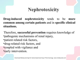
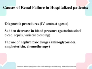
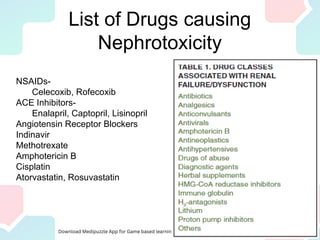
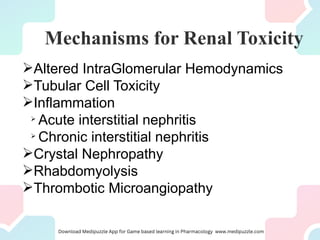
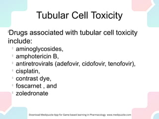
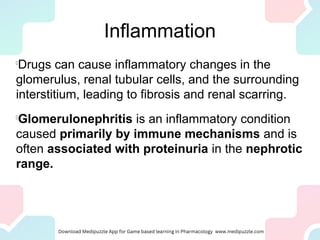
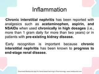
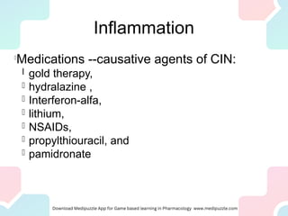
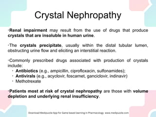
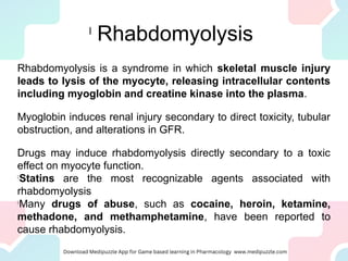
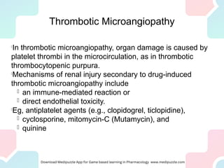
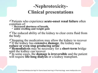
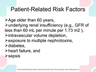
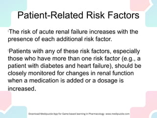
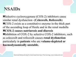
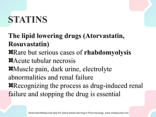
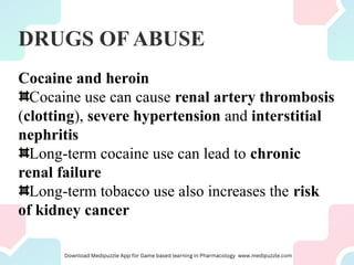
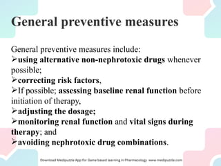
Nephrotoxic drugs can damage the kidneys by several mechanisms. Certain patient characteristics like older age, pre-existing kidney disease, and volume depletion increase risk. Common nephrotoxic drug classes include antibiotics, chemotherapy drugs, antivirals, NSAIDs, and statins. Drugs may cause acute kidney injury through effects on hemodynamics, tubular toxicity, inflammation, crystal formation, or rhabdomyolysis. Early recognition of rising creatinine and discontinuing the offending drug is important to reverse damage. Preventing nephrotoxicity requires assessing risks, monitoring for injury, and avoiding unnecessary nephrotoxic medications.
Urolithiasis is a common disease that is estimated to
produce medical costs of $2.1 billion per year in the United States alone.
Renal colic affects approximately 1.2 million people
each year in USA and accounts for approximately 1% of
all hospital admissions.
Most active emergency departments (EDs) manage
patients with acute renal colic every day.
Pyelonephritis
It is the inflammation of the kidney & upper urinary tract that usually results from the bacterial infection of the bladder.
Pyelonephritis can be classified in several different catagories:
-acute pyelonephritis
-chronic pyelonephritis
-xanthogranulomatous pyelonephritis
Hepatic encephalopathy is one of the deadly complication of liver diseases, occurs due to profound liver failure and from accumulation of ammonia and other toxic metabolites in blood.
Hepatic coma is advanced stage of hepatic encephalopathy.
Dr. Sachin Verma is a young, diligent and dynamic physician. He did his graduation from IGMC Shimla and MD in Internal Medicine from GSVM Medical College Kanpur. Then he did his Fellowship in Intensive Care Medicine (FICM) from Apollo Hospital Delhi. He has done fellowship in infectious diseases by Infectious Disease Society of America (IDSA). He has also done FCCS course and is certified Advance Cardiac Life support (ACLS) and Basic Life Support (BLS) provider by American Heart Association. He has also done a course in Cardiology by American College of Cardiology and a course in Diabetology by International Diabetes Centre. He specializes in the management of Infections, Multiorgan Dysfunctions and Critically ill patients and has many publications and presentations in various national conferences under his belt. He is currently working in NABH Approved Ivy super-specialty Hospital Mohali as Consultant Intensivists and Physician.
Acute kidney failure happens when your kidneys suddenly lose the ability to eliminate excess salts, fluids, and waste materials from the blood. Acute kidney failure is also called acute kidney injury or acute renal failure. It's common in people who are already in the hospital. It may develop rapidly over a few hours.
Peptic ulcers are sores that develop in the lining of the stomach, lower esophagus, or small intestine. They're usually formed as a result of inflammation caused by the bacteria H. pylori, as well as from erosion from stomach acids. Peptic ulcers are a fairly common health problem.
Chronic kidney disease (CKD) means your kidneys are damaged and can't filter blood the way they should. The disease is called “chronic” because the damage to your kidneys happens slowly over a long period of time.
Acute kidney failure occurs when your kidneys suddenly become unable to filter waste products from your blood. When your kidneys lose their filtering ability, dangerous levels of wastes may accumulate, and your blood's chemical makeup may get out of balance
In this presentation i have tried to thoroughly discuss about the concept of Drug induced kidney disease or injury, the mechanism behind it, its classification and how to access it.
Detailed mechanisms of certain antimicrobials that cause renal failure
ANTIMICROBIALS CAUSING RENAL FAILURE
Aminoglycosides
Amphotericin – B
Trimethoprim
B – lactam antibiotics
Fluoroquinolones
Vancomycin
Acyclovir
Tetracycline
Many medicines can cause acute kidney injury (which used to be called acute renal failure), such as:
Antibiotics. These include aminoglycosides, cephalosporins, amphotericin B, bacitracin, and vancomycin.
Antihypertensive: ACE inhibitors, such as lisinopril and ramipril; Angiotensin receptor blockers, such as candesartan and valsartan.
Anticancer drugs (chemotherapy): Examples are cisplatin, carboplatin, and methotrexate.
Dyes (contrast media):These are used in medical imaging tests.
Illegal drugs: Examples are heroin and methamphetamine.
Antiviral drugs: Examples are indinavir and ritonavir, acyclovir
Non-steroidal anti-inflammatory drugs: These include ibuprofen, ketoprofen, and naproxen.
Anti Ulcer medicines: One example is cimetidine.
Drug-induced kidney disease or nephrotoxicity (DIN) is a relatively common complication of several diagnostic and therapeutic agents.
Any drug in the blood will eventually reach the highly vascularized kidneys
It may potentially cause drug induced renal failure
If the drug is primarily cleared by the kidney, the drug will become increasingly concentrated as it moves from the renal artery into the smaller vasculature of the kidney
The drug may be filtered or secreted into the lumen of the renal tubules
The concentrated drug exposes the kidney tissue to far greater drug concentration per surface area
The kidney maintains the vital functions of clearing excess body fluid and removing metabolic and exogenous toxins from the blood
The kidney is particularly vulnerable to drugs and other agents that cause renal damage
The heart pumps approximately 25% of cardiac output into the kidneys
Any drug in the blood will eventually reach the highly vascularized kidneys
It may potentially cause drug induced renal failure
If the drug is primarily cleared by the kidney, the drug will become increasingly concentrated as it moves from the renal artery into the smaller vasculature of the kidney
The drug may be filtered or secreted into the lumen of the renal tubules
The concentrated drug exposes the kidney tissue to far greater drug concentration per surface area
Drug-induced kidney disease or nephrotoxicity (DIN) is a relatively common complication of several diagnostic and therapeutic agents.
Slides includes the introduction to drug induced renal disease, pathogenic mechanism by which drug acts on kidney, list of drugs and risk factors.
pathogenic mechanism like altered Interglomerular, Rhabdomyolysis, tubular toxicity, etc.
General Pharmacology Lecture Slides on Essential Drugs and Rational use of Medicines by Sanjaya Mani Dixit Assistant Professor of Pharmacology at Kathmandu Medical College
Dental Pharmacology Lecture Slides on Sialogogues and Antisialogogues by Sanjaya Mani Dixit Assistant Professor of Pharmacology at Kathmandu Medical College
Pharmacology Lecture Slides on Autonomic Nervous System Introduction by Sanjaya Mani Dixit Assistant Professor of Pharmacology at Kathmandu Medical College
Ozempic: Preoperative Management of Patients on GLP-1 Receptor Agonists Saeid Safari
Preoperative Management of Patients on GLP-1 Receptor Agonists like Ozempic and Semiglutide
ASA GUIDELINE
NYSORA Guideline
2 Case Reports of Gastric Ultrasound
Basavarajeeyam is an important text for ayurvedic physician belonging to andhra pradehs. It is a popular compendium in various parts of our country as well as in andhra pradesh. The content of the text was presented in sanskrit and telugu language (Bilingual). One of the most famous book in ayurvedic pharmaceutics and therapeutics. This book contains 25 chapters called as prakaranas. Many rasaoushadis were explained, pioneer of dhatu druti, nadi pareeksha, mutra pareeksha etc. Belongs to the period of 15-16 century. New diseases like upadamsha, phiranga rogas are explained.
Local Advanced Lung Cancer: Artificial Intelligence, Synergetics, Complex Sys...Oleg Kshivets
Overall life span (LS) was 1671.7±1721.6 days and cumulative 5YS reached 62.4%, 10 years – 50.4%, 20 years – 44.6%. 94 LCP lived more than 5 years without cancer (LS=2958.6±1723.6 days), 22 – more than 10 years (LS=5571±1841.8 days). 67 LCP died because of LC (LS=471.9±344 days). AT significantly improved 5YS (68% vs. 53.7%) (P=0.028 by log-rank test). Cox modeling displayed that 5YS of LCP significantly depended on: N0-N12, T3-4, blood cell circuit, cell ratio factors (ratio between cancer cells-CC and blood cells subpopulations), LC cell dynamics, recalcification time, heparin tolerance, prothrombin index, protein, AT, procedure type (P=0.000-0.031). Neural networks, genetic algorithm selection and bootstrap simulation revealed relationships between 5YS and N0-12 (rank=1), thrombocytes/CC (rank=2), segmented neutrophils/CC (3), eosinophils/CC (4), erythrocytes/CC (5), healthy cells/CC (6), lymphocytes/CC (7), stick neutrophils/CC (8), leucocytes/CC (9), monocytes/CC (10). Correct prediction of 5YS was 100% by neural networks computing (error=0.000; area under ROC curve=1.0).
Title: Sense of Taste
Presenter: Dr. Faiza, Assistant Professor of Physiology
Qualifications:
MBBS (Best Graduate, AIMC Lahore)
FCPS Physiology
ICMT, CHPE, DHPE (STMU)
MPH (GC University, Faisalabad)
MBA (Virtual University of Pakistan)
Learning Objectives:
Describe the structure and function of taste buds.
Describe the relationship between the taste threshold and taste index of common substances.
Explain the chemical basis and signal transduction of taste perception for each type of primary taste sensation.
Recognize different abnormalities of taste perception and their causes.
Key Topics:
Significance of Taste Sensation:
Differentiation between pleasant and harmful food
Influence on behavior
Selection of food based on metabolic needs
Receptors of Taste:
Taste buds on the tongue
Influence of sense of smell, texture of food, and pain stimulation (e.g., by pepper)
Primary and Secondary Taste Sensations:
Primary taste sensations: Sweet, Sour, Salty, Bitter, Umami
Chemical basis and signal transduction mechanisms for each taste
Taste Threshold and Index:
Taste threshold values for Sweet (sucrose), Salty (NaCl), Sour (HCl), and Bitter (Quinine)
Taste index relationship: Inversely proportional to taste threshold
Taste Blindness:
Inability to taste certain substances, particularly thiourea compounds
Example: Phenylthiocarbamide
Structure and Function of Taste Buds:
Composition: Epithelial cells, Sustentacular/Supporting cells, Taste cells, Basal cells
Features: Taste pores, Taste hairs/microvilli, and Taste nerve fibers
Location of Taste Buds:
Found in papillae of the tongue (Fungiform, Circumvallate, Foliate)
Also present on the palate, tonsillar pillars, epiglottis, and proximal esophagus
Mechanism of Taste Stimulation:
Interaction of taste substances with receptors on microvilli
Signal transduction pathways for Umami, Sweet, Bitter, Sour, and Salty tastes
Taste Sensitivity and Adaptation:
Decrease in sensitivity with age
Rapid adaptation of taste sensation
Role of Saliva in Taste:
Dissolution of tastants to reach receptors
Washing away the stimulus
Taste Preferences and Aversions:
Mechanisms behind taste preference and aversion
Influence of receptors and neural pathways
Impact of Sensory Nerve Damage:
Degeneration of taste buds if the sensory nerve fiber is cut
Abnormalities of Taste Detection:
Conditions: Ageusia, Hypogeusia, Dysgeusia (parageusia)
Causes: Nerve damage, neurological disorders, infections, poor oral hygiene, adverse drug effects, deficiencies, aging, tobacco use, altered neurotransmitter levels
Neurotransmitters and Taste Threshold:
Effects of serotonin (5-HT) and norepinephrine (NE) on taste sensitivity
Supertasters:
25% of the population with heightened sensitivity to taste, especially bitterness
Increased number of fungiform papillae
These lecture slides, by Dr Sidra Arshad, offer a quick overview of the physiological basis of a normal electrocardiogram.
Learning objectives:
1. Define an electrocardiogram (ECG) and electrocardiography
2. Describe how dipoles generated by the heart produce the waveforms of the ECG
3. Describe the components of a normal electrocardiogram of a typical bipolar lead (limb II)
4. Differentiate between intervals and segments
5. Enlist some common indications for obtaining an ECG
6. Describe the flow of current around the heart during the cardiac cycle
7. Discuss the placement and polarity of the leads of electrocardiograph
8. Describe the normal electrocardiograms recorded from the limb leads and explain the physiological basis of the different records that are obtained
9. Define mean electrical vector (axis) of the heart and give the normal range
10. Define the mean QRS vector
11. Describe the axes of leads (hexagonal reference system)
12. Comprehend the vectorial analysis of the normal ECG
13. Determine the mean electrical axis of the ventricular QRS and appreciate the mean axis deviation
14. Explain the concepts of current of injury, J point, and their significance
Study Resources:
1. Chapter 11, Guyton and Hall Textbook of Medical Physiology, 14th edition
2. Chapter 9, Human Physiology - From Cells to Systems, Lauralee Sherwood, 9th edition
3. Chapter 29, Ganong’s Review of Medical Physiology, 26th edition
4. Electrocardiogram, StatPearls - https://www.ncbi.nlm.nih.gov/books/NBK549803/
5. ECG in Medical Practice by ABM Abdullah, 4th edition
6. Chapter 3, Cardiology Explained, https://www.ncbi.nlm.nih.gov/books/NBK2214/
7. ECG Basics, http://www.nataliescasebook.com/tag/e-c-g-basics
ABDOMINAL TRAUMA in pediatrics part one.drhasanrajab
Abdominal trauma in pediatrics refers to injuries or damage to the abdominal organs in children. It can occur due to various causes such as falls, motor vehicle accidents, sports-related injuries, and physical abuse. Children are more vulnerable to abdominal trauma due to their unique anatomical and physiological characteristics. Signs and symptoms include abdominal pain, tenderness, distension, vomiting, and signs of shock. Diagnosis involves physical examination, imaging studies, and laboratory tests. Management depends on the severity and may involve conservative treatment or surgical intervention. Prevention is crucial in reducing the incidence of abdominal trauma in children.
Basavarajeeyam is a Sreshta Sangraha grantha (Compiled book ), written by Neelkanta kotturu Basavaraja Virachita. It contains 25 Prakaranas, First 24 Chapters related to Rogas& 25th to Rasadravyas.
263778731218 Abortion Clinic /Pills In Harare ,sisternakatoto
263778731218 Abortion Clinic /Pills In Harare ,ABORTION WOMEN’S CLINIC +27730423979 IN women clinic we believe that every woman should be able to make choices in her pregnancy. Our job is to provide compassionate care, safety,affordable and confidential services. That’s why we have won the trust from all generations of women all over the world. we use non surgical method(Abortion pills) to terminate…Dr.LISA +27730423979women Clinic is committed to providing the highest quality of obstetrical and gynecological care to women of all ages. Our dedicated staff aim to treat each patient and her health concerns with compassion and respect.Our dedicated group ABORTION WOMEN’S CLINIC +27730423979 IN women clinic we believe that every woman should be able to make choices in her pregnancy. Our job is to provide compassionate care, safety,affordable and confidential services. That’s why we have won the trust from all generations of women all over the world. we use non surgical method(Abortion pills) to terminate…Dr.LISA +27730423979women Clinic is committed to providing the highest quality of obstetrical and gynecological care to women of all ages. Our dedicated staff aim to treat each patient and her health concerns with compassion and respect.Our dedicated group of receptionists, nurses, and physicians have worked together as a teamof receptionists, nurses, and physicians have worked together as a team wwww.lisywomensclinic.co.za/
NVBDCP.pptx Nation vector borne disease control programSapna Thakur
NVBDCP was launched in 2003-2004 . Vector-Borne Disease: Disease that results from an infection transmitted to humans and other animals by blood-feeding arthropods, such as mosquitoes, ticks, and fleas. Examples of vector-borne diseases include Dengue fever, West Nile Virus, Lyme disease, and malaria.
These simplified slides by Dr. Sidra Arshad present an overview of the non-respiratory functions of the respiratory tract.
Learning objectives:
1. Enlist the non-respiratory functions of the respiratory tract
2. Briefly explain how these functions are carried out
3. Discuss the significance of dead space
4. Differentiate between minute ventilation and alveolar ventilation
5. Describe the cough and sneeze reflexes
Study Resources:
1. Chapter 39, Guyton and Hall Textbook of Medical Physiology, 14th edition
2. Chapter 34, Ganong’s Review of Medical Physiology, 26th edition
3. Chapter 17, Human Physiology by Lauralee Sherwood, 9th edition
4. Non-respiratory functions of the lungs https://academic.oup.com/bjaed/article/13/3/98/278874
2. Kidney and Drugs
The kidney maintains the vital functions of clearing excess body
fluid and removing metabolic and exogenous toxins from blood.
The heart pumps approximately 25% of blood into the
kidneys.
If the drug is primarily cleared by the kidney, the drug will
become increasingly concentrated as it is moves from the renal
artery into the smaller vasculature of the kidney.
The drug may be filtered or secreted into the lumen of the
renal tubules.
The concentrated drug exposes the kidney tissue to far greater
drug concentration per surface area.
The kidney is particularly vulnerable to drugs and other agents
that cause renal damage.
3. Nephrotoxicity
Drugs are a common source of acute kidney injury.
Drug-induced kidney failure is a major adverse event
associated with multiple medication classes .
Compared to a few decades earlier , the average patient
today is:
older,
has more comorbidities, and
is exposed to more diagnostic and therapeutic
procedures with the potential to harm kidney function.
4. Nephrotoxicity
Although renal impairment is
often reversible if the offending drug is
discontinued, the condition can be costly and
may require multiple interventions, including
hospitalization.
5. Drug-induced nephrotoxicity tends to be more
common among certain patients and in specific clinical
situations.
Therefore, successful prevention requires knowledge of
•pathogenic mechanisms of renal injury,
•patient related risk factors,
•drug-related risk factors, and
•coupled with vigilance and
•early intervention.
Nephrotoxicity
6. Causes of Renal Failure in Hospitalized patients:
l
Diagnostic procedures (IV contrast agents)
Sudden decrease in blood pressure (gastrointestinal
bleed, sepsis, variceal bleeding)
The use of nephrotoxic drugs (aminoglycosides,
amphotericin, chemotherapy)
7. List of Drugs causing
Nephrotoxicity
NSAIDs-
Celecoxib, Rofecoxib
ACE Inhibitors-
Enalapril, Captopril, Lisinopril
Angiotensin Receptor Blockers
Indinavir
Methotrexate
Amphotericin B
Cisplatin
Atorvastatin, Rosuvastatin
9. Altered IntraGlomerular
Hemodynamics
The amount of plasma cleared by the kidneys per minute is
120 ml, which corresponds to the GFR.
Kidney is known to auto-regulate the intraglomerular
pressure by changing the afferent and efferent arterial tone
to preserve GFR and urine output.
In case of volume depletion, following changes occur:
circulating prostaglandins vasodilate the afferent arterioles,
allowing more blood to flow through the glomerulus;
while angiotensin-II causes vasoconstriction of the efferent
arterioles
10. Altered IntraGlomerular
Hemodynamics
l
Some drug classes are known to interfere with kidneys
ability to autoregulate glomerular pressures and thereby
decrease GFR.
l
NSAIDs with antiprostaglandin activity
l
Drugs with anti-angiotensin-II activity (ACEIs &
ARBs)
l
Similarly, Calcineurin inhibitors Eg, cyclosporine,
tacrolimus, cause dose dependent vasoconstriction of
the afferent arterioles, leading to renal impairment in at-
risk patients.
11. Tubular Cell Toxicity
l
Renal tubular cells, in particular proximal tubule
cells, are vulnerable to the toxic effects of drugs
because their role in concentrating and
reabsorbing glomerular filtrate exposes them to
high levels of circulating toxins.
l
Drugs that cause tubular cell toxicity do so by:
impairing mitochondrial function,
interfering with tubular transport,
increasing oxidative stress, or
forming free radicals.
12. Tubular Cell Toxicity
l
Drugs associated with tubular cell toxicity
include:
l
aminoglycosides,
l
amphotericin B,
l
antiretrovirals (adefovir, cidofovir, tenofovir),
l
cisplatin,
l
contrast dye,
l
foscarnet , and
l
zoledronate
13. Inflammation
l
Drugs can cause inflammatory changes in the
glomerulus, renal tubular cells, and the surrounding
interstitium, leading to fibrosis and renal scarring.
l
Glomerulonephritis is an inflammatory condition
caused primarily by immune mechanisms and is
often associated with proteinuria in the nephrotic
range.
14. Inflammation
Acute interstitial nephritis, which can result from an allergic
response to a suspected drug, develops in an idiosyncratic,
non–dose-dependent fashion.
Numerous drugs have been implicated, including:
l
Allopurinol,
l
Antibiotics (beta lactams, quinolones, rifampin, sulfonamides, and
vancomycin)
l
Antivirals (acyclovir and indinavir)
l
Diuretics (loops, thiazides);
l
NSAIDs;
l
Phenytoin;
l
Proton pump inhibitors (omeprazole, pantoprazole, and
lansoprazole), and
l
Ranitidine.
15. Inflammation
l
Chronic interstitial nephritis has been reported with
analgesics such as acetaminophen, aspirin, and
NSAIDs when used chronically in high dosages (i.e.,
more than 1 gram daily for more than two years) or in
patients with pre-existing kidney disease.
l
Early recognition is important because chronic
interstitial nephritis has been known to progress to
end-stage renal disease.
17. Crystal Nephropathy
Renal impairment may result from the use of drugs that produce
crystals that are insoluble in human urine.
The crystals precipitate, usually within the distal tubular lumen,
obstructing urine flow and eliciting an interstitial reaction.
Commonly prescribed drugs associated with production of crystals
include:
Antibiotics (e.g., ampicillin, ciprofloxacin, sulfonamides);
Antivirals (e.g., acyclovir, foscarnet, ganciclovir, indinavir)
Methotrexate
Patients most at risk of crystal nephropathy are those with volume
depletion and underlying renal insufficiency.
18. l
Rhabdomyolysis
Rhabdomyolysis is a syndrome in which skeletal muscle injury
leads to lysis of the myocyte, releasing intracellular contents
including myoglobin and creatine kinase into the plasma.
Myoglobin induces renal injury secondary to direct toxicity, tubular
obstruction, and alterations in GFR.
Drugs may induce rhabdomyolysis directly secondary to a toxic
effect on myocyte function.
l
Statins are the most recognizable agents associated with
rhabdomyolysis
l
Many drugs of abuse, such as cocaine, heroin, ketamine,
methadone, and methamphetamine, have been reported to
cause rhabdomyolysis.
19. Thrombotic Microangiopathy
In thrombotic microangiopathy, organ damage is caused by
platelet thrombi in the microcirculation, as in thrombotic
thrombocytopenic purpura.
Mechanisms of renal injury secondary to drug-induced
thrombotic microangiopathy include
an immune-mediated reaction or
direct endothelial toxicity.
Eg, antiplatelet agents (e.g., clopidogrel, ticlopidine),
cyclosporine, mitomycin-C (Mutamycin), and
quinine
20. -Nephrotoxicity-
Clinical presentations
Patients who experience acute-onset renal failure often
complain of:
increased shortness of breath,
ankle swelling and weight gain
The reduced ability of the kidney to clear extra fluid from
the body.
Stopping the medication may allow the kidney to recover
If the kidney has extensive damage, the kidney may
reduce or even stop producing urine
Hemodialysis may be necessary for a short-term bridge
until the kidney can recover
In some cases, the damage is irreversible and the patient
will require life-long dialysis or a kidney transplant.
23. Patient-Related Risk Factors
Age older than 60 years,
underlying renal insufficiency (e.g., GFR of
less than 60 mL per minute per 1.73 m2 ),
intravascular volume depletion,
exposure to multiple nephrotoxins,
diabetes,
heart failure, and
sepsis
24. Patient-Related Risk Factors
l
The risk of acute renal failure increases with the
presence of each additional risk factor.
l
Patients with any of these risk factors, especially
those who have more than one risk factor (e.g., a
patient with diabetes and heart failure), should be
closely monitored for changes in renal function
when a medication is added or a dosage is
increased.
25. Patient-Related Risk Factors
l
Both “absolute” and “effective” intravascular volume depletion are
risk factors for drug-induced renal impairment.
l
Absolute intravascular volume depletion may occur in patients
who have gastroenteritis, chronic diarrhea, aggressive
diuresis, or poor oral intake.
l
Effective intravascular volume is the volume of blood
perceived by baroreceptors located in the right atrium and the
kidney.
l
Decreased effective circulating blood volume results from
sequestration of fluid into third-space compartments and is
associated with sepsis, heart failure, ascites, or pancreatitis.
26. ANTIBIOTICS
AIN is hypersensitivity or allergic reaction to drug
Up to 71% of all cases of acute interstitial nephritis
(AIN) are drug-induced
The most common antibiotic classes associated with AIN
are
Penicillins/cephalosporins and sulfonamides
Ciprofloxacin & Rifampin (Rifampicin)
Features
Acute renal failure, Skin rash, Increased eosinophils
27. ANTIBIOTICS
Aminoglycosides (tobramycin, gentamicin, amikacin) and
amphotericin B can cause ATN
Risk of aminoglycoside toxicity is associated with increased
dose, duration of therapy, dehydration and concurrent use of
nephrotoxic drugs, such as NSAIDs
Amphotericin B renal toxicity is related to cumulative dose,
concurrent use of nephrotoxic drugs, baseline abnormal creatinine
and concurrent use of diuretics
80% of patients receiving amphotericin B experiencing some
decrease of renal function
Saline hydration can decrease the toxicity
28. CHEMOTHERAPY DRUGS
Nephrotoxicity is the major dose-limiting toxicity for
Cisplatin
Both acute and late-onset toxicities occur
Aggressive replacement of magnesium (lost when the
proximal tubule is damaged), saline hydration or
mannitol infusion is required.
High dose Methotrexate : postrenal obstruction by
precipitating in the tubules of the nephron
Also known to cause direct toxicity
29. ANTIVIRALS &
IMMUNOSUPPRESSANT
ANTIVIRALS
Acyclovir, foscarnet, and interferons can cause ATN
Acyclovir can precipitate within the renal tubules
IMMUNOSUPPRESSANT
Cyclosporine and tacrolimus
Acute, dose-dependent reduction in renal
blood flow and chronic structural changes in the
kidney
30. NSAIDs
Long-term use can cause chronic renal insufficiency
Patients who experience ARF with NSAIDs have
underlying risk factors
Prolonged NSAID use can cause chronic kidney disease,
especially in the elderly
1-5 % of all end-stage renal disease (ESRD) patients
have analgesic-associated nephropathy
Risk factors for this nephropathy include
gender (women>men),
age (>50 years old) and
prolonged use of the analgesic
31. NSAIDs
Selective cyclooxygenase (COX-2) inhibitors cause
similar renal dysfunction -(Celecoxib, Rofecoxib)
COX-2 exists as a constitutive enzyme in the thick part
of the ascending loop of Henle and in the renal medulla
COX-2 causes natriuresis and diuresis
Inhibition of COX-2 by selective COX-2 inhibitors, such
as celecoxib and rofecoxib causes renal dysfunction
particularly in patients who are volume-depleted or
haemodynamically unstable.
32. STATINS
The lipid lowering drugs (Atorvastatin,
Rosuvastatin)
Rare but serious cases of rhabdomyolysis
Acute tubular necrosis
Muscle pain, dark urine, electrolyte
abnormalities and renal failure
Recognizing the process as drug-induced renal
failure and stopping the drug is essential
33. DRUGS OF ABUSE
Cocaine and heroin
Cocaine use can cause renal artery thrombosis
(clotting), severe hypertension and interstitial
nephritis
Long-term cocaine use can lead to chronic
renal failure
Long-term tobacco use also increases the risk
of kidney cancer
34. General preventive measures
General preventive measures include:
using alternative non-nephrotoxic drugs whenever
possible;
correcting risk factors,
If possible; assessing baseline renal function before
initiation of therapy,
adjusting the dosage;
monitoring renal function and vital signs during
therapy; and
avoiding nephrotoxic drug combinations.
35. Recognition and Early
Intervention
l
Most episodes of drug-induced renal
impairment are reversible.
l
Renal function generally returns to baseline
provided the impairment is recognized early
and the offending medication is discontinued.
36. Recognition and Early
Intervention
A decrease in renal function as evidenced by a rise
in serum creatinine levels following the initiation of a
drug signals the possibility of drug-induced renal
injury.
An exception to this is an increase in serum
creatinine following the initiation of cimetidine or
trimethoprim, because they compete with creatinine
for tubular secretion and are not associated with
kidney damage or urine abnormalities.
37. Recognition and Early
Intervention
Serum creatinine levels- Acute renal failure
A 50 percent rise from baseline, an increase
of 0.5 mg per dL or more when baseline
serum creatinine is less than 2 mg per dL or
an increase of 1 mg per dL or more if
baseline creatinine is greater than 2 mg per
dL have been used as biochemical criteria of
acute renal failure.
38. Recognition and Early
Intervention
At the first sign of renal dysfunction, the patient’s
medication list should be reviewed to identify offending
agents.
If multiple medications are present and the patient is
clinically stable, physicians should start by discontinuing
the drug most recently added to the patient’s medication
regimen.
Attention should then be directed at avoiding further renal
insults by:
supporting blood pressure,
maintaining adequate hydration, and
temporarily discontinuing all other possible nephrotoxins.