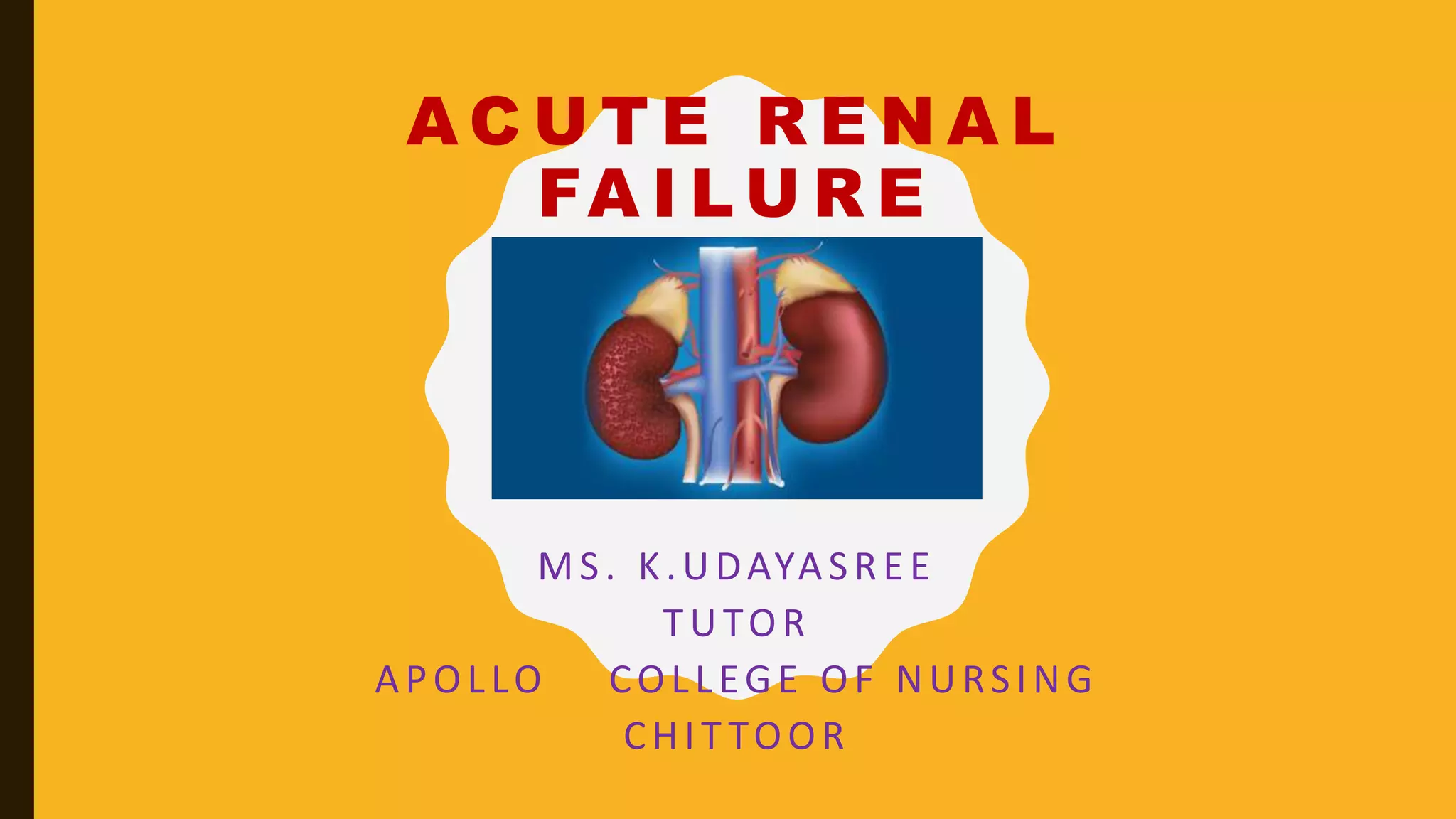
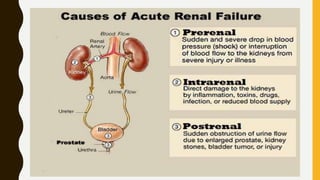
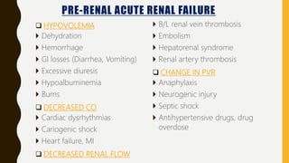
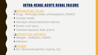
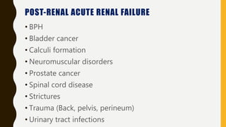
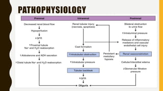
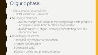
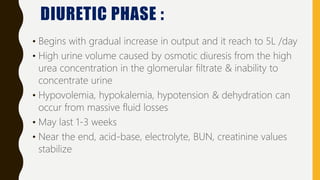
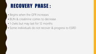
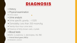
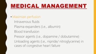
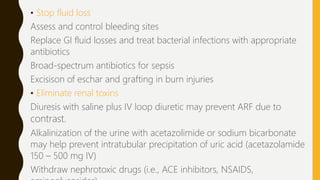
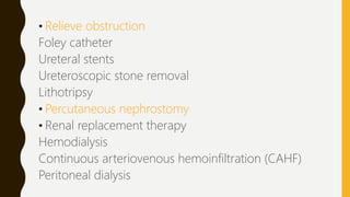
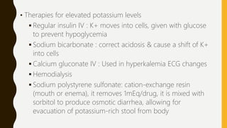
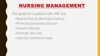
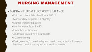
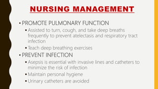
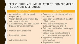
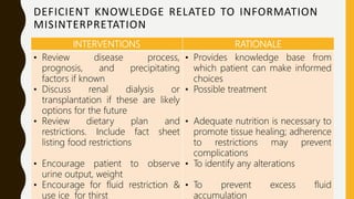
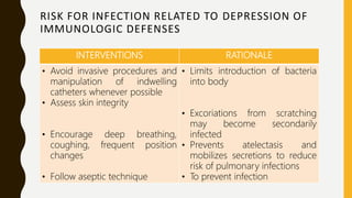
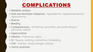
A C U T E R E N A L
FA I L U R E
MS. K.UDAYASREE
TUTOR
APOLLO COLLEGE OF NURSING
CHIT TOOR
Abrupt decrease in renal function resulting in the
accumulation of nitrogenous compounds such as urea
and creatinine
ARF is a sudden decline in glomerular filtration rate (GFR)
often accompanied by a drop in urine output. Decreased
GFR results in accumulation of nitrogenous wastes and
alterations in fluid and electrolyte balance.
DEFINITION
INCIDENCE
• The incidence rises to approx. 20% in critically ill
patients. Renal replacement therapy (RRT) may be
necessary, depending on the severity of renal failure
and related clinical conditions
Type Incidence
Prerenal 35%
Intrarenal 55%
Postrenal 10%
CLASSIFICATION
• Prerenal failure: circumstances or conditions that
decrease blood flow to the kidneys
• Post renal failure: conditions that obstruct urine
outflow from the kidney
• Intrarenal failure: conditions affecting internal
structures of the kidney; most often, acute tubular
necrosis (ATN) , which develops when a toxin or
ischemia damages the renal tubules.
PRE-RENAL ACUTE RENAL FAILURE
 HYPOVOLEMIA
 Dehydration
 Hemorrhage
 GI losses (Diarrhea, Vomiting)
 Excessive diuresis
 Hypoalbuminemia
 Burns
 DECREASED CO
 Cardiac dysrhythmias
 Cariogenic shock
 Heart failure, MI
 DECREASED RENAL FLOW
 B/L renal vein thrombosis
 Embolism
 Hepatorenal syndrome
 Renal artery thrombosis
 CHANGE IN PVR
 Anaphylaxis
 Neurogenic injury
 Septic shock
 Antihypertensive drugs, drug
overdose
INTRA-RENAL ACUTE RENAL FAILURE
NEPHROTOXIC INJURY
 Drugs : Aminoglycosides, antineoplastics, NSAIDS
 Contrast media
 Hemolytic blood transfusion reaction
 Severe crush injury
 Chemical exposure :lead, arsenic
INTERSTITIAL NEPHRITIS
 Allergies : Antibiotics, NSAIDs
 Infections
OTHERS
 A/C Glomerulonephritis, toxemia, SLE
POST-RENAL ACUTE RENAL FAILURE
• BPH
• Bladder cancer
• Calculi formation
• Neuromuscular disorders
• Prostate cancer
• Spinal cord disease
• Strictures
• Trauma (Back, pelvis, perineum)
• Urinary tract infections
PATHOPHYSIOLOGY
PHASES
There are four phases of ARF
• INITIATION : The initiation period begins with the initial
and ends when oliguria develops
• OLIGURIA : The oliguria period is accompanied by an increase
increase in the serum concentration of substances usually
excreted by kidneys
• DIURESIS : The diuresis period is marked by a gradual increase
increase in urine output, which signals that glomerular
filtration has started to recover
• RECOVERY : The recovery period signals the improvement of
renal function and may take 3 to 12 months
CLINICAL FEATURES
•Oliguric phase
• Urinary changes :
– <400 ml/day
– Occurs within 7 days of injury(depending on cause and tissue injury)
– Lasts for about 10-14 days
– Longer it lasts, poor prognosis
• Fluid volume :
– Treat with fluid replacement
– If oliguria/ anuria fluid retention results which can lead to fluid excess
– Results in increased JVP
, bounding pulse, edema, hypertension
– It can lead to complications such as HF, pulmonary edema, pericardial
pleural effusion
• Metabolic acidosis:
– Impaired excretion of hydrogen ions and metabolic waste
products
– Decreased production of bicarbonate ions
– May develop Kussmaul respiration
• Sodium balance:
– Sodium may be below normal levels
• Potassium excess:
– Hyperkalemia
– Bleeding, blood transfusion & metabolic acidosis worsens
hyperkalemia
 Waste product accumulation :
o BUN, creatinine - elevated
Neurologic disorders :
oNeuro changes can occur as the nitrogenous waste products
accumulate in the brain & other nervous tissue
oManifestations : Fatigue, difficulty concentrating, seizures,
stupor & coma
Hematologic disorders :
oImpaired erythropoietin production
oPlatelet abnormalities
oDecreased WBC
oCalcium deficit and phosphate excess
DIURETIC PHASE :
• Begins with gradual increase in output and it reach to 5L /day
• High urine volume caused by osmotic diuresis from the high
urea concentration in the glomerular filtrate & inability to
concentrate urine
• Hypovolemia, hypokalemia, hypotension & dehydration can
occur from massive fluid losses
• May last 1-3 weeks
• Near the end, acid-base, electrolyte, BUN, creatinine values
stabilize
RECOVERY PHASE :
• Begins when the GFR increases
• BUN & creatinine comes to decrease
• 1-2wks but may last for 12 months
• Some individuals do not recover & progress to ESRD
DIAGNOSIS
• History
• Physical examination
 Urine output
• Urine analysis
 Urine specific gravity : <1.020
 Osmolality: Less than 350 mosm/kg
 Twenty–four–hour urine tests
 Presence of dirty brown casts, crystals
• Blood tests
 BUN, S.creatinine, S. electrolytes, Hb, RBC
• Arterial blood gases (ABGs)
 Metabolic acidosis
DIAGNOSIS
 Kidney, ureter, bladder (KUB) X-ray :
 Demonstrates size of kidneys/ureters/bladder,
presence of cysts, tumors, stones
 Retrograde pyelogram:
 Outlines abnormalities of renal pelvis and ureters
 Renal arteriogram:
 Voiding cystoureterogram:
Shows bladder size, reflux into ureters,
retention
 Renal ultrasound
DIAGNOSIS
 Nonnuclear computed tomography (CT) scan: Cross-
sectional view of kidney and urinary tract detects
presence/extent of disease
 Magnetic resonance imaging (MRI): Provides information
about soft tissue damage
 Excretory urography (intravenous urogram
or pyelogram): Radiopaque contrast
concentrates in urine and facilitates
visualization of KUB.
DIAGNOSIS
• Endourology: Direct visualization may be done of urethra,
bladder, ureters, and kidney to diagnose problems, biopsy,
and remove small lesions and/or calculi
• Electrocardiogram (ECG): May be abnormal, reflecting
electrolyte and acid-base imbalances
• Renal biopsy : A sample of the kidney tissue (biopsy) is
sometimes required in cases in which the cause of the kidney
disease is unclear
MEDICAL MANAGEMENT
• Treat the underlying disease
• Strictly monitor I/O, weight
• Monitor serum electrolytes
• Adjust medication dosages according to GFR
• Avoid highly nephrotoxic drugs
• Determine adequate CO to ensure kidney perfusion
MEDICAL MANAGEMENT
• Maximize perfusion
Intravenous fluids
Plasma expanders (i.e., albumin)
Blood transfusion
Pressor agents (i.e., dopamine / dobutamine)
Unloading agents (i.e., nipride/ nitroglycerine) in
cases of congestive heart failure
• Stop fluid loss
Assess and control bleeding sites
Replace GI fluid losses and treat bacterial infections with appropriate
antibiotics
Broad-spectrum antibiotics for sepsis
Excisison of eschar and grafting in burn injuries
• Eliminate renal toxins
Diuresis with saline plus IV loop diuretic may prevent ARF due to
contrast.
Alkalinization of the urine with acetazolimide or sodium bicarbonate
may help prevent intratubular precipitation of uric acid (acetazolamide
150 – 500 mg IV)
Withdraw nephrotoxic drugs (i.e., ACE inhibitors, NSAIDS,
• Relieve obstruction
Foley catheter
Ureteral stents
Ureteroscopic stone removal
Lithotripsy
• Percutaneous nephrostomy
• Renal replacement therapy
Hemodialysis
Continuous arteriovenous hemoinfiltration (CAHF)
Peritoneal dialysis
• Therapies for elevated potassium levels
 Regular insulin IV : K+ moves into cells, given with glucose
to prevent hypoglycemia
 Sodium bicarbonate : correct acidosis & cause a shift of K+
into cells
 Calcium gluconate IV : Used in hyperkalemia ECG changes
 Hemodialysis
 Sodium polystyrene sulfonate: cation-exchange resin
(mouth or enema), it removes 1mEq/drug, it is mixed with
sorbitol to produce osmotic diarrhea, allowing for
evacuation of potassium-rich stool from body
NUTRITIONAL THERAPY
• Potassium intake is limited to 40mEq/day
• Low-protein, high-calorie diet : Caloric intake – 30-35kcal/kg &
0.8 – 1g of protein/kg of body weight to prevent breakdown of
body protein
• Sodium is restricted (To prevent edema, hypertension, HF)
• Dietary fat intake is increased (To prevent ketosis & endogenous
fat breakdown)
• Fat emulsion IV infusion
• TPN
• Potassium or phosphorus diet are restricted
NURSING MANAGEMENT
The goals for a patient with ARF are:
–Restore fluid & electrolyte balance
–Promote pulmonary function
–Prevent infection
–Promote skin care
–Improve nutritional intake
NURSING MANAGEMENT
• MAINTAIN FLUID & ELECTROLYTE BALANCE
 Fluid restriction- 24hrs fluid loss + 600ml
 Monitor daily weight (0.2-0.5kg/day)
 Diuretic therapy (Eg: Lasix)
 Monitor electrolytes & ABG
 Electrolyte replacement
 Acidosis is treated with bicarbonate
 ECG monitoring
 Dark green veg’s, unrefined grains, seeds, nuts, antacids & osmotic
laxatives containing magnesium should be avoided
NURSING MANAGEMENT
• PROMOTE PULMONARY FUNCTION
 Assisted to turn, cough, and take deep breaths
frequently to prevent atelectasis and respiratory tract
infection
 Teach deep breathing exercises
• PREVENT INFECTION
 Asepsis is essential with invasive lines and catheters to
minimize the risk of infection
 Maintain personal hygiene
 Urinary catheters are avoided
NURSING MANAGEMENT
PROVIDE SKIN CARE
 Bathing the patient with cool water
 Frequent turning
 keep the skin clean and well moisturized
 keep the fingernails trimmed to avoid excoriation and prevent
skin breakdown
IMPROVE NUTRITIONAL INTAKE
 Low-protein, high-calorie diet
 Sodium, potassium & phosphorus restricted diet
EXCESS FLUID VOLUME RELATED TO COMPROMISED
REGULATORY MECHANISM
INTERVENTIONS RATIONALE
• Accurately record intake and
output (I&O)
• Weigh daily at same time of day,
with same equipment
• Auscultate lung and heart sounds
• Monitor serum electrolytes,
• Monitor BUN, creatinine
• Restrict fluid intake
• Administer diuretics
• Decrease in output may indicate
acute failure
• Daily body weight is best monitor
of fluid status
• Fluid overload may lead to
pulmonary edema and HF
• Fluid overload leads to electrolyte
imbalances
• Lack of renal excretion leads to
accumulation of waste products
• To prevent further fluid
accumulation
• To promote diuresis
DEFICIENT KNOWLEDGE RELATED TO INFORMATION
MISINTERPRETATION
INTERVENTIONS RATIONALE
• Review disease process,
prognosis, and precipitating
factors if known
• Discuss renal dialysis or
transplantation if these are likely
options for the future
• Review dietary plan and
restrictions. Include fact sheet
listing food restrictions
• Encourage patient to observe
urine output, weight
• Encourage for fluid restriction &
use ice for thirst
• Provides knowledge base from
which patient can make informed
choices
• Possible treatment
• Adequate nutrition is necessary to
promote tissue healing; adherence
to restrictions may prevent
complications
• To identify any alterations
• To prevent excess fluid
accumulation
RISK FOR INFECTION RELATED TO DEPRESSION OF
IMMUNOLOGIC DEFENSES
INTERVENTIONS RATIONALE
• Avoid invasive procedures and
manipulation of indwelling
catheters whenever possible
• Assess skin integrity
• Encourage deep breathing,
coughing, frequent position
changes
• Follow aseptic technique
• Limits introduction of bacteria
into body
• Excoriations from scratching
may become secondarily
infected
• Prevents atelectasis and
mobilizes secretions to reduce
risk of pulmonary infections
• To prevent infection
COMPLICATIONS
• Metabolic acidosis
• Fluid and electrolyte imbalances : Hyperkalemia, Hyperphosphatemia,
Hypocalcemia
• Anemia
• Bleeding
• Cardiopulmonary : Arrhythmia, pericarditis, pericardial effusion,
pulmonary edema
• Hyperuricemia
• Infection : Pneumonia, sepsis
• GI : Nausea, vomiting, malnutrition, GI bleeding
• CNS : Asterixis, mental changes, seizures
• Uremic syndrome
REFERENCES
• Brunner and suddarth’s, “Textbook of medical surgical nursing”, volume
2, 13th edition, wolter Kluwer publications.
• Lewis ‘s, “Medical surgical nursing assessment and management of
clinical problems”,7th edition, Elsevier publications.
• BT.Basavanthappa, “Medical surgical nursing”, 1st edition, Jaypee
brother medical publishers.
• Joyce M. Black, Jane Hokanson Hawks, “Medical surgical nursing clinical
management for positive outcomes”, volume-1, 7th edition, Elsevier
publications.
• Cambell - Walsh, “Text book of urology”, volume-2, 10th edition, Elsevier publications.
.

Acute renal failure

  • 1.
    A C UT E R E N A L FA I L U R E MS. K.UDAYASREE TUTOR APOLLO COLLEGE OF NURSING CHIT TOOR
  • 2.
    Abrupt decrease inrenal function resulting in the accumulation of nitrogenous compounds such as urea and creatinine ARF is a sudden decline in glomerular filtration rate (GFR) often accompanied by a drop in urine output. Decreased GFR results in accumulation of nitrogenous wastes and alterations in fluid and electrolyte balance. DEFINITION
  • 3.
    INCIDENCE • The incidencerises to approx. 20% in critically ill patients. Renal replacement therapy (RRT) may be necessary, depending on the severity of renal failure and related clinical conditions Type Incidence Prerenal 35% Intrarenal 55% Postrenal 10%
  • 4.
    CLASSIFICATION • Prerenal failure:circumstances or conditions that decrease blood flow to the kidneys • Post renal failure: conditions that obstruct urine outflow from the kidney • Intrarenal failure: conditions affecting internal structures of the kidney; most often, acute tubular necrosis (ATN) , which develops when a toxin or ischemia damages the renal tubules.
  • 6.
    PRE-RENAL ACUTE RENALFAILURE  HYPOVOLEMIA  Dehydration  Hemorrhage  GI losses (Diarrhea, Vomiting)  Excessive diuresis  Hypoalbuminemia  Burns  DECREASED CO  Cardiac dysrhythmias  Cariogenic shock  Heart failure, MI  DECREASED RENAL FLOW  B/L renal vein thrombosis  Embolism  Hepatorenal syndrome  Renal artery thrombosis  CHANGE IN PVR  Anaphylaxis  Neurogenic injury  Septic shock  Antihypertensive drugs, drug overdose
  • 7.
    INTRA-RENAL ACUTE RENALFAILURE NEPHROTOXIC INJURY  Drugs : Aminoglycosides, antineoplastics, NSAIDS  Contrast media  Hemolytic blood transfusion reaction  Severe crush injury  Chemical exposure :lead, arsenic INTERSTITIAL NEPHRITIS  Allergies : Antibiotics, NSAIDs  Infections OTHERS  A/C Glomerulonephritis, toxemia, SLE
  • 8.
    POST-RENAL ACUTE RENALFAILURE • BPH • Bladder cancer • Calculi formation • Neuromuscular disorders • Prostate cancer • Spinal cord disease • Strictures • Trauma (Back, pelvis, perineum) • Urinary tract infections
  • 9.
  • 10.
    PHASES There are fourphases of ARF • INITIATION : The initiation period begins with the initial and ends when oliguria develops • OLIGURIA : The oliguria period is accompanied by an increase increase in the serum concentration of substances usually excreted by kidneys • DIURESIS : The diuresis period is marked by a gradual increase increase in urine output, which signals that glomerular filtration has started to recover • RECOVERY : The recovery period signals the improvement of renal function and may take 3 to 12 months
  • 11.
    CLINICAL FEATURES •Oliguric phase •Urinary changes : – <400 ml/day – Occurs within 7 days of injury(depending on cause and tissue injury) – Lasts for about 10-14 days – Longer it lasts, poor prognosis • Fluid volume : – Treat with fluid replacement – If oliguria/ anuria fluid retention results which can lead to fluid excess – Results in increased JVP , bounding pulse, edema, hypertension – It can lead to complications such as HF, pulmonary edema, pericardial pleural effusion
  • 12.
    • Metabolic acidosis: –Impaired excretion of hydrogen ions and metabolic waste products – Decreased production of bicarbonate ions – May develop Kussmaul respiration • Sodium balance: – Sodium may be below normal levels • Potassium excess: – Hyperkalemia – Bleeding, blood transfusion & metabolic acidosis worsens hyperkalemia
  • 13.
     Waste productaccumulation : o BUN, creatinine - elevated Neurologic disorders : oNeuro changes can occur as the nitrogenous waste products accumulate in the brain & other nervous tissue oManifestations : Fatigue, difficulty concentrating, seizures, stupor & coma Hematologic disorders : oImpaired erythropoietin production oPlatelet abnormalities oDecreased WBC oCalcium deficit and phosphate excess
  • 14.
    DIURETIC PHASE : •Begins with gradual increase in output and it reach to 5L /day • High urine volume caused by osmotic diuresis from the high urea concentration in the glomerular filtrate & inability to concentrate urine • Hypovolemia, hypokalemia, hypotension & dehydration can occur from massive fluid losses • May last 1-3 weeks • Near the end, acid-base, electrolyte, BUN, creatinine values stabilize
  • 15.
    RECOVERY PHASE : •Begins when the GFR increases • BUN & creatinine comes to decrease • 1-2wks but may last for 12 months • Some individuals do not recover & progress to ESRD
  • 16.
    DIAGNOSIS • History • Physicalexamination  Urine output • Urine analysis  Urine specific gravity : <1.020  Osmolality: Less than 350 mosm/kg  Twenty–four–hour urine tests  Presence of dirty brown casts, crystals • Blood tests  BUN, S.creatinine, S. electrolytes, Hb, RBC • Arterial blood gases (ABGs)  Metabolic acidosis
  • 17.
    DIAGNOSIS  Kidney, ureter,bladder (KUB) X-ray :  Demonstrates size of kidneys/ureters/bladder, presence of cysts, tumors, stones  Retrograde pyelogram:  Outlines abnormalities of renal pelvis and ureters  Renal arteriogram:  Voiding cystoureterogram: Shows bladder size, reflux into ureters, retention  Renal ultrasound
  • 18.
    DIAGNOSIS  Nonnuclear computedtomography (CT) scan: Cross- sectional view of kidney and urinary tract detects presence/extent of disease  Magnetic resonance imaging (MRI): Provides information about soft tissue damage  Excretory urography (intravenous urogram or pyelogram): Radiopaque contrast concentrates in urine and facilitates visualization of KUB.
  • 19.
    DIAGNOSIS • Endourology: Directvisualization may be done of urethra, bladder, ureters, and kidney to diagnose problems, biopsy, and remove small lesions and/or calculi • Electrocardiogram (ECG): May be abnormal, reflecting electrolyte and acid-base imbalances • Renal biopsy : A sample of the kidney tissue (biopsy) is sometimes required in cases in which the cause of the kidney disease is unclear
  • 20.
    MEDICAL MANAGEMENT • Treatthe underlying disease • Strictly monitor I/O, weight • Monitor serum electrolytes • Adjust medication dosages according to GFR • Avoid highly nephrotoxic drugs • Determine adequate CO to ensure kidney perfusion
  • 21.
    MEDICAL MANAGEMENT • Maximizeperfusion Intravenous fluids Plasma expanders (i.e., albumin) Blood transfusion Pressor agents (i.e., dopamine / dobutamine) Unloading agents (i.e., nipride/ nitroglycerine) in cases of congestive heart failure
  • 22.
    • Stop fluidloss Assess and control bleeding sites Replace GI fluid losses and treat bacterial infections with appropriate antibiotics Broad-spectrum antibiotics for sepsis Excisison of eschar and grafting in burn injuries • Eliminate renal toxins Diuresis with saline plus IV loop diuretic may prevent ARF due to contrast. Alkalinization of the urine with acetazolimide or sodium bicarbonate may help prevent intratubular precipitation of uric acid (acetazolamide 150 – 500 mg IV) Withdraw nephrotoxic drugs (i.e., ACE inhibitors, NSAIDS,
  • 23.
    • Relieve obstruction Foleycatheter Ureteral stents Ureteroscopic stone removal Lithotripsy • Percutaneous nephrostomy • Renal replacement therapy Hemodialysis Continuous arteriovenous hemoinfiltration (CAHF) Peritoneal dialysis
  • 24.
    • Therapies forelevated potassium levels  Regular insulin IV : K+ moves into cells, given with glucose to prevent hypoglycemia  Sodium bicarbonate : correct acidosis & cause a shift of K+ into cells  Calcium gluconate IV : Used in hyperkalemia ECG changes  Hemodialysis  Sodium polystyrene sulfonate: cation-exchange resin (mouth or enema), it removes 1mEq/drug, it is mixed with sorbitol to produce osmotic diarrhea, allowing for evacuation of potassium-rich stool from body
  • 25.
    NUTRITIONAL THERAPY • Potassiumintake is limited to 40mEq/day • Low-protein, high-calorie diet : Caloric intake – 30-35kcal/kg & 0.8 – 1g of protein/kg of body weight to prevent breakdown of body protein • Sodium is restricted (To prevent edema, hypertension, HF) • Dietary fat intake is increased (To prevent ketosis & endogenous fat breakdown) • Fat emulsion IV infusion • TPN • Potassium or phosphorus diet are restricted
  • 26.
    NURSING MANAGEMENT The goalsfor a patient with ARF are: –Restore fluid & electrolyte balance –Promote pulmonary function –Prevent infection –Promote skin care –Improve nutritional intake
  • 27.
    NURSING MANAGEMENT • MAINTAINFLUID & ELECTROLYTE BALANCE  Fluid restriction- 24hrs fluid loss + 600ml  Monitor daily weight (0.2-0.5kg/day)  Diuretic therapy (Eg: Lasix)  Monitor electrolytes & ABG  Electrolyte replacement  Acidosis is treated with bicarbonate  ECG monitoring  Dark green veg’s, unrefined grains, seeds, nuts, antacids & osmotic laxatives containing magnesium should be avoided
  • 28.
    NURSING MANAGEMENT • PROMOTEPULMONARY FUNCTION  Assisted to turn, cough, and take deep breaths frequently to prevent atelectasis and respiratory tract infection  Teach deep breathing exercises • PREVENT INFECTION  Asepsis is essential with invasive lines and catheters to minimize the risk of infection  Maintain personal hygiene  Urinary catheters are avoided
  • 29.
    NURSING MANAGEMENT PROVIDE SKINCARE  Bathing the patient with cool water  Frequent turning  keep the skin clean and well moisturized  keep the fingernails trimmed to avoid excoriation and prevent skin breakdown IMPROVE NUTRITIONAL INTAKE  Low-protein, high-calorie diet  Sodium, potassium & phosphorus restricted diet
  • 30.
    EXCESS FLUID VOLUMERELATED TO COMPROMISED REGULATORY MECHANISM INTERVENTIONS RATIONALE • Accurately record intake and output (I&O) • Weigh daily at same time of day, with same equipment • Auscultate lung and heart sounds • Monitor serum electrolytes, • Monitor BUN, creatinine • Restrict fluid intake • Administer diuretics • Decrease in output may indicate acute failure • Daily body weight is best monitor of fluid status • Fluid overload may lead to pulmonary edema and HF • Fluid overload leads to electrolyte imbalances • Lack of renal excretion leads to accumulation of waste products • To prevent further fluid accumulation • To promote diuresis
  • 31.
    DEFICIENT KNOWLEDGE RELATEDTO INFORMATION MISINTERPRETATION INTERVENTIONS RATIONALE • Review disease process, prognosis, and precipitating factors if known • Discuss renal dialysis or transplantation if these are likely options for the future • Review dietary plan and restrictions. Include fact sheet listing food restrictions • Encourage patient to observe urine output, weight • Encourage for fluid restriction & use ice for thirst • Provides knowledge base from which patient can make informed choices • Possible treatment • Adequate nutrition is necessary to promote tissue healing; adherence to restrictions may prevent complications • To identify any alterations • To prevent excess fluid accumulation
  • 32.
    RISK FOR INFECTIONRELATED TO DEPRESSION OF IMMUNOLOGIC DEFENSES INTERVENTIONS RATIONALE • Avoid invasive procedures and manipulation of indwelling catheters whenever possible • Assess skin integrity • Encourage deep breathing, coughing, frequent position changes • Follow aseptic technique • Limits introduction of bacteria into body • Excoriations from scratching may become secondarily infected • Prevents atelectasis and mobilizes secretions to reduce risk of pulmonary infections • To prevent infection
  • 33.
    COMPLICATIONS • Metabolic acidosis •Fluid and electrolyte imbalances : Hyperkalemia, Hyperphosphatemia, Hypocalcemia • Anemia • Bleeding • Cardiopulmonary : Arrhythmia, pericarditis, pericardial effusion, pulmonary edema • Hyperuricemia • Infection : Pneumonia, sepsis • GI : Nausea, vomiting, malnutrition, GI bleeding • CNS : Asterixis, mental changes, seizures • Uremic syndrome
  • 34.
    REFERENCES • Brunner andsuddarth’s, “Textbook of medical surgical nursing”, volume 2, 13th edition, wolter Kluwer publications. • Lewis ‘s, “Medical surgical nursing assessment and management of clinical problems”,7th edition, Elsevier publications. • BT.Basavanthappa, “Medical surgical nursing”, 1st edition, Jaypee brother medical publishers. • Joyce M. Black, Jane Hokanson Hawks, “Medical surgical nursing clinical management for positive outcomes”, volume-1, 7th edition, Elsevier publications. • Cambell - Walsh, “Text book of urology”, volume-2, 10th edition, Elsevier publications. .