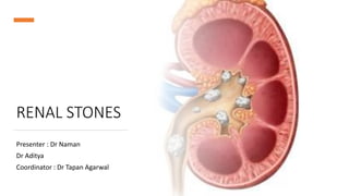
RENAL STONES
Presenter : Dr Naman
Dr Aditya
Coordinator : Dr Tapan Agarwal
Anatomy
• B/L bean shaped organs, reddish brown color, Retroperitoneal organ.
• They extend from T12-L3 vertebrae.
• Encased in 4 layers :- renal capsule
perirenal fat
Gerota’s fascia
pararenal fat
• At the hilum, the structures from top to bottom are renal vein, artery,
then pelvis.
• Right renal vein is shorter than left.
• Left renal vein is occasionally retro aortic.
• Right adrenal and gonadal veins drain into IVC whereas left adrenal
and gonadal veins drain into left renal vein.
• Gonadal veins can be easily confused with renal pelvis. These
structures can be easily distinguished by pinching the ureter which
contracts (VERMICULATION).
Renal stone
• Renal stone (Nephrolithiasis/Urolithiasis) are hard deposits made of
minerals and salts that form inside kidneys.
• Renal stones is common condition across world with prevalence of
12% worldwide (15% in india).
• They can both form as kidney or ureteric stones.
• Around 80% of stones are made of Calcium, as either Calcium Oxalate
or Calcium Phosphate.
• Remaining stone composition include Struvite stone, Urate stone,
and Cystine stone.
Pathophysiology
• The basis for formation of renal stone is over saturation of urine
followed by crystallization of urine.
• High levels of Purine in the blood leads to urate stone.
• Cystine stone formation is associated with homocystinuria.
• Most stones <5mm will pass spontaneously.
• Most common in males
Location of ureteric stones
• There are 3 natural narrowed points where stones are likely to
impact:
• 1) Pelviureteric junction
• 2) Iliac vessels crossing at pelvic brim
• 3) Vesicoureteric junction
Aetiology
• Idiopathic calcium urolithiasis
• Hypercalcemic disorders :- Primary hyperparathyroidism
Prolonged immobilization
Milk alkali syndrome
• Renal tubular syndromes :- Renal tubular acidosis
Cystinuria
• Uric acid lithiasis
• Enzyme disorders :- Primary hyperoxaluria
Xanthinuria
2,8-Dihydroadenuria
• Secondary lithiasis :- Secondary hyperoxaluria
Dietary excess
Infection
Obstruction and stasis
Drugs(Acetazolamide, Allopurinol, Thiazide)
• Other factors :- Geography
Less Water intake
Diet
Occupation (sedentary jobs in hot environment)
Clinical features
• The most common presenting symptom of ureteric stone
is pain*, termed ureteric colic, which occurs from the increased
peristalsis from around the site of obstruction. The pain
is sudden onset, severe, and radiating from flank to
pelvis(termed “loin to groin”), often associated with nausea and
vomiting.
• Haematuria occurs in around 90% cases; this is typically non-
visible.
• Concurrent infection should be assessed for, with symptoms
such as rigors, fevers, or lethargy.
• Examination is typically unremarkable, only demonstrating
some tenderness in the affected flank. There may be signs of
dehydration, from reduced fluid intake secondary to associated
vomiting.
• It is possible to have no pain with a stone, especially if the stone
is non-obstructing.
Differential Diagnosis
• Pyelonephritis
• Ruptured AAA
• Biliary pathology
• Bowel obstruction
• Lower lobe pneumonia
• Musculoskeletal related pain
Imaging
1. The gold standard for diagnosis of renal stones is NCCT
KUB. The
benefit of the CT KUB as an imaging modality is the high
sensitivity and specificity.
• NCCT KUB gives Hounsfield unit which can differentiate
calcium stones from others.
2. X-Ray abdomen
Radiopaque stones are seen.
3. Intravenous Pyelography
IV contrast is given to patient and
series of x rays are taken to look for
filling defect.
4. USG KUB
Conservative Management
• Ureteric stone 4-5mm in size have 50% chance of passage,
whereas stone >6mm have 15% chance.
• Adequate fluid resuscitation as Patients with renal stones will
often be dehydrated.
• NSAID
• Dissolution agents like oral alkalising agents
• Antibiotics for significant infection.
EUA guidelines for surgical management
Extracorporeal Shock Wave
Lithotripsy (ESWL)
• This is a non invasive method of breaking stones by generating
shockwaves outside the body which are focused on stones.
• Different method for generating shockwaves include spark gap,
electromagnetic, piezoelectric and microexpulsive.
• Shock produced @ 2/sec . 1000-4000 shocks are required to break
stones.
Dornier Lithotripter
It is used for ESWL.
Stone is located by C-arm then waves are
applied followed by flushing of
fragmented stone.
3-4 sittings are needed for optimal
results.
• Advantages:-
No anesthesia required
day care surgery
upto 2.5cm stones can be fragmented
oxalate stones are better eliminated
• Complication:-
severe hematuria, Renal hematoma
accumulation of fragments in ureter leading to blockage
injury to surrounding structures
UTI
• Contraindication:-
coagulopathies
pregnancy
obesity
sepsis & renal failure
abdominal aneurysm
Cysteine stones
stone density >1000 HU
Percutaneous Nephrolithotomy (PCNL)
• Gold standard technique
• Indications:-
Staghorn calculus
stones >2cm at lower calyces
difficult to break by ESWL
malformed kidney with decreased possibility of fragment passage
Obesity
• Contraindications:-
Uncorrected coagulopathy
Active UTI
Antiplatelet meds
• Preop CT :- spatial relation of kidney & stone, HU of stone
• Antibiotic prophylaxis :- 1st & 2nd gen Cephalosporins
Metronidazole
Steps of PCNL
1. Cystoscopy done and ureteric stent is placed and renal pelvicalyceal
system identified under C-arm guidance.
2. Under C-arm guidance, needle puncture is made in loin
percutaneously. Through kidney & calyx , pelvis is approached by
Guide wire.
3. Graduated dilators are passed and track is widened.
4. Through that a Nephroscope is inserted.
5. Stones are fragmented by different methods (Laser, pneumatic,
ultrasonic or electrohydraulic) , small stones are taken out by basket
6. Nephrostomy tube is placed.
• Role of nephrostomy tube:- aid in healing nephrostomy tract
prevent urinary extravasation
allow reentry if required
drain infection
• Complication of PCNL:- Hemorrhage
Sepsis
Urinary extravasation
Colon/Pleura injury
URS/RIRS
• Ureterorenoscopic removal of stone (URS) is used for ureteric stones
• Retrograde Intrarenal surgery (RIRS) is used for kidney stones
• Flexible ureteroscope is passed through Urethra➡ Bladder➡ Ureter
• Indications:- stone >5-8mm
IVU showing deterioration of function
coexisting infection
impacted stone in ureter with persistent symptoms
• Contraindication:- UTI
Uncorrected coagulopathy
Phimosis
Stricture
Prostate adenoma
Steps
1. Patient in lithotomy position
2. Cystoscopy done with continuous irrigation.
3. Ureteroscopy done and guide wire inserted.
4. Stone visualized
5. Small stones are extracted using Basket / forceps
Large stones are to be fragmented and then taken out or flushed
6. DJ stenting may be done if significant residual fragments present or
there is injury to ureter.
• Complication:- Bleeding/Hematuria
Perforation of ureter/pelvic wall
Ureteric avulsion
UTI
Ureteric striture
Open Pyelolithotomy
• Open renal surgeries are rarely used nowadays.
• Used For Extra renal pelvis stones
• Steps :- Loin incision given
Kidney is approached
Incision is given on pelvis ,Stone removed
DJ stent is placed if necessary
Pelvis is sutured closed with 3-0 Vicryl
Drain placed and skin closed
Position for loin incision Loin incision given
Kidney
approached
Incision on pelvis given
Stone removed
Pelvis sutured with vicryl
Skin closed
THANK YOU

Renal stone.pptx

  • 1.
    RENAL STONES Presenter :Dr Naman Dr Aditya Coordinator : Dr Tapan Agarwal
  • 2.
    Anatomy • B/L beanshaped organs, reddish brown color, Retroperitoneal organ. • They extend from T12-L3 vertebrae. • Encased in 4 layers :- renal capsule perirenal fat Gerota’s fascia pararenal fat
  • 3.
    • At thehilum, the structures from top to bottom are renal vein, artery, then pelvis. • Right renal vein is shorter than left. • Left renal vein is occasionally retro aortic. • Right adrenal and gonadal veins drain into IVC whereas left adrenal and gonadal veins drain into left renal vein. • Gonadal veins can be easily confused with renal pelvis. These structures can be easily distinguished by pinching the ureter which contracts (VERMICULATION).
  • 4.
    Renal stone • Renalstone (Nephrolithiasis/Urolithiasis) are hard deposits made of minerals and salts that form inside kidneys. • Renal stones is common condition across world with prevalence of 12% worldwide (15% in india). • They can both form as kidney or ureteric stones. • Around 80% of stones are made of Calcium, as either Calcium Oxalate or Calcium Phosphate. • Remaining stone composition include Struvite stone, Urate stone, and Cystine stone.
  • 5.
    Pathophysiology • The basisfor formation of renal stone is over saturation of urine followed by crystallization of urine. • High levels of Purine in the blood leads to urate stone. • Cystine stone formation is associated with homocystinuria. • Most stones <5mm will pass spontaneously. • Most common in males
  • 6.
    Location of uretericstones • There are 3 natural narrowed points where stones are likely to impact: • 1) Pelviureteric junction • 2) Iliac vessels crossing at pelvic brim • 3) Vesicoureteric junction
  • 7.
    Aetiology • Idiopathic calciumurolithiasis • Hypercalcemic disorders :- Primary hyperparathyroidism Prolonged immobilization Milk alkali syndrome • Renal tubular syndromes :- Renal tubular acidosis Cystinuria • Uric acid lithiasis
  • 8.
    • Enzyme disorders:- Primary hyperoxaluria Xanthinuria 2,8-Dihydroadenuria • Secondary lithiasis :- Secondary hyperoxaluria Dietary excess Infection Obstruction and stasis Drugs(Acetazolamide, Allopurinol, Thiazide) • Other factors :- Geography Less Water intake Diet Occupation (sedentary jobs in hot environment)
  • 9.
    Clinical features • Themost common presenting symptom of ureteric stone is pain*, termed ureteric colic, which occurs from the increased peristalsis from around the site of obstruction. The pain is sudden onset, severe, and radiating from flank to pelvis(termed “loin to groin”), often associated with nausea and vomiting. • Haematuria occurs in around 90% cases; this is typically non- visible. • Concurrent infection should be assessed for, with symptoms such as rigors, fevers, or lethargy.
  • 10.
    • Examination istypically unremarkable, only demonstrating some tenderness in the affected flank. There may be signs of dehydration, from reduced fluid intake secondary to associated vomiting. • It is possible to have no pain with a stone, especially if the stone is non-obstructing.
  • 11.
    Differential Diagnosis • Pyelonephritis •Ruptured AAA • Biliary pathology • Bowel obstruction • Lower lobe pneumonia • Musculoskeletal related pain
  • 12.
    Imaging 1. The goldstandard for diagnosis of renal stones is NCCT KUB. The benefit of the CT KUB as an imaging modality is the high sensitivity and specificity. • NCCT KUB gives Hounsfield unit which can differentiate calcium stones from others.
  • 13.
    2. X-Ray abdomen Radiopaquestones are seen. 3. Intravenous Pyelography IV contrast is given to patient and series of x rays are taken to look for filling defect.
  • 14.
  • 15.
    Conservative Management • Uretericstone 4-5mm in size have 50% chance of passage, whereas stone >6mm have 15% chance. • Adequate fluid resuscitation as Patients with renal stones will often be dehydrated. • NSAID • Dissolution agents like oral alkalising agents • Antibiotics for significant infection.
  • 16.
    EUA guidelines forsurgical management
  • 18.
    Extracorporeal Shock Wave Lithotripsy(ESWL) • This is a non invasive method of breaking stones by generating shockwaves outside the body which are focused on stones. • Different method for generating shockwaves include spark gap, electromagnetic, piezoelectric and microexpulsive. • Shock produced @ 2/sec . 1000-4000 shocks are required to break stones.
  • 19.
    Dornier Lithotripter It isused for ESWL. Stone is located by C-arm then waves are applied followed by flushing of fragmented stone. 3-4 sittings are needed for optimal results.
  • 20.
    • Advantages:- No anesthesiarequired day care surgery upto 2.5cm stones can be fragmented oxalate stones are better eliminated • Complication:- severe hematuria, Renal hematoma accumulation of fragments in ureter leading to blockage injury to surrounding structures UTI
  • 21.
    • Contraindication:- coagulopathies pregnancy obesity sepsis &renal failure abdominal aneurysm Cysteine stones stone density >1000 HU
  • 22.
    Percutaneous Nephrolithotomy (PCNL) •Gold standard technique • Indications:- Staghorn calculus stones >2cm at lower calyces difficult to break by ESWL malformed kidney with decreased possibility of fragment passage Obesity
  • 23.
    • Contraindications:- Uncorrected coagulopathy ActiveUTI Antiplatelet meds • Preop CT :- spatial relation of kidney & stone, HU of stone • Antibiotic prophylaxis :- 1st & 2nd gen Cephalosporins Metronidazole
  • 24.
    Steps of PCNL 1.Cystoscopy done and ureteric stent is placed and renal pelvicalyceal system identified under C-arm guidance. 2. Under C-arm guidance, needle puncture is made in loin percutaneously. Through kidney & calyx , pelvis is approached by Guide wire. 3. Graduated dilators are passed and track is widened. 4. Through that a Nephroscope is inserted. 5. Stones are fragmented by different methods (Laser, pneumatic, ultrasonic or electrohydraulic) , small stones are taken out by basket 6. Nephrostomy tube is placed.
  • 25.
    • Role ofnephrostomy tube:- aid in healing nephrostomy tract prevent urinary extravasation allow reentry if required drain infection • Complication of PCNL:- Hemorrhage Sepsis Urinary extravasation Colon/Pleura injury
  • 26.
    URS/RIRS • Ureterorenoscopic removalof stone (URS) is used for ureteric stones • Retrograde Intrarenal surgery (RIRS) is used for kidney stones • Flexible ureteroscope is passed through Urethra➡ Bladder➡ Ureter
  • 27.
    • Indications:- stone>5-8mm IVU showing deterioration of function coexisting infection impacted stone in ureter with persistent symptoms • Contraindication:- UTI Uncorrected coagulopathy Phimosis Stricture Prostate adenoma
  • 28.
    Steps 1. Patient inlithotomy position 2. Cystoscopy done with continuous irrigation. 3. Ureteroscopy done and guide wire inserted. 4. Stone visualized 5. Small stones are extracted using Basket / forceps Large stones are to be fragmented and then taken out or flushed 6. DJ stenting may be done if significant residual fragments present or there is injury to ureter.
  • 29.
    • Complication:- Bleeding/Hematuria Perforationof ureter/pelvic wall Ureteric avulsion UTI Ureteric striture
  • 30.
    Open Pyelolithotomy • Openrenal surgeries are rarely used nowadays. • Used For Extra renal pelvis stones • Steps :- Loin incision given Kidney is approached Incision is given on pelvis ,Stone removed DJ stent is placed if necessary Pelvis is sutured closed with 3-0 Vicryl Drain placed and skin closed
  • 31.
    Position for loinincision Loin incision given Kidney approached
  • 32.
    Incision on pelvisgiven Stone removed
  • 33.
    Pelvis sutured withvicryl Skin closed
  • 34.