Downloaded 184 times
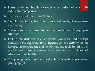
![ Counterstaining e.g. with toluidine blue, shows the
histological details of the tissue. The staining must be able to
penetrate, but not have an adverse affect on the emulsion
Alternatively, pre-staining of the entire block of tissue can be
done (e.g. with Osmium on plastic sections coated with
stripping film [or dipping emulsion] as in papers by
McGeachie and Grounds) before exposure to the
photographic emulsion. This avoids the need for individually
(post-) staining each slide.](https://image.slidesharecdn.com/receptordown-regulation-151128190638-lva1-app6891/85/Receptor-down-regulation-16-320.jpg)


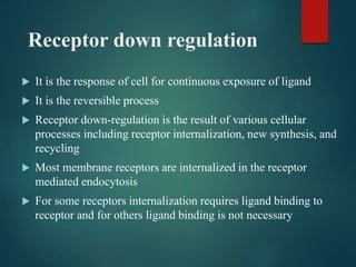
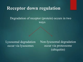
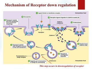
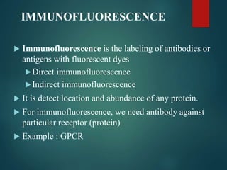
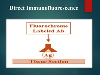
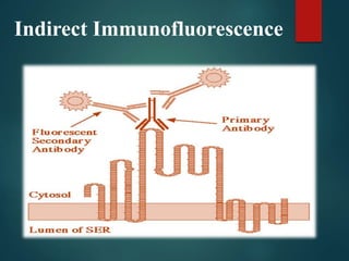
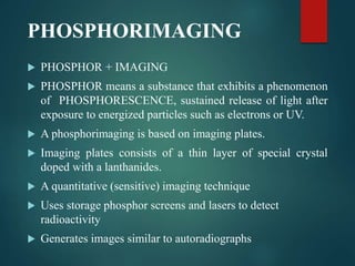
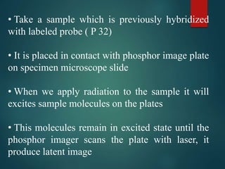
This document discusses receptor down-regulation, a cellular response to continuous exposure of ligands such as hormones and drugs. It details the mechanisms involved in receptor down-regulation, including receptor internalization and degradation via lysosomal and non-lysosomal pathways. Additionally, it describes techniques for determining receptor down-regulation, such as immunofluorescence, phosphorimaging, and autoradiography.


