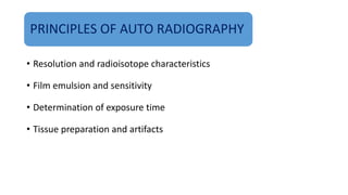
MICRO
AUTORADIOGRAPHY
S.MURALI
2016602005
Autoradiography
Autoradiography is the bio-analytical technique used to visualize the distribution of radioactive
labelled substance with radioisotope in a biological sample.
It is a method by which a radioactive material can be localized within a particular tissue, cell, cell
organelles or even biomolecules.
 It is a very sensitive technique and is being used in a wide variety of biological experiments.
 Autoradiography, although used to locate the radioactive substances, it can also be used for
quantitative estimation by using densitometer.
HISTORY
 The first autoradiography was obtained accidently around 1867 when a blackening was produced on
emulsions of silver chloride and iodide by uranium salts observed by Niepce de St. Victor.
 In 1924 first biological experiment involving autoradiography traced the distribution of polonium in
biological specimens.
 The development of autoradiography as a biological technique really started to happen after World war II
with the development of photographic emulsions and then stripping made of silver halide.
 Radioactivity is now no longer the property of a few rare elements of minor biological interest (such as
radium, thorium or uranium) as now any biological compound can be labelled with radioactive isotopes
opening up many possibilities in the study of living systems.
MICRO AUTORADIOGRAPHY
 Micro autoradiography, has been developed for studying subcellular structures,
even those as small as individual strands of deoxyribonucleic acid (DNA).
Much interesting information has been learned about the mechanisms of
cell division and other processes in cell biology.
The cells being studied are given a nutrient solution containing molecules that
have been labeled, usually with radioactive tritium, carbon, or phosphorus.
PRINCIPLES OF AUTO RADIOGRAPHY
• Resolution and radioisotope characteristics
• Film emulsion and sensitivity
• Determination of exposure time
• Tissue preparation and artifacts
PRINCIPLE
Incubate tissue with
radioactive ligand
Expose to film
or emulsion
Isotope will emit
radiation (usually beta)
Radiation will hit silver grains in emulsion and expose them
BASIC COMPONENTS
• Specimen
• Tracer
• Detector
MICRO AUTORADIOGRAPHY PROCEDURE
Autoradiography is based upon the ability of radioactive substance to expose the photographic
film by ionizing it.
 In this technique a radioactive substance is put in direct contact with a thick layer of a
photographic emulsion (thickness of 5-50 mm) having gelatin substances and silver halide
crystals.
This emulsion differs from the standard photographic film in terms of having higher ratio of silver
halide to gelatin and small size of grain.
 It is then left in dark for several days for proper exposure.
The silver halide crystals are exposed to the radiation which chemically converts silver halide into
metallic silver (reduced) giving a dark colour band.
The resulting radiography is viewed by electron microscope, pre flashed screen, intensifying
screen, electrophoresis, digital scanners etc.
RECEPTOR MICRO-AUTORADIOGRAPHY
Sequence of steps
APPLICATION of radiolabelled compound with high specific activity (including competition)
EXCISION
of multiple tissues and positioning on holders
FREEZE-MOUNTING
on tissue holders
LIQUID NITROGEN STORAGE
of mounted tissues
CRYO-SECTIONING
of 4 lm (or less) sections
THAW-MOUNTING on emulsion-coated slides
PHOTOGRAPHIC EXPOSURE in desiccator boxes
PHOTOGRAPHIC PROCESSING
STAINING with MBBF or MGP
AIR-DRYING and COVER-SLIPPING
EVALUATION
qualitative and quantitative
Applications
To find and investigate the various properties of DNA
To find the location and amount of particular substance within a cell
including cell organelle, metabolites etc.
Tissue localization of radioactive substance.
To find the site and performance of targeted drug.
To locate the metabolic activity site in the cell.
 Radioactive precursors of DNA and RNA, [3H]-thymidine and [3H]-
uridine respectively, may be to determine the timing of several
phases of the cell cycle. introduced to living cells
RNA or DNA viral sequences can also be located in this fashion. These
probes are usually labelled with 32P, 33P, or 35S.
Practical
Applications
In agricultural research, the effectiveness of herbicides, insecticides, and
fertilizers is studied to determine which ones can increase productivity without
causing serious environmental problems. Radioactive phosphorus can be used in
this regard to study plant metabolism.
The uptake of iron or zinc from the soil and their circulation in a plant can be
studied to ascertain the effect of soil acidity and chemical form. Sometimes the
presence or absence of other elements can inhibit translocation of an essential
nutrient.
New plant growth regulators may move from one plant through the soil to
a nearby untreated plant. Autoradiography is an important analytical
technique for observing the route of micronutrients and discovering what
factors can change their mobility in a plant.
The sequence of bases in DNA molecules can be decoded by using
electrophoresis combined with autoradiography, and the study of DNA
sequences is crucial to research in many diverse areas of biology. Although
alternatives to using autoradiography in DNA sequencing are now
common, autoradiography is still a standard technique used in many other
aspects of molecular biology.
Conclusion
MARG is a High Resolution Histological Tool to investigate spatial localization of radiolabelled
drugs at a tissue, and cellular level.
Ex vivo and exsanguination occurs
 Numerous elaborations on the techniques
Cryo-preservation required for soluble compounds. Liquid tissue fixation (formalin) often
solubilizes and relocates diffusible test articles. Exception for receptor-bound TA.
Not Quantitative – No standards used, prone to artefacts, lack of control on detection media and
section thickness.
References :
 Adamczyk, J., M. Hesselhoe, N. Iversen, M. Horn, A. Lehner, P.H. Nielsen, M. Scholter, P. Roslev, and
M. Wagner. 2003. The isotope array, a new tool that employs substrate mediated labelling of rRNA for
determination of microbial community structure and function. Appl. Environ. Microbiol. 69:6875–
6887.
 Thiel, R., and M. Blaut. 2005. An improved method for the automated enumeration of fluorescently
labelled bacteria in human faeces. J. Microbiol. Methods 61:369–379.
 Stumpf W E, Sar M, Joshi S G. Estrogen target cells in the skin. Experientia 1974: 30: 196–198.
THANK YOU

Micro autoradiography pptx

  • 1.
  • 2.
    Autoradiography Autoradiography is thebio-analytical technique used to visualize the distribution of radioactive labelled substance with radioisotope in a biological sample. It is a method by which a radioactive material can be localized within a particular tissue, cell, cell organelles or even biomolecules.  It is a very sensitive technique and is being used in a wide variety of biological experiments.  Autoradiography, although used to locate the radioactive substances, it can also be used for quantitative estimation by using densitometer.
  • 3.
    HISTORY  The firstautoradiography was obtained accidently around 1867 when a blackening was produced on emulsions of silver chloride and iodide by uranium salts observed by Niepce de St. Victor.  In 1924 first biological experiment involving autoradiography traced the distribution of polonium in biological specimens.  The development of autoradiography as a biological technique really started to happen after World war II with the development of photographic emulsions and then stripping made of silver halide.  Radioactivity is now no longer the property of a few rare elements of minor biological interest (such as radium, thorium or uranium) as now any biological compound can be labelled with radioactive isotopes opening up many possibilities in the study of living systems.
  • 4.
    MICRO AUTORADIOGRAPHY  Microautoradiography, has been developed for studying subcellular structures, even those as small as individual strands of deoxyribonucleic acid (DNA). Much interesting information has been learned about the mechanisms of cell division and other processes in cell biology. The cells being studied are given a nutrient solution containing molecules that have been labeled, usually with radioactive tritium, carbon, or phosphorus.
  • 5.
    PRINCIPLES OF AUTORADIOGRAPHY • Resolution and radioisotope characteristics • Film emulsion and sensitivity • Determination of exposure time • Tissue preparation and artifacts
  • 6.
    PRINCIPLE Incubate tissue with radioactiveligand Expose to film or emulsion Isotope will emit radiation (usually beta) Radiation will hit silver grains in emulsion and expose them
  • 7.
  • 8.
    MICRO AUTORADIOGRAPHY PROCEDURE Autoradiographyis based upon the ability of radioactive substance to expose the photographic film by ionizing it.  In this technique a radioactive substance is put in direct contact with a thick layer of a photographic emulsion (thickness of 5-50 mm) having gelatin substances and silver halide crystals. This emulsion differs from the standard photographic film in terms of having higher ratio of silver halide to gelatin and small size of grain.  It is then left in dark for several days for proper exposure. The silver halide crystals are exposed to the radiation which chemically converts silver halide into metallic silver (reduced) giving a dark colour band. The resulting radiography is viewed by electron microscope, pre flashed screen, intensifying screen, electrophoresis, digital scanners etc.
  • 11.
    RECEPTOR MICRO-AUTORADIOGRAPHY Sequence ofsteps APPLICATION of radiolabelled compound with high specific activity (including competition) EXCISION of multiple tissues and positioning on holders FREEZE-MOUNTING on tissue holders LIQUID NITROGEN STORAGE of mounted tissues CRYO-SECTIONING of 4 lm (or less) sections THAW-MOUNTING on emulsion-coated slides
  • 12.
    PHOTOGRAPHIC EXPOSURE indesiccator boxes PHOTOGRAPHIC PROCESSING STAINING with MBBF or MGP AIR-DRYING and COVER-SLIPPING EVALUATION qualitative and quantitative
  • 13.
    Applications To find andinvestigate the various properties of DNA To find the location and amount of particular substance within a cell including cell organelle, metabolites etc. Tissue localization of radioactive substance. To find the site and performance of targeted drug. To locate the metabolic activity site in the cell.
  • 14.
     Radioactive precursorsof DNA and RNA, [3H]-thymidine and [3H]- uridine respectively, may be to determine the timing of several phases of the cell cycle. introduced to living cells RNA or DNA viral sequences can also be located in this fashion. These probes are usually labelled with 32P, 33P, or 35S.
  • 15.
    Practical Applications In agricultural research,the effectiveness of herbicides, insecticides, and fertilizers is studied to determine which ones can increase productivity without causing serious environmental problems. Radioactive phosphorus can be used in this regard to study plant metabolism. The uptake of iron or zinc from the soil and their circulation in a plant can be studied to ascertain the effect of soil acidity and chemical form. Sometimes the presence or absence of other elements can inhibit translocation of an essential nutrient.
  • 16.
    New plant growthregulators may move from one plant through the soil to a nearby untreated plant. Autoradiography is an important analytical technique for observing the route of micronutrients and discovering what factors can change their mobility in a plant. The sequence of bases in DNA molecules can be decoded by using electrophoresis combined with autoradiography, and the study of DNA sequences is crucial to research in many diverse areas of biology. Although alternatives to using autoradiography in DNA sequencing are now common, autoradiography is still a standard technique used in many other aspects of molecular biology.
  • 17.
    Conclusion MARG is aHigh Resolution Histological Tool to investigate spatial localization of radiolabelled drugs at a tissue, and cellular level. Ex vivo and exsanguination occurs  Numerous elaborations on the techniques Cryo-preservation required for soluble compounds. Liquid tissue fixation (formalin) often solubilizes and relocates diffusible test articles. Exception for receptor-bound TA. Not Quantitative – No standards used, prone to artefacts, lack of control on detection media and section thickness.
  • 18.
    References :  Adamczyk,J., M. Hesselhoe, N. Iversen, M. Horn, A. Lehner, P.H. Nielsen, M. Scholter, P. Roslev, and M. Wagner. 2003. The isotope array, a new tool that employs substrate mediated labelling of rRNA for determination of microbial community structure and function. Appl. Environ. Microbiol. 69:6875– 6887.  Thiel, R., and M. Blaut. 2005. An improved method for the automated enumeration of fluorescently labelled bacteria in human faeces. J. Microbiol. Methods 61:369–379.  Stumpf W E, Sar M, Joshi S G. Estrogen target cells in the skin. Experientia 1974: 30: 196–198.
  • 19.

Editor's Notes

  • #7 Biological tissue or a living organism is injected with a radiolabelled substance with the purpose of investigating its distribution in the organism. At the end of experiment thin tissue sections are placed in contact with a photographic emulsion.the ionic radiation will convert the emulsion as dark spots which makes it possible to localize the radioactive compound (or its metabolites) in the section. The distribution of radioactive material may be investigated as a function of time after the injection of the radiolabelled compound (”pulse labelling”).