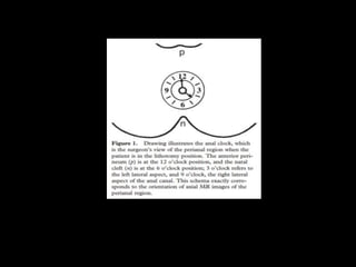
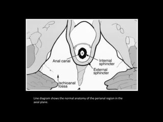
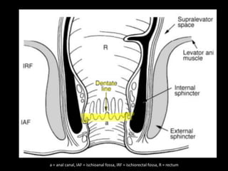
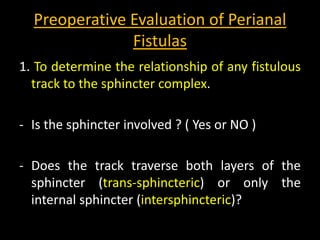
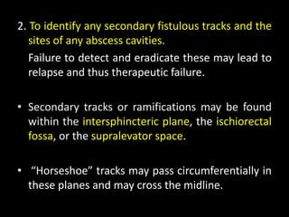
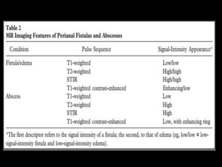
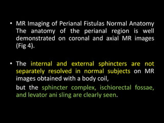
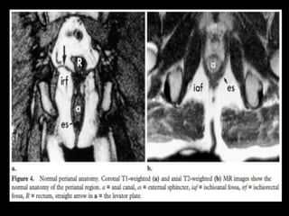
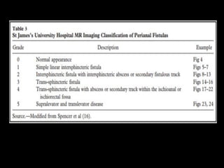
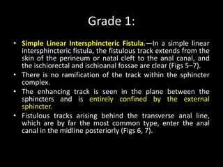
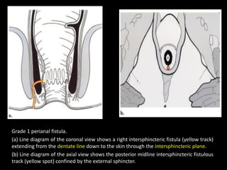
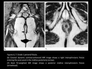
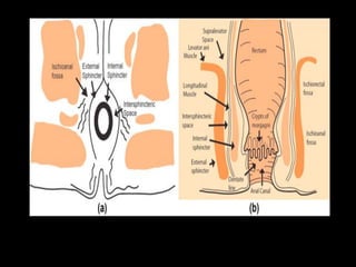
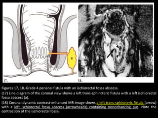
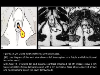
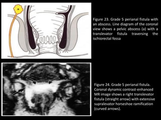
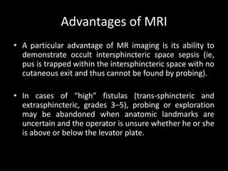
This document discusses MR imaging techniques for evaluating perianal fistulas. It begins by describing the anatomy of the perianal region and anal sphincter mechanism. It then discusses the aims and implications of MR imaging-based grading of fistulas. Various fistula classifications and types are defined based on their relationship to anal anatomy. The document outlines normal anal region anatomy as seen on MR images and techniques for identifying fistulous tracks, abscesses, and complications. It emphasizes the importance of MR imaging in surgical planning and treatment of perianal fistulas.
radiological anatomy of retroperitoneum powerpointDactarAdhikari
brief and concise on radiological anatomy of retroperitoneum
includes topic like pararenal space,perirenal space,fascial plane,retroperitoneum hematoma and sign of mass origin
Hepatobiliary system radiology revision notesTONY SCARIA
hepatobiliary system
hepatic segments
image based questions
last minute revision
radiology radiodiagnosis
hepatic investigations
based image based questions f
fistula-in-ano, or anal fistula, is a chronic abnormal communication extending from the anorectal lumen (the internal opening) to an external opening on the skin of the perineum or buttock.
Majority are idiopathic.
ARTIFICIAL INTELLIGENCE IN HEALTHCARE.pdfAnujkumaranit
Artificial intelligence (AI) refers to the simulation of human intelligence processes by machines, especially computer systems. It encompasses tasks such as learning, reasoning, problem-solving, perception, and language understanding. AI technologies are revolutionizing various fields, from healthcare to finance, by enabling machines to perform tasks that typically require human intelligence.
HOT NEW PRODUCT! BIG SALES FAST SHIPPING NOW FROM CHINA!! EU KU DB BK substit...GL Anaacs
Contact us if you are interested:
Email / Skype : kefaya1771@gmail.com
Threema: PXHY5PDH
New BATCH Ku !!! MUCH IN DEMAND FAST SALE EVERY BATCH HAPPY GOOD EFFECT BIG BATCH !
Contact me on Threema or skype to start big business!!
Hot-sale products:
NEW HOT EUTYLONE WHITE CRYSTAL!!
5cl-adba precursor (semi finished )
5cl-adba raw materials
ADBB precursor (semi finished )
ADBB raw materials
APVP powder
5fadb/4f-adb
Jwh018 / Jwh210
Eutylone crystal
Protonitazene (hydrochloride) CAS: 119276-01-6
Flubrotizolam CAS: 57801-95-3
Metonitazene CAS: 14680-51-4
Payment terms: Western Union,MoneyGram,Bitcoin or USDT.
Deliver Time: Usually 7-15days
Shipping method: FedEx, TNT, DHL,UPS etc.Our deliveries are 100% safe, fast, reliable and discreet.
Samples will be sent for your evaluation!If you are interested in, please contact me, let's talk details.
We specializes in exporting high quality Research chemical, medical intermediate, Pharmaceutical chemicals and so on. Products are exported to USA, Canada, France, Korea, Japan,Russia, Southeast Asia and other countries.
TEST BANK for Operations Management, 14th Edition by William J. Stevenson, Ve...kevinkariuki227
TEST BANK for Operations Management, 14th Edition by William J. Stevenson, Verified Chapters 1 - 19, Complete Newest Version.pdf
micro teaching on communication m.sc nursing.pdfAnurag Sharma
Microteaching is a unique model of practice teaching. It is a viable instrument for the. desired change in the teaching behavior or the behavior potential which, in specified types of real. classroom situations, tends to facilitate the achievement of specified types of objectives.
MANAGEMENT OF ATRIOVENTRICULAR CONDUCTION BLOCK.pdfJim Jacob Roy
Cardiac conduction defects can occur due to various causes.
Atrioventricular conduction blocks ( AV blocks ) are classified into 3 types.
This document describes the acute management of AV block.
Ozempic: Preoperative Management of Patients on GLP-1 Receptor Agonists Saeid Safari
Preoperative Management of Patients on GLP-1 Receptor Agonists like Ozempic and Semiglutide
ASA GUIDELINE
NYSORA Guideline
2 Case Reports of Gastric Ultrasound
Report Back from SGO 2024: What’s the Latest in Cervical Cancer?bkling
Are you curious about what’s new in cervical cancer research or unsure what the findings mean? Join Dr. Emily Ko, a gynecologic oncologist at Penn Medicine, to learn about the latest updates from the Society of Gynecologic Oncology (SGO) 2024 Annual Meeting on Women’s Cancer. Dr. Ko will discuss what the research presented at the conference means for you and answer your questions about the new developments.
2. AIM
• Demonstrate accurately the anatomy of the perianal region.
• The anal sphincter mechanism, MR imaging clearly shows the
relationship of fistulas to the pelvic diaphragm (levator plate)
and the ischiorectal fossae.
• This relationship has important implications for surgical
management and outcome and has been classified into five
MR imaging–based grades.
3. 1. If the ischio-anal and ischiorectal fossae are unaffected,
disease is likely confined to the sphincter complex (simple
intersphincteric fistulization, grade 1 or 2), and outcome
following simple surgical management is favorable.
2. Involvement of the ischio-anal or ischiorectal fossa by a
fistulous track or abscess indicates complex disease
related to trans-sphincteric or suprasphincteric disease
(grade 3 or 4). Correspondingly more complex surgery
may be required that may threaten continence or may
require colostomy to allow healing.
3. If the track traverses the levator plate, a translevator
fistula (grade 5) is present, and a source of pelvic sepsis
should be sought.
4. • Much of our understanding of perianal fistulas
comes from the work of surgeons at St Mark’s
Hospital: Dr Salmon, who operated on Charles
Dickens.
• Goodsall:- who described the course of
fistulous tracks from the skin to the anus.
• Parks:- whose classification of fistulas in
relation to anal anatomy is widely used in
surgical practice.
5. • Goodsall described the relationship of the cutaneous
opening to the expected site of the enteric opening.
• The rule states that cutaneous openings –
Anterior to the transverse anal line are associated with
Direct radial fistulous tracks into the anal canal,
• whereas openings Posterior to the line have tracks that
enter the canal in the midline posteriorly.
•
6. • The cutaneous opening is evident to the surgeon and
thus of little importance for the radiologist to
demonstrate.
• The surgeon passes blunt probes along the track from
the cutaneous opening and may determine the enteric
opening of the fistula at proctoscopy.
• The challenge in the management of fistulas is to
define the course of the track between these openings
so that the appropriate surgical option can be used.
7. • MR imaging–based grading system for
perianal fistulas (the St James’s University
Hospital classification) that has been validated
by surgically proved cases with documented
long-term outcome.
8. • Anatomy of the Anal Region :-
• Surgeons describe the site and direction of fistulous tracks
by referring to the “anal clock” ,that is, the view of the anal
region with the patient in the lithotomy position usually
used for fistula surgery.
• At 12 o’clock is the anterior perineum and at 6 o’clock, the
natal cleft; 3 o’clock refers to the left lateral aspect, and 9
o’clock, to the right of the anal canal.
• **Fortunately these descriptions correspond exactly with
the view of the anal canal on axial MR images, and it is
helpful for surgical colleagues if the radiologists relate the
MR imaging findings to the anal clock.
9. • To understand the surgical options for treating fistulous disease,
one must first consider the anatomy and function of the anal
sphincters and the causes of perianal fistulas.
• The internal sphincter is involuntary and is composed of smooth
muscle continuous with the circular smooth muscle of the rectum.
It is responsible for 85% of resting anal tone. In most individuals, it
can be divided (OPERATED) without causing a loss of continence.
• The external sphincter is composed of striated muscle and is
continuous superiorly with the puborectalis and levator Ani
muscles.
• It contributes only 15% of resting anal tone, but its strong voluntary
contractions resist defecation.
• A division of the external sphincter can lead to incontinence.
10.
11. a = anal canal, IAF = ischioanal fossa, IRF = ischiorectal fossa, R = rectum
12. Line diagram shows the normal anatomy of the perianal region in the
axial plane.
13. • Causes of Perianal Fistulas:-
• Idiopathic fistulas are generally believed to represent
the chronic phase of intramuscular anal gland sepsis
(ie, the crypto glandular hypothesis).
• However, perianal fistulas may also be caused by other
conditions and events, including
- Crohn disease,
- tuberculosis,
- trauma during childbirth,
- pelvic infection,
- pelvic malignancy, and
- radiation therapy.
14. • Anal glands lie at the level of the dentate line in
the midanal canal and can penetrate the internal
sphincter to lie in the intersphincteric plane.
• From this space, the infection may track down
the intersphincteric plane to the skin, and about
70% of fistulas behave in this way.
• Alternatively, infection may pass through both
layers of the anal sphincter (trans-sphincteric
fistulization) to enter the ischiorectal fossa; this
development pattern occurs in about 20% of
cases.
15. a = anal canal, IAF = ischioanal fossa, IRF = ischiorectal fossa, R = rectum
16. • Abscess cavities may develop along the course of
fistulous tracks. Characteristically, the abscesses
associated with intersphincteric fistulas are perianal or
indeed encysted within the intersphincteric space.
• Trans-sphincteric fistulas are typically associated with
ischiorectal fossa abscesses.
• In rare cases, infection tracks upward over the
sphincter complex to enter the ischiorectal fossa
(suprasphincteric fistulization).
• Sepsis arising within the pelvis may track down to the
skin through the ischiorectal fossa, resulting in fistulas
that are referred to as extrasphincteric or translevator.
17. • Surgical Approaches / Surgical management of
perianal fistulas depends on the
- nature of the primary fistula and
- any secondary fistulous tracks or
- associated abscesses.
• For simple intersphincteric fistulas, the surgeon
performs a fistulotomy or fistulectomy, in which the
internal sphincter is divided to lay open the track.
• Alternatively, in patients with perianal abscess, the
surgeon performs a simple incision and drainage first.
18. • Preservation of fecal continence is the paramount
consideration, and treatment strategies aim to
preserve the integrity of the external sphincter.
• Over the years, surgeons have been acutely
aware of the damage to their reputations that
has resulted from rendering patients incontinent.
Surgical papers abound with quotes, such as “if
this ring be cut, loss of control surely follows”
19. Preoperative Evaluation of Perianal
Fistulas
1. To determine the relationship of any fistulous
track to the sphincter complex.
- Is the sphincter involved ? ( Yes or NO )
- Does the track traverse both layers of the
sphincter (trans-sphincteric) or only the
internal sphincter (intersphincteric)?
20. 2. To identify any secondary fistulous tracks and the
sites of any abscess cavities.
Failure to detect and eradicate these may lead to
relapse and thus therapeutic failure.
• Secondary tracks or ramifications may be found
within the intersphincteric plane, the ischiorectal
fossa, or the supralevator space.
• “Horseshoe” tracks may pass circumferentially in
these planes and may cross the midline.
21.
22. • MR imaging examinations performed with a body coil
require no patient preparation and are well tolerated.
• Use of endoanal coils was initially hoped to further improve
the MR imaging evaluation of perianal fistulas (14).
• However, this technique is poorly tolerated in symptomatic
patients, and although it provides excellent anatomic detail
of the anal sphincters, it fails to provide the overview
required for surgical management (15).
• It seems likely that use of pelvic surface coils may further
improve image quality.
23.
24. • MR Imaging of Perianal Fistulas Normal Anatomy
The anatomy of the perianal region is well
demonstrated on coronal and axial MR images
(Fig 4).
• The internal and external sphincters are not
separately resolved in normal subjects on MR
images obtained with a body coil,
but the sphincter complex, ischiorectal fossae,
and levator ani sling are clearly seen.
25.
26. • Techniques for Imaging Perianal Fistulas Unenhanced T1-weighted
images provide an excellent anatomic overview of the sphincter
complex, levator plate, and the ischiorectal fossae.
• Fistulous tracks, inflammation, and abscesses, however, appear as
areas of low to intermediate signal intensity and may not be
distinguished from normal structures such as the sphincters and
levator ani muscles.
• On T2-weighted and STIR images, pathologic processes including
fistulas, secondary fistulous tracks, and fluid collections are clearly
depicted (Table 2).
• They appear as areas of high signal intensity in contrast with the
lower signal intensity of the sphincters, muscles, and fat (especially
on STIR images). The only comparative study of imaging sequences
suggested for use in this condition showed that STIR imaging has
certain limitations (10).
27. • In some cases, STIR imaging failed to
demonstrate secondary tracks, and in others it
did not reveal small residual abscesses within
edematous inflammatory change. Furthermore,
spurious incidences of high signal intensity in
inactive tracks were also observed.
• Fat suppression techniques used with T2-
weighted imaging have also been proposed (11)
28.
29. • We use gradient-echo T1-weighted dynamic
intravenous contrast material–enhanced MR
imaging combined with T2-weighted imaging to
assess perianal fistulas and their complications .
• With use of this technique, active fistulous tracks,
secondary ramifications, and abscesses are
clearly demonstrated.
• The tracks brilliantly enhance as do the walls of
abscess cavities.
• Retained pus remains unenhanced, with resulting
ring enhancement, an appearance that is typical
of abscess formation elsewhere in the body.
30. • Unenhanced T1-weighted imaging may be helpful in
postoperative assessment.
• Firstly, if MR imaging is performed in the immediate
postoperative period, hemorrhage will appear
hyperintense and may thus be differentiated from a
residual track.
• Secondly, fat-containing “grafts” may be placed to fill
cavities and resection voids in restorative surgery.
These hyperintense structures are readily identified on
unenhanced T1- weighted images and can be
distinguished from active disease, which appears
hyperintense only after enhancement with gadolinium.
31. St James’s University Hospital
Classification
• the demonstration of the primary fistulous
track but also with secondary ramifications
and associated abscesses.
32. Grade 1:
• Simple Linear Intersphincteric Fistula.—In a simple linear
intersphincteric fistula, the fistulous track extends from the
skin of the perineum or natal cleft to the anal canal, and
the ischiorectal and ischioanal fossae are clear (Figs 5–7).
• There is no ramification of the track within the sphincter
complex.
• The enhancing track is seen in the plane between the
sphincters and is entirely confined by the external
sphincter.
• Fistulous tracks arising behind the transverse anal line,
which are by far the most common type, enter the anal
canal in the midline posteriorly (Figs 6, 7).
33. Grade 1 perianal fistula.
(a) Line diagram of the coronal view shows a right intersphincteric fistula (yellow track)
extending from the dentate line down to the skin through the intersphincteric plane.
(b) Line diagram of the axial view shows the posterior midline intersphincteric fistulous
track (yellow spot) confined by the external sphincter.
34. Figures 6, 7. Grade 1 perianal fistula.
(6) Coronal dynamic contrast-enhanced MR image shows a right intersphincteric fistula
entering the anal canal in the midline posteriorly (arrow).
(7) Axial T2-weighted MR image shows a posterior midline intersphincteric fistula
(arrowhead).
35. Grade 2:
• Intersphincteric Fistula with Abscess or Secondary Track.—
Intersphincteric fistulas with an abscess or secondary track
are also bounded by the external sphincter.
• Secondary fistulous tracks may be of the horseshoe type,
crossing the midline (Figs 8, 9), or they may ramify in the
ipsilateral intersphincteric plane (Figs 10, 11).
• Even when there is abscess formation, this process is
confined within the sphincter complex regardless of
imaging plane or sequence (Fig 12).
36. • On T2-weighted images, pus has high signal intensity and thus
cannot be reliably distinguished from edema and inflammation,
• but gas within abscesses has a low signal intensity similar to that of
the anorectal lumen (Fig 13).
• Intersphincteric abscesses and secondary fistulous tracks are well
shown by dynamic contrast-enhanced MR imaging.
• On these contrast-enhanced images, the pus in the central cavity
has low signal intensity and is surrounded by a brightly enhancing
rim (Figs 11, 12).
• A horseshoe fistula, in which the process extends to the opposite
side, is best demonstrated in the axial plane (Figs 10, 12b).
37. Figures 8, 9. Grade 2 horseshoe perianal
fistula.
(8) Line diagram of the axial view shows an
intersphincteric horseshoe fistula (yellow track,
arrow) confined by the external sphincter.
(9) Axial T2-weighted image shows an
intersphincteric horseshoe fistula (arrow).
38. Figures 10, 11. Grade 2 perianal
fistula with an abscess.
(10) Line diagram of the coronal view
shows a left intersphincteric abscess
(a) .
(11) Coronal dynamic contrast-
enhanced MR image shows a left
intersphincteric abscess cavity
(arrowhead) above the primary
intersphincteric track (curved arrow).
The enteric entry point is suggested
by a medial track (straight arrow).
39. Grade 3: Trans-sphincteric Fistula.
• Instead of tracking down the intersphincteric plane to the skin, the trans-
sphincteric fistula pierces through both layers of the sphincter complex
and then arcs down to the skin through the ischiorectal and ischioanal
fossae (Fig 14).
• Thus, a transsphincteric fistula may disrupt the normal fat of the
ischiorectal and ischioanal fossae with secondary edema and hyperemia
(Figs 15, 16).
• These fistulas are distinguished by the site of the enteric entry point in the
middle third of the anal canal (ie, corresponding to the position of the
dentate line), as seen on coronal images.
• Because these fistulas disrupt the integrity of the sphincter mechanism,
their tracks must be excised by dividing both layers of the sphincter, thus
risking fecal incontinence.
40. Figures 15, 16. GRADE 3 PERIANAL FISTULA.
(15) Coronal dynamic contrast-enhanced MR image shows a right trans-sphincteric fistula (arrow) and
inflammatory change in the right ischiorectal fossa. Note the entry site in the middle third of the anal
canal.
(16) Axial dynamic contrast-enhanced MR image shows a left trans-sphincteric fistula within the
ischiorectal fossa and piercing the external sphincter (arrow).
41. Grade 4:
• Grade 4: Trans-sphincteric Fistula with Abscess or Secondary Track
within the Ischiorectal Fossa.—
• A trans-sphincteric fistula can be complicated by sepsis in the
ischiorectal or ischioanal fossa (Figs 17, 18).
• Such an abscess may manifest as an expansion along the primary
track or as a structure distorting or filling the ischiorectal fossa.
• Axial and coronal dynamic contrast-enhanced MR imaging clearly
depicts a trans-sphincteric abscess, which characteristically has a
central focus of low-signal-intensity pus (Figs 18–20).
• As with grade 3 lesions, the key anatomic discriminator of a grade 4
fistula is the track crossing the external sphincter (Figs 21, 22).
42. Grade 5:
• Grade 5: Supralevator and Translevator Disease.—
• In rare cases, perianal fistulous disease extends above the insertion
of the levator ani muscle.
• Suprasphincteric fistulas extend upward in the intersphincteric
plane and over the top of the levator ani to pierce downward
through the ischiorectal fossa.
• Extrasphincteric fistulas reflect extension of primary pelvic disease
down through the levator plate (Fig 23). These fistulas pose
problems for management because further assessment is needed
to detect pelvic sepsis. Coronal dynamic contrast-enhanced MR
imaging elegantly demonstrates breaches of the levator plate,
which is clearly shown in this plane. In some translevator fistulas,
horseshoe ramifications to the contralateral side may occur (Fig 24).
43.
44. Figures 17, 18. Grade 4 perianal fistula with an ischiorectal fossa abscess.
(17) Line diagram of the coronal view shows a left trans-sphincteric fistula with a left ischiorectal
fossa abscess (a).
(18) Coronal dynamic contrast-enhanced MR image shows a left trans-sphincteric fistula (arrow)
with a left ischiorectal fossa abscess (arrowheads) containing nonenhancing pus. Note the
contraction of the ischiorectal fossa.
45. Figures 19, 20. Grade 4 perianal fistula with an abscess.
(19) Line diagram of the axial view shows a left trans-sphincteric fistula and left ischioanal
fossa abscess (a).
(20) Axial T2- weighted (a) and dynamic contrast enhanced (b) MR images show a left
trans-sphincteric fistula (straight arrow) with a left ischioanal fossa abscess (curved arrow)
and nonenhancing pus in the cavity (arrowhead).
46. Figure 23. Grade 5 perianal fistula with
an abscess. Line diagram of the coronal
view shows a pelvic abscess (a) with a
translevator fistula traversing the
ischiorectal fossa
Figure 24. Grade 5 perianal fistula.
Coronal dynamic contrast-enhanced
MR image shows a right translevator
fistula (straight arrow) with extensive
supralevator horseshoe ramification
(curved arrows).
47. Advantages of MRI
• A particular advantage of MR imaging is its ability to
demonstrate occult intersphincteric space sepsis (ie,
pus is trapped within the intersphincteric space with no
cutaneous exit and thus cannot be found by probing).
• In cases of “high” fistulas (trans-sphincteric and
extrasphincteric, grades 3–5), probing or exploration
may be abandoned when anatomic landmarks are
uncertain and the operator is unsure whether he or she
is above or below the levator plate.
48. Teaching points
• 1. If the ischioanal and ischiorectal fossae are unaffected at MR
imaging, disease is likely confined to the sphincter complex
(intersphincteric fistulization, grade 1 or 2). Outcome following
simple surgical management is favorable.
• 2. If there is any track or abscess within the ischiorectal fossa, it is
usually related to a complex perianal fistula (typically trans-
sphincteric fistulization, grade 3 or 4). Correspondingly more
complex surgery may be required that may threaten continence or
may require fecal diversion (colostomy) to allow healing.
• 3. When the track traverses the levator plate, a translevator fistula
(grade 5) is present, and a source of pelvic sepsis should be sought.