Downloaded 22 times


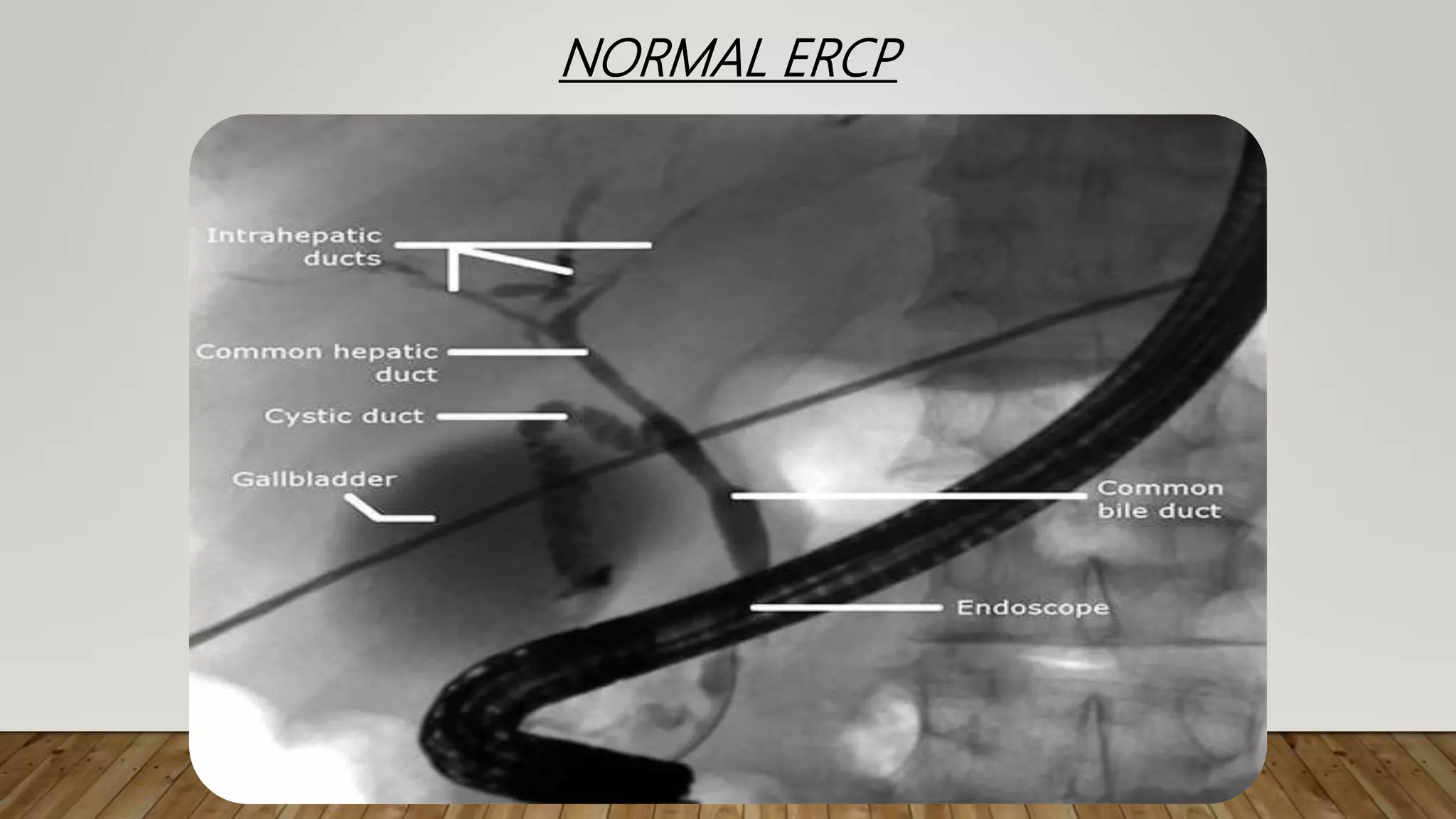
![ENDOSCOPIC RETROGRADE CHOLANGIO-
PANCREATICOGRAPHY [ERCP]](https://image.slidesharecdn.com/radiologicalanatomyofbiliarysystem-200916130946/75/Radiological-anatomy-of-biliary-system-21-2048.jpg)

![MAGNETIC RESONANCE CHOLANGIO-
PANCRETICOGRAPHY[MRCP]](https://image.slidesharecdn.com/radiologicalanatomyofbiliarysystem-200916130946/75/Radiological-anatomy-of-biliary-system-27-2048.jpg)


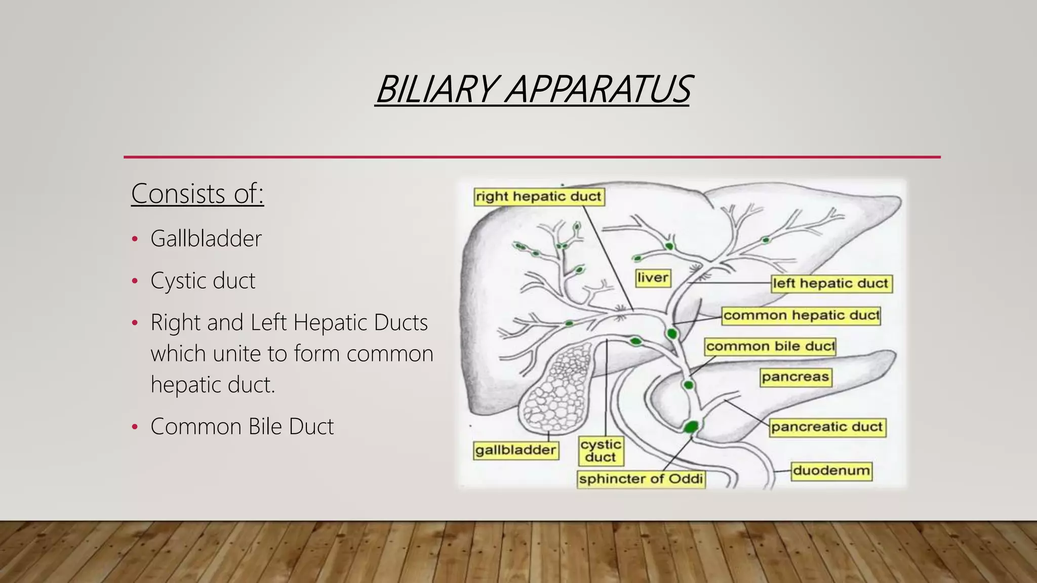
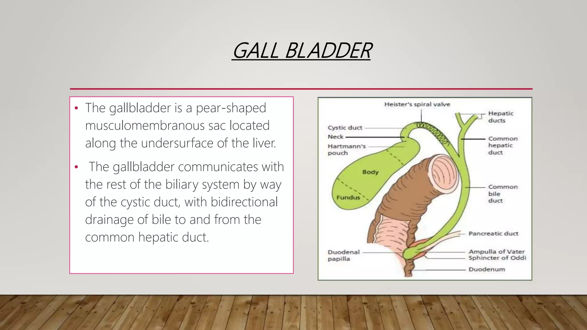
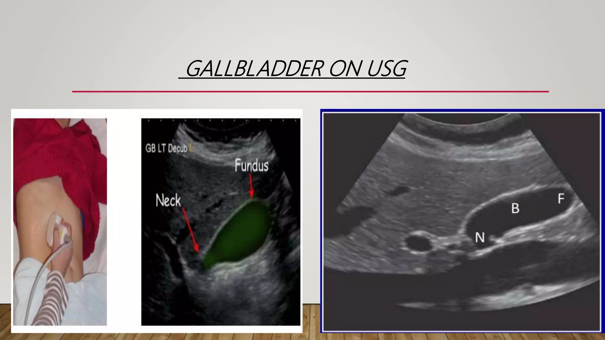
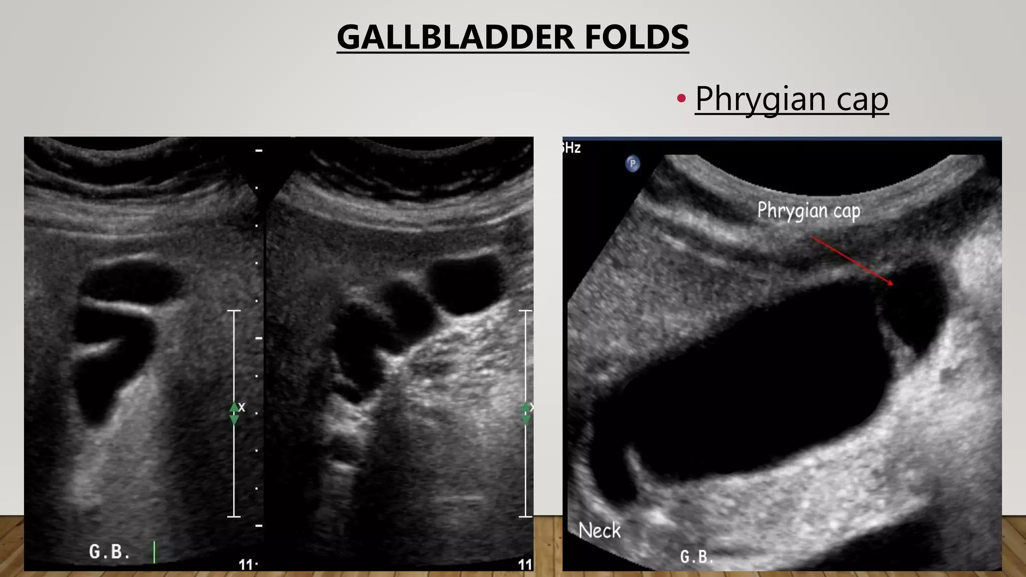
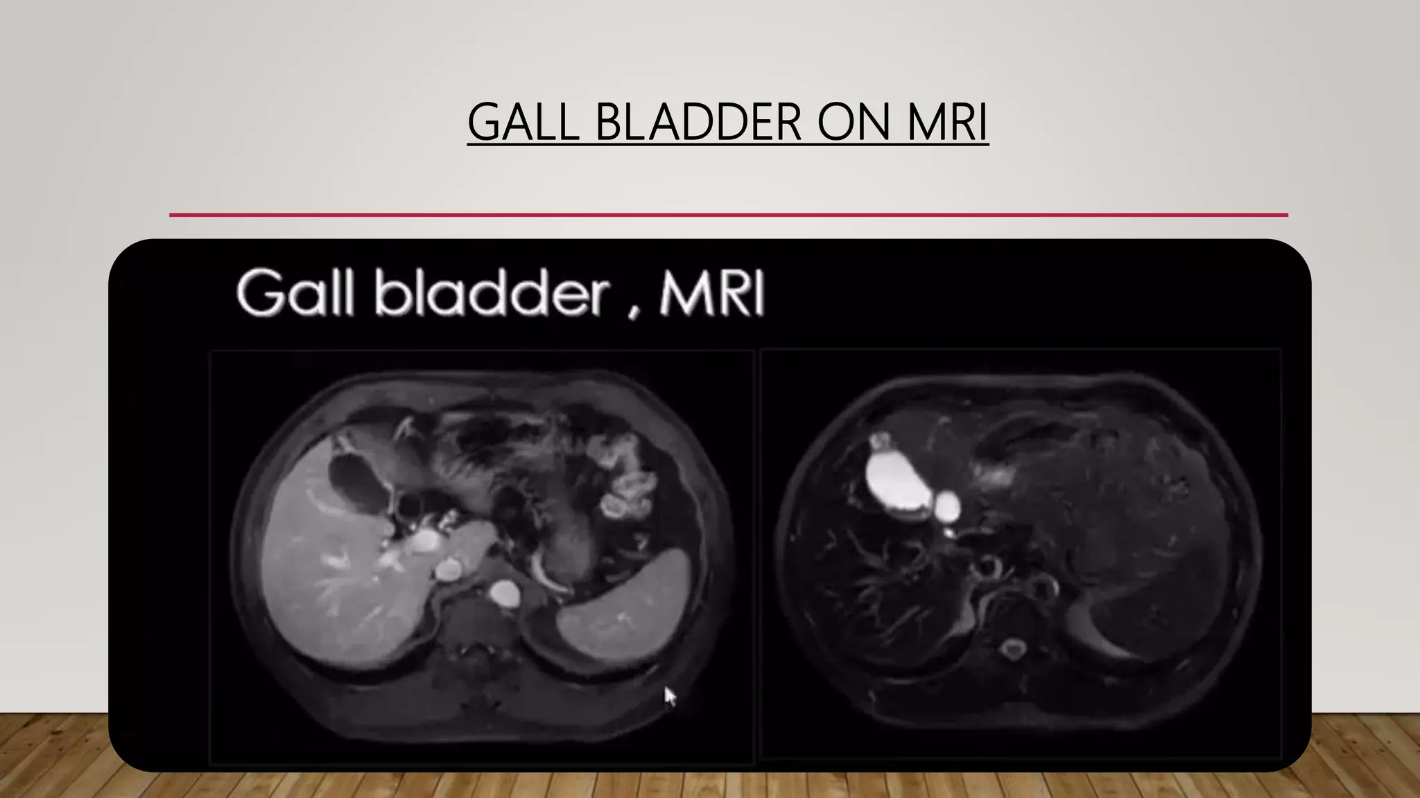
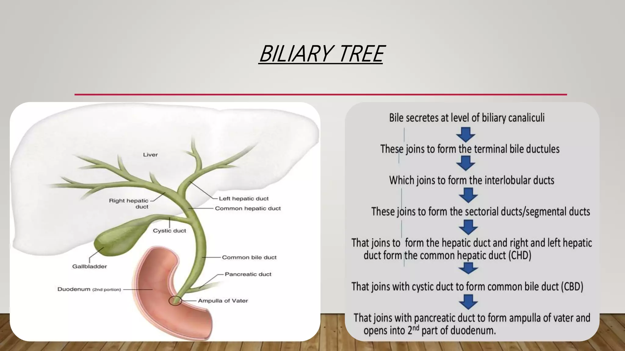
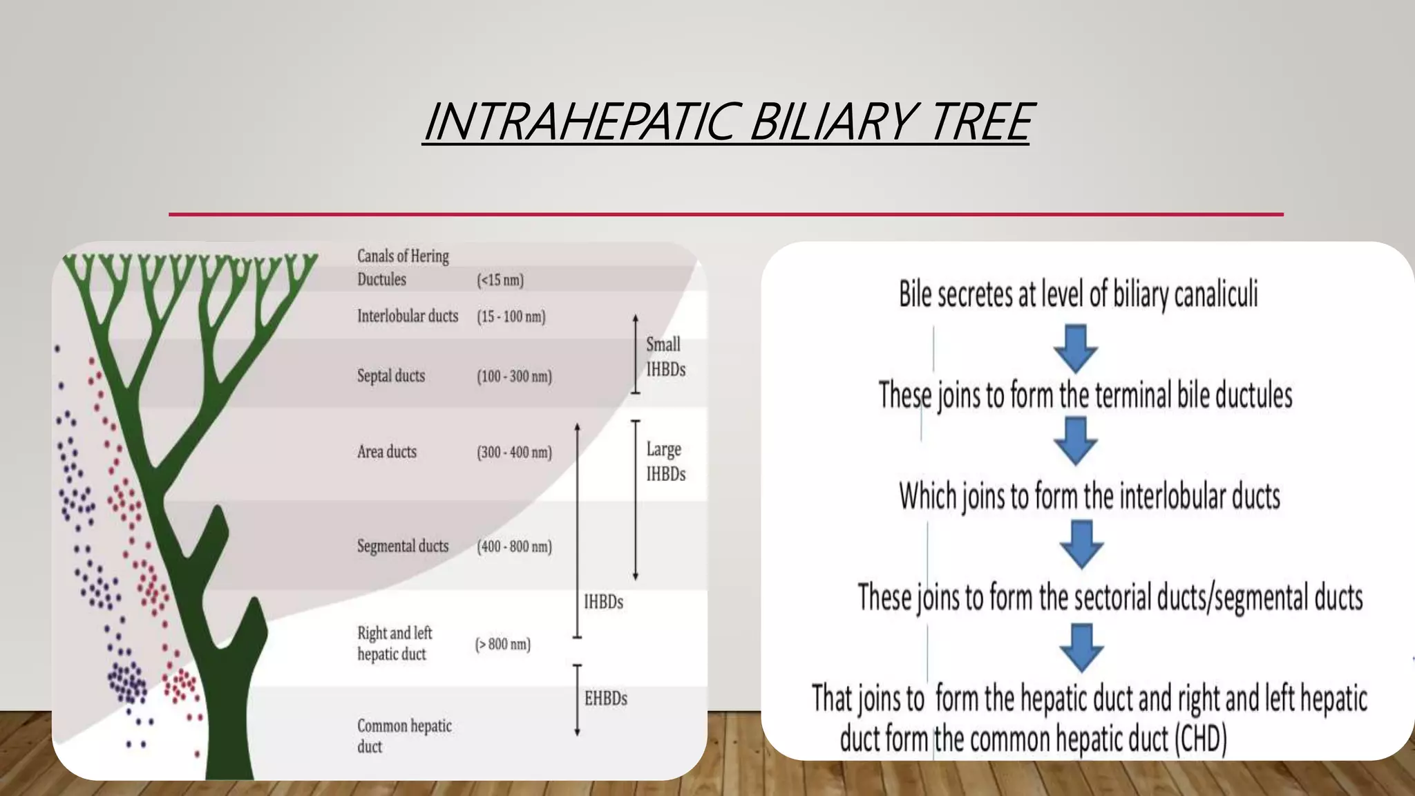
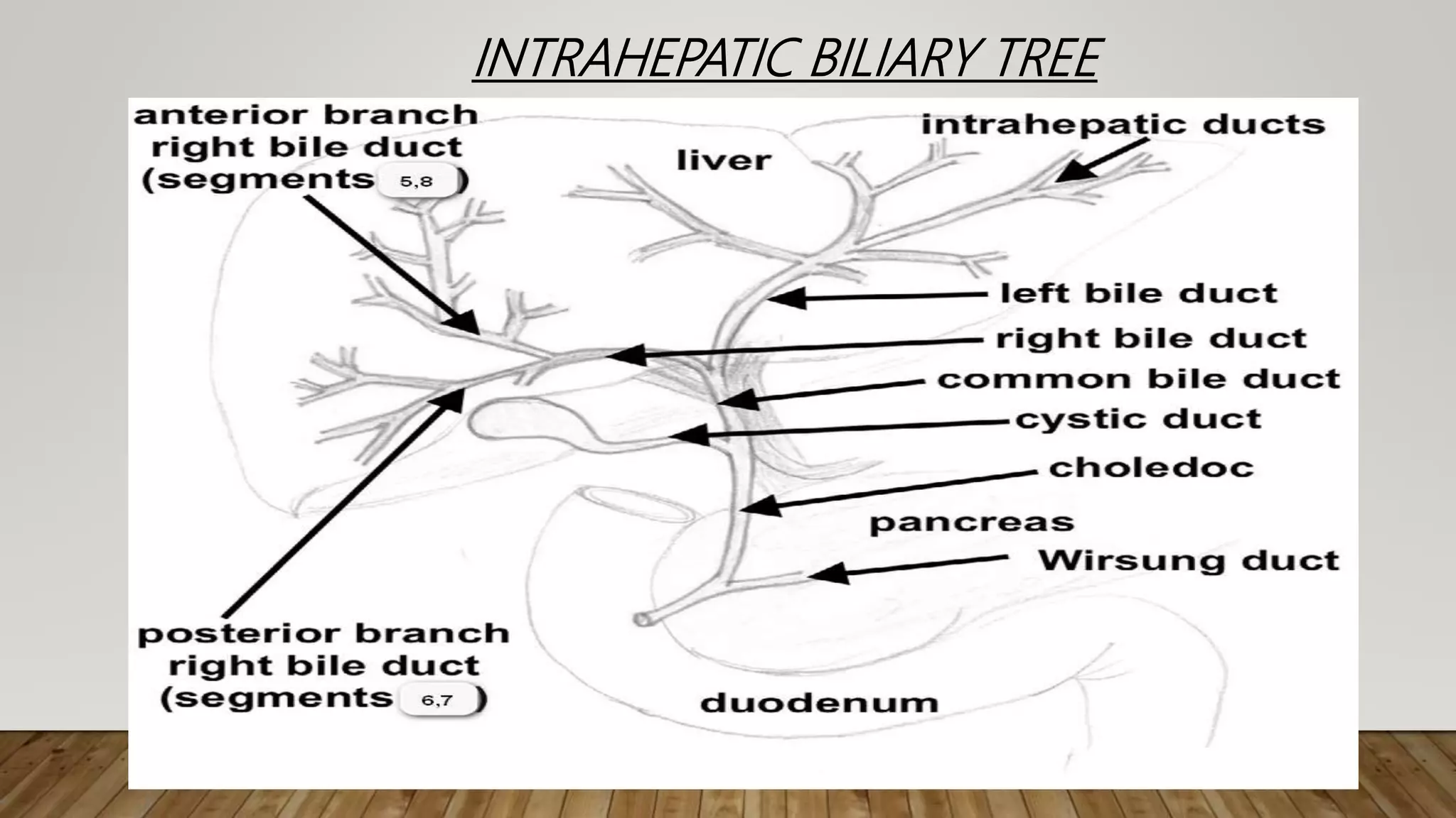
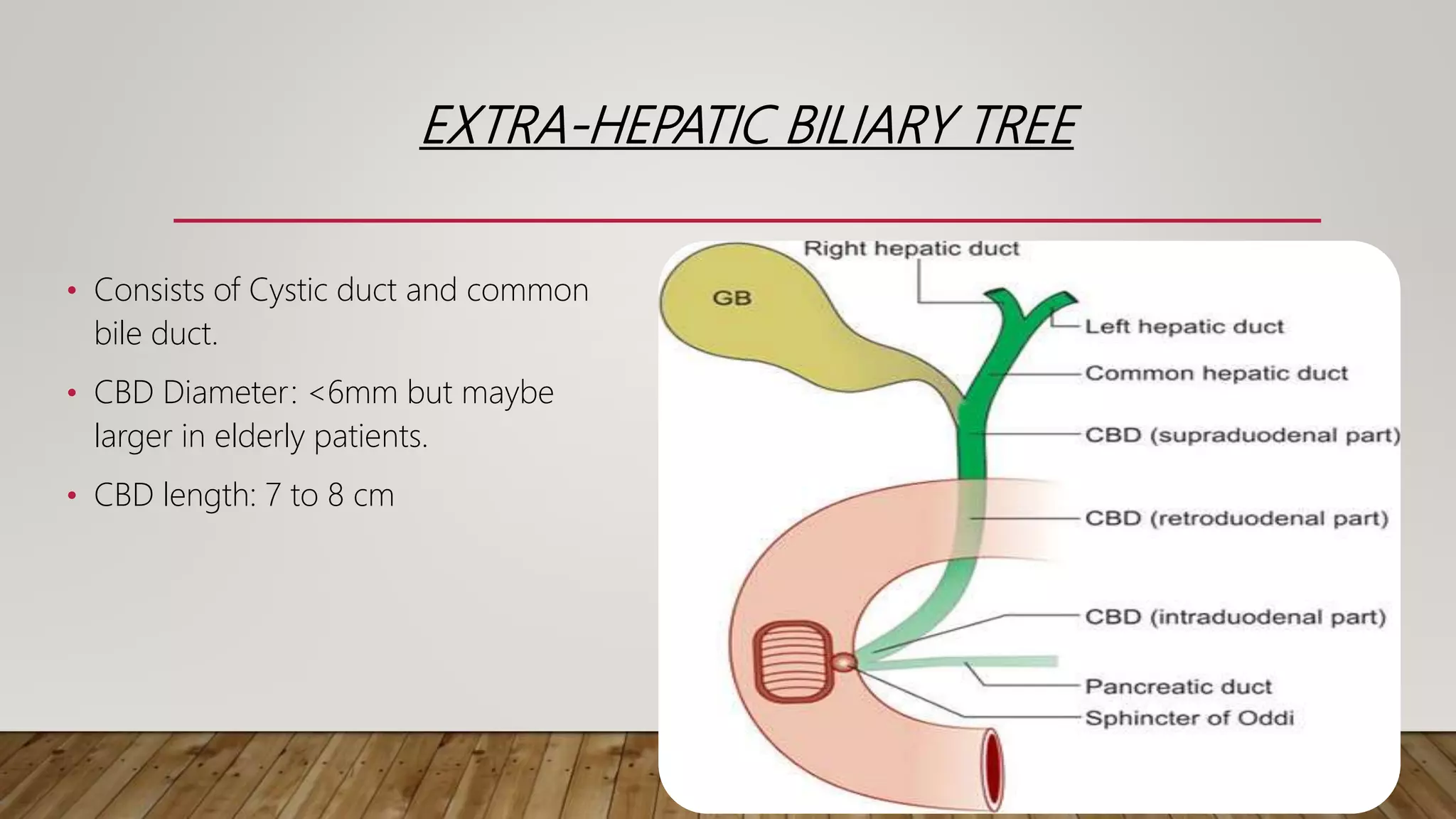
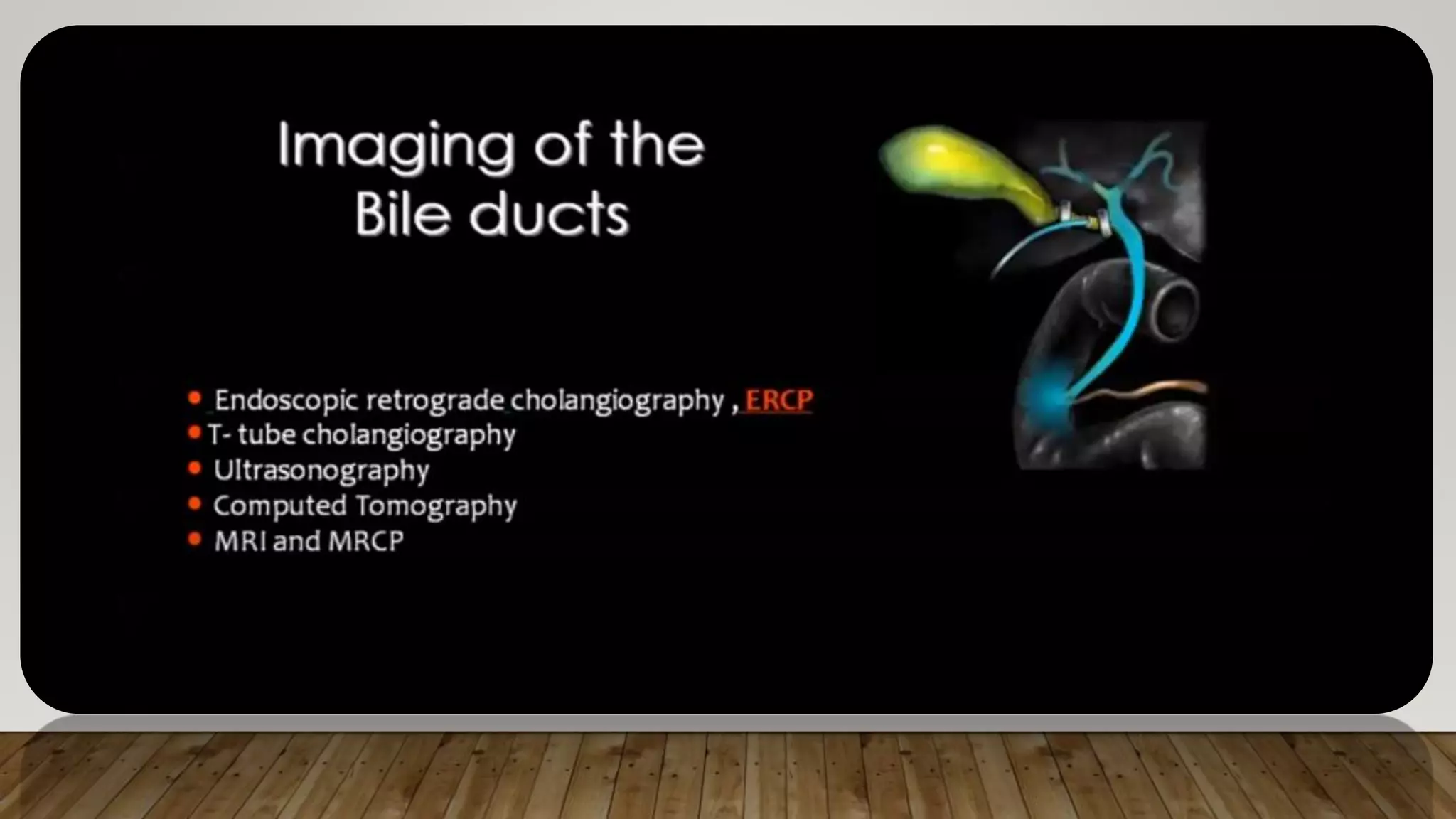
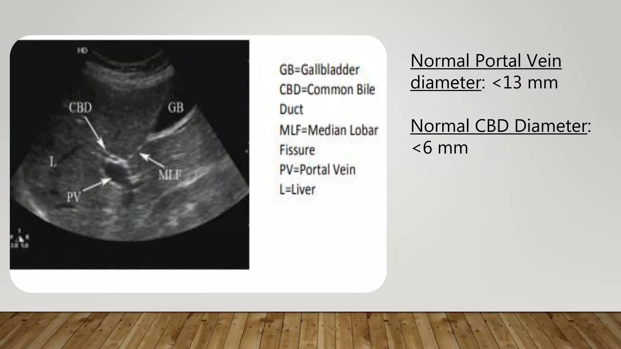
This document provides an overview of the radiological anatomy of the biliary system. It describes the components of the biliary apparatus including the gallbladder, cystic duct, hepatic ducts and common bile duct. It details the blood supply, nerve supply and lymphatic drainage of the gallbladder. It also describes how the biliary tree appears on various medical imaging modalities like ultrasound, MRI, CT, ERCP, and cholangiography. These modalities can be used to visualize the intrahepatic and extrahepatic biliary structures.


![Radiological anatomy of_abdomen[1]](https://cdn.slidesharecdn.com/ss_thumbnails/radiologicalanatomyofabdomen1-170830125353-thumbnail.jpg?width=640&height=640&fit=bounds)


