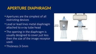
Prepared by :
Sajan Bohara
BSc. MIT (1st
Year)
NMCTH
Basic Introduction
• Xray- a form of high-energy electromagnetic radiation ( 0.01 to 10
nanometers)which invisible and penetrates materials (like soft tissues),
and is absorbed by denser substances (like bones)
• Beam- A focused stream of particles or waves moving in a specific
direction
• X-rays are the type of radiation, while the beam is how they are delivered
• X-ray Beam- A controlled stream of X-ray photons emitted from an X-ray
tube. Unlike scattered light, the beam is collimated (narrowed) to target
a specific area.
X-ray Beam
• Characterized mainly by quality and quantity
• Quality is the overall energy of x-ray beam
• Quantity is the number of photon in x-ray beam
• Beam Quality is the primary factor that effects the image quality in radiography
• Beam Quantity determines the intensity of the image
• Changes in quality and quantity affect the image quality and radiation dose.
X-ray emission spectrum
• refers to the energy distribution of the radiation
produced in an x-ray exposure
• In x-ray spectra curve, Bremsstrahlung and
characteristics radiation is represented by
continuous spectrum and line spectrum respectively
• It contain information about quality and quantity of
beam
• The area under the curve represents the quantity,
and the peak of the curve implies the average
energy
• The general shape of an emission spectrum curve is
always the same, but its relative position along the
energy axis can alter
• the farther to the right a spectrum is, the higher the effective enegry or the quality of x ray
• the larger the area under the curve, the higher the x ray quantity
fig:- general form of x-ray emission spectrum
Beam Centering
• Beam centering is the process of aligning the central ray of the X-ray beam
with the center of the area of interest on the patient’s body and the image
receptor.
• It ensures the accurate visualization of the area of interest with minimal
distortion and optimal image quality.
• It is important to limit the size of the primary beam and prevent exposure
of body region which are not required in image formation.
• Large X-ray field result in unnecessary exposure, thus scatter radiation is
increased and image contrast is decreased.
OFF FOCUS RADIATION
• X-Ray tube are designed so that the
projectile electrons interact with the target
• Some of the electrons bounce off the focal
spot and interact with other areas of target,
causing X-Ray production outside
of the focal spot.
SCATTARED RADIATION
• Type of radiation that occurs when a
primary radiation beam interacts with
matter, causing it to scatter in various
directions.
• In diagnostic radiology scatter radiation
leads to Fog in the X-Ray image.
• Two kinds of x-rays are responsible for
the optical density and contrast on
radiograph image
• Those that pass through the patient
without interacting called remnant ray.
• Those that are scattered through
Compton interaction
FACTORS AFFECTING SCATTERED RADIATION
Three factors contribute to an increase in scatter radiation:
• KVp
-:Higher kVp → More Compton scatter, less absorption → Lower contrast,
less patient dose
-:Lower kVp → More photoelectric effect→ Higher contrast, more patient dose
• Field Size
-:Larger field size → More scatter radiation → Increased image density
-:Smaller field size→Less scatter radiation→Reduced image density and better
image quality
•Patient thickness
-:More scatter results from imaging thick body parts compared to thin body parts
-:As tissue thickness increases, more of the rays go through multiple scattering
The relationship between collimation (field size) and
quantity of scatter radiation
• Collimated beam means
decreasing field size,
and decreasing
collimation means
increasing field size.
• Collimation and
scatter radiation are
Inversely proportional.
Beam Restriction:
• The beam restriction and collimation
are refer to decrease in the size and
shape of the projected radiation field.
• Collimation of beam decreases the risk
of radiation and minimizes scattered
radiation with better contrast.
BEAM RESTRICTING DEVICES
• Beam restrictors is a type of device attach to the opening in
Xray Tube housing to limit the size of the x-ray field to only
the anatomical structures of interest.
• There are mainly two reasons to restrict any x-ray beam.
Only the tissue being examined should be exposed.
Larger x-ray field results unnecessary patient exposure. So to
reduce patient dose.
Importance of beam limiting devices
:
Beam restriction serves for three purposes:
• Limiting patient exposure
• Reducing the amount of scatter radiation
• Focus on the correct anatomy .
• Improve image quality.
Limitation:
Reduce intensity by blocking incoming radiation.
BEAM RESTRICTORS
All beam-restricting devices are made of metal or a
combination of metals that readily absorb x-rays.
Classified into 3 categories:
• Aperture Diaphragm
• Cones & Cylinders
• Collimators
APERTURE DIAPHRAGM
• Apertures are the simplest of all
restricting devices
• Lead or lead lines metal diaphragm
attached to x-ray tube head
• The opening in the diaphragm is
usually designed to cover just less
then the size of the image receptor
used.
• Thickness 3-5mm
Applications of Aperture Diaphragm
• Used in dental radiography with rectangular collimation.
• Also, used in trauma and chest radiography.
DISADVANTAGE
•Size and shape is fixed
•Produce large penumbra
•Reduces sharpness and resolution
CONES AND CYLINDERS
• Cones and cylinders are modifications to the
aperture
• Heavy metal base like lead
• Copper lined barrel
• Cones and cylinders are shaped differently.
• Both have extended metal structures (10-
20inch) that restrict the useful circular beam
to require size.
• Positioning and size of the distal end
determines the field size.
Application:
• Designed to operate most effectively at a
designed SID
• Sinuses
• Dental x ray and treatment
ADVANTAGES
• Better restriction to x-ray field
• Produce less penumbra than
aperture diaphragm
• Round image on a rectangular film
• Reduce scatter radiation
• If the x ray source, film and cone are not aligned properly, then one
side of the film may not be exposed, which is called cone cutting.
• These systems provide only limited number of field sizes.
Disadvantages
Collimators
• The most useful, and accepted beam-restricting device is the
collimator.
• The terms collimation and beam restriction are used
interchangeably.
It has two advantages over the other types:
 it provides an infinite variety of rectangular x-ray fields
 a light beam shows the center and exact configuration of the x-ray field.
Structure of the collimator:
• 2 sets of shutters control the dimensions.
• Each shutter contains 4 or more lead plates of 3mm thickness, which
move in independent pairs.
• When the shutters are closed, they meet at the center.
• Collimator also has a light and mirror arrangement to create radiation
and optical coincidence.
• The light beam is deflected by a mirror mounted in the path of x ray
beam at an angle of 45 degree.
• The target of x ray tube and the light bulb should be the exactly
same distance from the center of the mirror.
• The light field and radiation field should match exactly with each
other.
VARIABLE APERTURE COLLIMATOR
•Proper collimation of the x-ray beam has the primary
effect of reducing patient dose by restricting the
volume of tissue irradiated.
•Proper collimation also reduces scatter radiation that
improves contrast.
LIGHT LOCALIZING COLLIMATOR
• The light localizing variable aperture
collimator is the most common beam
restricting device in diagnostic
radiography
COLLIMATOR
Two stages of shutter to control the beam
• First stage shutters protrude into the tube
housing to control the off-focus radiation.
• Adjustable second stage shutter pairs are
used to restrict the beam.
• Light localization is accomplished by a small projector
lamp and mirror to project the setting of the shutters on
the patient
• The light field and x-ray beam should match to avoid
collimator cut-off
• A scale on the collimator is used to match the beam to the
film size at fixed target film distance.
Automatic Collimator or Positive Beam
Limiting Devices
• A sensor in the Bucky and the motor used to automatically
collimate the image to film size called a positive-beam limiting
(PBL) device
• When film loaded cassette is inserted in the Bucky tray and
clamped into place sensing devices in the tray identify the
size and alignment of the cassette.
• An electric signal is transmitted to the collimator housing, and
accurate the synchronous motors that drive the collimator
leave to a pre-calibrated position
COLLIMATION RULES
• Requires 4 borders of collimation to be seen on the film.
• Collimation must be slightly less than film size or to the area
of clinical interest, whichever is smaller.
• Any exposure beyond the film is unnecessary patient
exposure
• Collimate the area within the patient not beyond the body
Quality assurance :
• Do collimator –beam alignment check at least every six months
• Do check of alignment of center of x ray beam at least every six
months.
FILTERS
•When the X ray beam is produced many energies of
photons exit (polychromatic). Many are of such low
energies that they were offered nothing to production
of that radiograph. So, removal of low energy x rays
from beam spectrum is called filtration. And the device
for that filtration is called Filters.
• It is placed in the path of x-ray beam.
WHY WE USE FILTERS IN THE DIAGNOSTIC
RADIOLOGY?
In diagnostic radiology, if the filters are not used,
• The patient acts as a filter, which will result in absorption of X rays
by the tissues.
• Unnecessary dosage of radiation to the patient.
• Also, to Increase the tube age
Preferred metal:
Most commonly preferred in diagnostic radiology
• Copper (Z= 29)
• Aluminum (Z=13)
low atomic number therefore excellent material for absorbing soft
rays.
low energy X ray photons low in weight therefore make the extra
tube lighter in weight and easily handling
Types of Filtration
Mainly 3 types:
a. Inherent
b. Added
c .Compensating
INHERENT FILTRATION
• A type of filtration that results from the
composition(aluminum of about 0.5-1 mm thickness) of
the tube and housing
ADDED FILTRATION
• Any filtration that occurs outside the tube and housing and before the
image receptor. Added to the port of the x-ray tube to absorb as many
low-energy photons as possible.
• Thickness 1- 2mm
Copper : pediatric applications
Aluminum : general radiography x ray tubes
Rhodium : mammography with Rh anode
Molybdenum : Mammography
Silver : mammography with tungsten anode
COMPENSATING FILTRATION
• They are filters designed to solve a problem involving
unequal subject densities
• Wedge Filter-Most common kind of compensating filter
which is used to radiograph body parts that vary
considerably, such as the foot. (1-3mm)
• Trough Filter-Compensating filter used mainly for chest
radiography. (Around 3mm)
CONE FILTERS
• Concave or convex shaped, used in applications in digital
fluoroscopy, where image-intensifier tubes and receptors
are round
1. Chesneys' Equipment for Student Radiographers, 4th Edition
2. The Physics of Radiology and imaging by K.Thayalan
3. Christensen’s Physics of Diagnostic Radiology
4. Handbook of medical radiography C Ramamohan
REFERENCE
Beam centering and Beam Limiting Device Sajan.pptx

Beam centering and Beam Limiting Device Sajan.pptx

  • 1.
    Prepared by : SajanBohara BSc. MIT (1st Year) NMCTH
  • 2.
    Basic Introduction • Xray-a form of high-energy electromagnetic radiation ( 0.01 to 10 nanometers)which invisible and penetrates materials (like soft tissues), and is absorbed by denser substances (like bones) • Beam- A focused stream of particles or waves moving in a specific direction • X-rays are the type of radiation, while the beam is how they are delivered • X-ray Beam- A controlled stream of X-ray photons emitted from an X-ray tube. Unlike scattered light, the beam is collimated (narrowed) to target a specific area.
  • 3.
    X-ray Beam • Characterizedmainly by quality and quantity • Quality is the overall energy of x-ray beam • Quantity is the number of photon in x-ray beam • Beam Quality is the primary factor that effects the image quality in radiography • Beam Quantity determines the intensity of the image • Changes in quality and quantity affect the image quality and radiation dose.
  • 4.
    X-ray emission spectrum •refers to the energy distribution of the radiation produced in an x-ray exposure • In x-ray spectra curve, Bremsstrahlung and characteristics radiation is represented by continuous spectrum and line spectrum respectively • It contain information about quality and quantity of beam • The area under the curve represents the quantity, and the peak of the curve implies the average energy • The general shape of an emission spectrum curve is always the same, but its relative position along the energy axis can alter • the farther to the right a spectrum is, the higher the effective enegry or the quality of x ray • the larger the area under the curve, the higher the x ray quantity fig:- general form of x-ray emission spectrum
  • 5.
    Beam Centering • Beamcentering is the process of aligning the central ray of the X-ray beam with the center of the area of interest on the patient’s body and the image receptor. • It ensures the accurate visualization of the area of interest with minimal distortion and optimal image quality. • It is important to limit the size of the primary beam and prevent exposure of body region which are not required in image formation. • Large X-ray field result in unnecessary exposure, thus scatter radiation is increased and image contrast is decreased.
  • 6.
    OFF FOCUS RADIATION •X-Ray tube are designed so that the projectile electrons interact with the target • Some of the electrons bounce off the focal spot and interact with other areas of target, causing X-Ray production outside of the focal spot.
  • 7.
    SCATTARED RADIATION • Typeof radiation that occurs when a primary radiation beam interacts with matter, causing it to scatter in various directions. • In diagnostic radiology scatter radiation leads to Fog in the X-Ray image. • Two kinds of x-rays are responsible for the optical density and contrast on radiograph image • Those that pass through the patient without interacting called remnant ray. • Those that are scattered through Compton interaction
  • 8.
    FACTORS AFFECTING SCATTEREDRADIATION Three factors contribute to an increase in scatter radiation: • KVp -:Higher kVp → More Compton scatter, less absorption → Lower contrast, less patient dose -:Lower kVp → More photoelectric effect→ Higher contrast, more patient dose • Field Size -:Larger field size → More scatter radiation → Increased image density -:Smaller field size→Less scatter radiation→Reduced image density and better image quality •Patient thickness -:More scatter results from imaging thick body parts compared to thin body parts -:As tissue thickness increases, more of the rays go through multiple scattering
  • 9.
    The relationship betweencollimation (field size) and quantity of scatter radiation • Collimated beam means decreasing field size, and decreasing collimation means increasing field size. • Collimation and scatter radiation are Inversely proportional.
  • 10.
    Beam Restriction: • Thebeam restriction and collimation are refer to decrease in the size and shape of the projected radiation field. • Collimation of beam decreases the risk of radiation and minimizes scattered radiation with better contrast.
  • 11.
    BEAM RESTRICTING DEVICES •Beam restrictors is a type of device attach to the opening in Xray Tube housing to limit the size of the x-ray field to only the anatomical structures of interest. • There are mainly two reasons to restrict any x-ray beam. Only the tissue being examined should be exposed. Larger x-ray field results unnecessary patient exposure. So to reduce patient dose.
  • 12.
    Importance of beamlimiting devices : Beam restriction serves for three purposes: • Limiting patient exposure • Reducing the amount of scatter radiation • Focus on the correct anatomy . • Improve image quality. Limitation: Reduce intensity by blocking incoming radiation.
  • 13.
    BEAM RESTRICTORS All beam-restrictingdevices are made of metal or a combination of metals that readily absorb x-rays. Classified into 3 categories: • Aperture Diaphragm • Cones & Cylinders • Collimators
  • 14.
    APERTURE DIAPHRAGM • Aperturesare the simplest of all restricting devices • Lead or lead lines metal diaphragm attached to x-ray tube head • The opening in the diaphragm is usually designed to cover just less then the size of the image receptor used. • Thickness 3-5mm
  • 15.
    Applications of ApertureDiaphragm • Used in dental radiography with rectangular collimation. • Also, used in trauma and chest radiography.
  • 16.
    DISADVANTAGE •Size and shapeis fixed •Produce large penumbra •Reduces sharpness and resolution
  • 17.
    CONES AND CYLINDERS •Cones and cylinders are modifications to the aperture • Heavy metal base like lead • Copper lined barrel • Cones and cylinders are shaped differently. • Both have extended metal structures (10- 20inch) that restrict the useful circular beam to require size. • Positioning and size of the distal end determines the field size.
  • 18.
    Application: • Designed tooperate most effectively at a designed SID • Sinuses • Dental x ray and treatment
  • 19.
    ADVANTAGES • Better restrictionto x-ray field • Produce less penumbra than aperture diaphragm • Round image on a rectangular film • Reduce scatter radiation
  • 20.
    • If thex ray source, film and cone are not aligned properly, then one side of the film may not be exposed, which is called cone cutting. • These systems provide only limited number of field sizes. Disadvantages
  • 21.
    Collimators • The mostuseful, and accepted beam-restricting device is the collimator. • The terms collimation and beam restriction are used interchangeably. It has two advantages over the other types:  it provides an infinite variety of rectangular x-ray fields  a light beam shows the center and exact configuration of the x-ray field.
  • 22.
    Structure of thecollimator: • 2 sets of shutters control the dimensions. • Each shutter contains 4 or more lead plates of 3mm thickness, which move in independent pairs. • When the shutters are closed, they meet at the center. • Collimator also has a light and mirror arrangement to create radiation and optical coincidence.
  • 23.
    • The lightbeam is deflected by a mirror mounted in the path of x ray beam at an angle of 45 degree. • The target of x ray tube and the light bulb should be the exactly same distance from the center of the mirror. • The light field and radiation field should match exactly with each other.
  • 26.
    VARIABLE APERTURE COLLIMATOR •Propercollimation of the x-ray beam has the primary effect of reducing patient dose by restricting the volume of tissue irradiated. •Proper collimation also reduces scatter radiation that improves contrast.
  • 27.
    LIGHT LOCALIZING COLLIMATOR •The light localizing variable aperture collimator is the most common beam restricting device in diagnostic radiography
  • 28.
    COLLIMATOR Two stages ofshutter to control the beam • First stage shutters protrude into the tube housing to control the off-focus radiation. • Adjustable second stage shutter pairs are used to restrict the beam.
  • 29.
    • Light localizationis accomplished by a small projector lamp and mirror to project the setting of the shutters on the patient • The light field and x-ray beam should match to avoid collimator cut-off • A scale on the collimator is used to match the beam to the film size at fixed target film distance.
  • 30.
    Automatic Collimator orPositive Beam Limiting Devices • A sensor in the Bucky and the motor used to automatically collimate the image to film size called a positive-beam limiting (PBL) device • When film loaded cassette is inserted in the Bucky tray and clamped into place sensing devices in the tray identify the size and alignment of the cassette. • An electric signal is transmitted to the collimator housing, and accurate the synchronous motors that drive the collimator leave to a pre-calibrated position
  • 31.
    COLLIMATION RULES • Requires4 borders of collimation to be seen on the film. • Collimation must be slightly less than film size or to the area of clinical interest, whichever is smaller. • Any exposure beyond the film is unnecessary patient exposure • Collimate the area within the patient not beyond the body
  • 32.
    Quality assurance : •Do collimator –beam alignment check at least every six months • Do check of alignment of center of x ray beam at least every six months.
  • 33.
    FILTERS •When the Xray beam is produced many energies of photons exit (polychromatic). Many are of such low energies that they were offered nothing to production of that radiograph. So, removal of low energy x rays from beam spectrum is called filtration. And the device for that filtration is called Filters. • It is placed in the path of x-ray beam.
  • 34.
    WHY WE USEFILTERS IN THE DIAGNOSTIC RADIOLOGY? In diagnostic radiology, if the filters are not used, • The patient acts as a filter, which will result in absorption of X rays by the tissues. • Unnecessary dosage of radiation to the patient. • Also, to Increase the tube age
  • 35.
    Preferred metal: Most commonlypreferred in diagnostic radiology • Copper (Z= 29) • Aluminum (Z=13) low atomic number therefore excellent material for absorbing soft rays. low energy X ray photons low in weight therefore make the extra tube lighter in weight and easily handling
  • 36.
    Types of Filtration Mainly3 types: a. Inherent b. Added c .Compensating
  • 37.
    INHERENT FILTRATION • Atype of filtration that results from the composition(aluminum of about 0.5-1 mm thickness) of the tube and housing
  • 38.
    ADDED FILTRATION • Anyfiltration that occurs outside the tube and housing and before the image receptor. Added to the port of the x-ray tube to absorb as many low-energy photons as possible. • Thickness 1- 2mm Copper : pediatric applications Aluminum : general radiography x ray tubes Rhodium : mammography with Rh anode Molybdenum : Mammography Silver : mammography with tungsten anode
  • 39.
    COMPENSATING FILTRATION • Theyare filters designed to solve a problem involving unequal subject densities • Wedge Filter-Most common kind of compensating filter which is used to radiograph body parts that vary considerably, such as the foot. (1-3mm) • Trough Filter-Compensating filter used mainly for chest radiography. (Around 3mm)
  • 40.
    CONE FILTERS • Concaveor convex shaped, used in applications in digital fluoroscopy, where image-intensifier tubes and receptors are round
  • 41.
    1. Chesneys' Equipmentfor Student Radiographers, 4th Edition 2. The Physics of Radiology and imaging by K.Thayalan 3. Christensen’s Physics of Diagnostic Radiology 4. Handbook of medical radiography C Ramamohan REFERENCE

Editor's Notes

  • #4 Integration =determination of total x-ray
  • #6 It reduces image contrast and in crease unnecessary patient dose
  • #14 an unexposed border is visible on each edge of the radiograph.
  • #16 The width or size of the aperture is fixed and can not be adjusted
  • #17 Most commonly used type is cylinder Cones are typically used in dental radiography
  • #19 Frontal sinuses with and without cone
  • #26 Most affective beam restrictng devices
  • #27 Modern automatic variable aperature collimator
  • #28 To control the off focus radiation ,first stage entrance shuttering…and second stage shutter are usally made of lead at least 3mm thick and they work in pairs and are independently control,allowing for bboth rectangular and square field
  • #29 Many newer collimators a bright slit of light is provided to properly center the beam and the film. Collimator lamp and mirror should adjusted so that the projected light field coincide with x-ray beam
  • #30 Today nearly all light localizing collimators manufacture for fixed radiographic equipment are automatic 3..so the x-ray beam is restricted to the image receptor in use
  • #38 Added filtration is made of interchangale metal sheet made of aluminum ,copper and its thickness range from 1-2mm…total filtyration 2.5mm