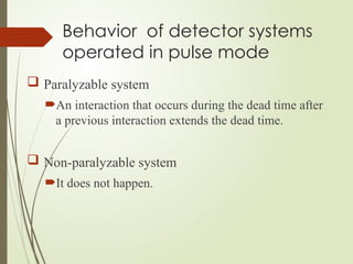
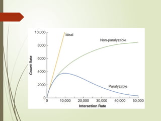
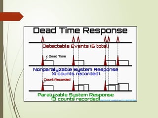
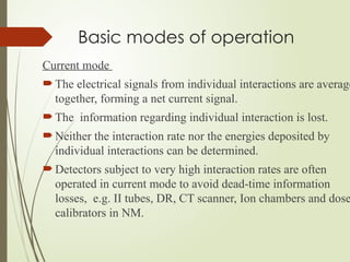
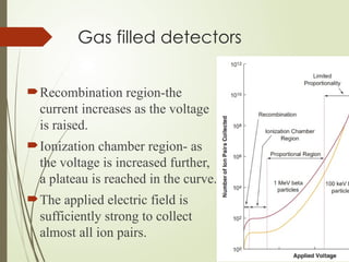
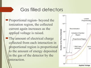
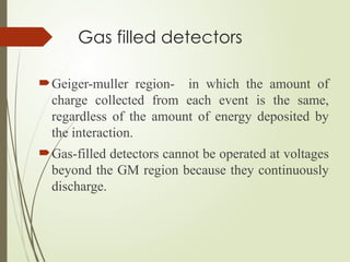
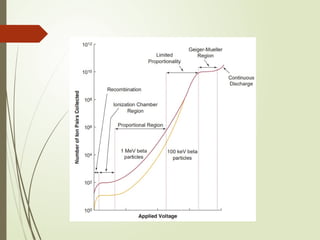
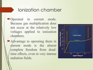
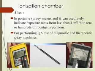
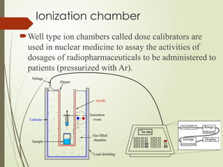
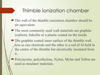
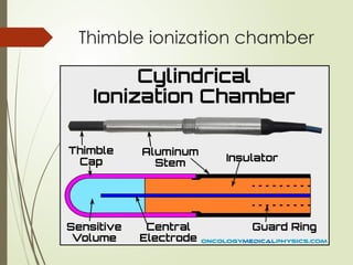
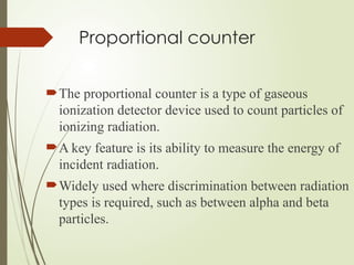
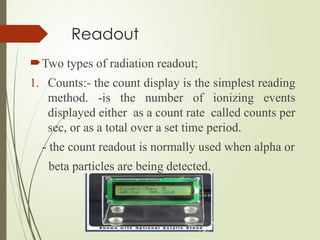
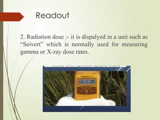
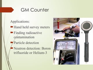
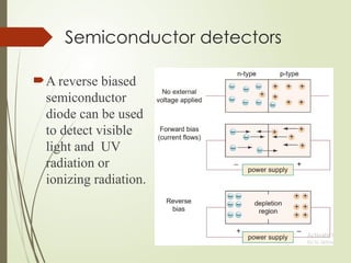
Radiation detection and measurement
Ram Datt Joshi
M..Sc. MIT Final year
IOM, Kathmandu,
Nepal
Introduction
The detection and measurement of ionizing
radiation are the basis for the majority of
diagnostic imaging and radiotherapy.
All detectors of ionizing radiation require the
interaction of the radiation with matter.
Radiation detection
Most energy deposited by ionizing radiation is
ultimately converted into thermal energy.
For e.g., a 140-keV gamma ray deposits 2.24 × 10⁻¹⁴
Joules if completely absorbed.
To raise the temperature of 1 g of water by 1˚C (i.e., 1
calorie) would require the complete absorption of 187
trillions ( 187 × 1012
) photons.
Types of detectors
 Detection method
Gas filled
Scintillators
Semiconductors
Types of information
Counters
Spectrometers
Dosimeters
Basic modes of operation
There are two basic modes of operation of detectors,
1. Pulse mode
2. Current mode
Basic modes of operation
Pulse mode
The signal from each interaction is processed
individually.
Two interactions must be separated by a finite amount of
time to produce distinct signals.
This interval is called the dead time of the system.
If a second interaction is close enough in time to the first
interaction; it may even distort the signal from the 1st
interaction. e.g. GM counters.
Effect of interaction on detectors
operated in pulse mode
Dead time
The fraction of counts lost from
dead-time effects is smallest at low
interaction rates and vice-versa.
The dead times of different types
of systems vary widely.
e.g.GM counters have dead times
ranging from tens to hundreds of
microseconds, whereas most other
systems have dead times of less
than a few microseconds.
Behavior of detector systems
operated in pulse mode
 Paralyzable system
An interaction that occurs during the dead time after
a previous interaction extends the dead time.
 Non-paralyzable system
It does not happen.
Basic modes of operation
Current mode
 The electrical signals from individual interactions are average
together, forming a net current signal.
 The information regarding individual interaction is lost.
 Neither the interaction rate nor the energies deposited by
individual interactions can be determined.
 Detectors subject to very high interaction rates are often
operated in current mode to avoid dead-time information
losses, e.g. II tubes, DR, CT scanner, Ion chambers and dose
calibrators in NM.
Detection efficiency
The efficiency (sensitivity) of a detector is a measure
of its ability to detect radiation.
Efficiency = no. detected / no. emitted
Efficiency = geometric efficiency x intrinsic
efficiency
Gas filled detectors- basic
principle
A gas-filled detector consists of a volume of gas between
two electrodes, with an electric potential difference
(voltage) applied between the electrodes.
Gas filled detectors- basic
principle
The cathode is the wall of the container that holds the
gas or a conductive coating on the inside of the wall,
and the anode is a wire inside the container
Gas filled detectors
There are three types of gas-filled detectors in
common use:-
1. Ionization chambers,
2. Proportional counters, and
3. GM counters
The type of detector is determined by the voltage
applied between the electrodes.
Gas filled detectors
Recombination region-the
current increases as the voltage
is raised.
Ionization chamber region- as
the voltage is increased further,
a plateau is reached in the curve.
The applied electric field is
sufficiently strong to collect
almost all ion pairs.
Gas filled detectors
Proportional region- beyond the
ionization region, the collected
current again increases as the
applied voltage is raised.
The amount of electrical charge
collected from each interaction in
proportional region is proportional
to the amount of energy deposited
in the gas of the detector by the
interaction.
Gas filled detectors
Geiger-muller region- in which the amount of
charge collected from each event is the same,
regardless of the amount of energy deposited by
the interaction.
Gas-filled detectors cannot be operated at voltages
beyond the GM region because they continuously
discharge.
Ionization chamber
Operated in current mode.
Because gas multiplication does
not occur at the relatively low
voltages applied to ionization
chambers.
Advantage to operating them in
current mode is the almost
complete freedom from dead-
time effects, even in very intense
radiation fields.
Ionization chamber
Almost any gas can be used to fill the chamber.
If the gas is air and the walls of the chamber are of a
material whose effective atomic number is similar to
air, the amount of current produced is proportional to
the exposure rate.
The sensitivity of ion chambers to x-rays and gamma
rays can be enhanced by filling them with a gas that
has a high atomic number, such as argon (Z=18) or
xenon (Z=54), and pressurizing the gas to increase its
density.
Ionization chamber
Uses :
In portable survey meters and it can accurately
indicate exposure rates from less than 1 mR/h to tens
or hundreds of roentgens per hour.
For performing QA test of diagnostic and therapeutic
x-ray machines.
Ionization chamber
Well type ion chambers called dose calibrators are
used in nuclear medicine to assay the activities of
dosages of radiopharmaceuticals to be administered to
patients (pressurized with Ar).
Thimble ionization chamber
The chamber wall is shaped like sewing thimble.
The high voltage, usually 200-500V is applied to
the thimble wall with the central electrode
connected to the electrometer input at or near the
ground potential.
The guard ring encircling the insulator is also at
ground potential.
Thimble ionization chamber
The wall of the thimble ionization chamber should be
air equivalent.
The most commonly used wall materials are graphite
(carbon), bakelite or a plastic coated on the inside.
The graphite coated inner surface of the thimble wall
acts as one electrode and the other is a rod of Al held in
the centre of the thimble but electrically insulated from
it.
Polystyrene, polyethylene, Nylon, Mylar and Teflon are
used as insulator materials.
Thimble ionization chamber
Proportional counter
The proportional counter is a type of gaseous
ionization detector device used to count particles of
ionizing radiation.
A key feature is its ability to measure the energy of
incident radiation.
Widely used where discrimination between radiation
types is required, such as between alpha and beta
particles.
Proportional counter
A proportional counter uses a combination of the
mechanisms of a Geiger-Muller tube and an
ionization chamber, and operates in an
intermediate voltage region between these.
In this region, electrons approaching the anode are
accelerated to such high kinetic energies that they
cause additional ionization, called gas
multiplication, which amplifies the collected
current.
The amount of amplification increases as the
applied voltage is raised.
Proportional counter
In a proportional counter, the fill gas of the chamber is
an inert gas which is ionized by incident radiation, and
a quench gas to ensure each pulse discharge terminates.
 A common mixture is 90% argon, 10% methane,
known as P-10.
GM counter- principle of
operation
A Geiger counter consists of a Geiger-Muller tube,
the sensing element which detects the radiation and
the processing electronics, which display the result.
The Geiger Muller tube is filled with an inert gas
such as helium, neon, or argon at low pressure, to
which a high voltage is applied.
The tube briefly conducts electrical charge when a
particle or photon of incident radiation makes the
gas conductive by ionization.
GM Counter
The ionization is considerably amplified within the
tube by the Townsend discharge effect to produce an
easily measured detection pulse, which is fed to
processing and display electronics.
Readout
Two types of radiation readout;
1. Counts:- the count display is the simplest reading
method. -is the number of ionizing events
displayed either as a count rate called counts per
sec, or as a total over a set time period.
- the count readout is normally used when alpha or
beta particles are being detected.
Readout
2. Radiation dose :- it is dispalyed in a unit such as
“Seivert” which is normally used for measuring
gamma or X-ray dose rates.
GM Counter
Applications:
Hand held survey meters
Finding radioactive
contamination
Particle detection
Neutron detection: Boron
trifluoride or Helium-3
GM Counter
Advantages :
They are relatively inexpensive.
They are durable and easily portable.
They can detect all types of radiation.
GM counter
Disadvantages:
They cannot differentiate which type of radiation
is being detected.
They cannot be used to determine the exact
energy of the detected radiation.
They have very low efficiency.
They have extremely long dead time.
Scintillation detectors-basic
principle
Emit visible light or ultraviolet radiation after the
interaction of ionizing radiation with the material.
Oldest type of radiation detectors.
Scintillators are used in conventional film-screen
radiography, many direct digital radiographic image
receptors, fluoroscopy, scintillation cameras, CT
scanners, and positron emission tomography (PET)
scanners.
Scintillation detectors- basic
principle
When ionizing radiation interacts with a
scintillator, electrons are raised to an excited
energy level.
Ultimately, these electrons fall back to a lower
energy state, with the emission of visible light or
ultraviolet radiation.
In all scintillators, the amount of light emitted
after an interaction increases with the energy
deposited by the interaction.
Scintillation detectors- basic
principle
Many organic and inorganic materials are there
that can scintillate.
Organic scintillators are not used for medical
imaging because the low atomic numbers of their
constituent elements and their low densities make
them poor x-ray and gamma-ray detectors.
In the inorganic materials, the scintillation is a
property of crystalline structure.
Scintillation detectors
Properties:
The conversion efficiency, should be high.
The decay times of excited states should be short.
The material should be transparent to its own emissions.
The frequency spectrum (color) of emitted light or UV
radiation should match the spectral sensitivity of the
light receptor (PMT, photodiode or film).
If used for x-ray and gamma-ray detection, the
attenuation coefficient (μ) should be large.
The material should be rugged, unaffected by moisture,
and inexpensive to manufacture.
Scintillation detectors in
radiology
Conversion of light into an
electrical signal
Photomultiplier tube(PMT)- perform two functions;
Conversion of ultraviolet and visible light photons into
an electrical signal and signal amplification.
When a scintillator is coupled to a PMT, an optical
coupling material is placed between the two
components to minimize reflection losses.
Conversion of light into an
electrical signal
Photodiodes- they are semiconductor diodes that convert
light into electrical signals.
Photodiodes are reverse biased.
When the photodiode is exposed to light, an electrical
current is generated that is proportional to the intensity
of the light.
Photodiodes produce more electrical noise than PMTs
do, but they are smaller and less expensive.
Do not amplify the signal, however, a type of photodiode
called an avalanche photodiode does provide signal
amplification.
Semiconductor detectors
A crystal of a semiconductor material can be used
as a radiation detector.
The semiconductor crystal is “doped” with a trace
amount of impurities so that it acts as a diode.
A voltage is placed between two terminals on
opposite sides of the crystal.
When ionizing radiation interacts with the detector,
electrons in the crystal are raised to an excited
state, permitting an electrical current to flow.
Semiconductor detectors
In semiconductors, valence-band electrons can be raised
to the conduction band by ionizing radiation, leaving
vacancy called hole.
Semiconductor detectors
Can be :
 n- type (electron donor) or
 p- type (hole forming)
A semiconductor diode consists of a crystal of
semiconductor material with a region of n-
type material that forms a junction with a
region of p-type material.
Semiconductor detectors
A reverse biased
semiconductor
diode can be used
to detect visible
light and UV
radiation or
ionizing radiation.
Semiconductor detectors
Liquid nitrogen–cooled germanium detectors are
widely used for the identification of individual
gamma ray–emitting radionuclides in mixed
radionuclide samples.
Semiconductor detectors are seldom used for
medical imaging devices because of high expense
and low quantum detection efficiencies.
Cadmium zinc telluride(CZT) is used as radiation
detector in photon counting CT.
Thank you

Radiation detection and measurement.pptx

  • 1.
    Radiation detection andmeasurement Ram Datt Joshi M..Sc. MIT Final year IOM, Kathmandu, Nepal
  • 2.
    Introduction The detection andmeasurement of ionizing radiation are the basis for the majority of diagnostic imaging and radiotherapy. All detectors of ionizing radiation require the interaction of the radiation with matter.
  • 4.
    Radiation detection Most energydeposited by ionizing radiation is ultimately converted into thermal energy. For e.g., a 140-keV gamma ray deposits 2.24 × 10⁻¹⁴ Joules if completely absorbed. To raise the temperature of 1 g of water by 1˚C (i.e., 1 calorie) would require the complete absorption of 187 trillions ( 187 × 1012 ) photons.
  • 5.
    Types of detectors Detection method Gas filled Scintillators Semiconductors Types of information Counters Spectrometers Dosimeters
  • 6.
    Basic modes ofoperation There are two basic modes of operation of detectors, 1. Pulse mode 2. Current mode
  • 7.
    Basic modes ofoperation Pulse mode The signal from each interaction is processed individually. Two interactions must be separated by a finite amount of time to produce distinct signals. This interval is called the dead time of the system. If a second interaction is close enough in time to the first interaction; it may even distort the signal from the 1st interaction. e.g. GM counters.
  • 8.
    Effect of interactionon detectors operated in pulse mode Dead time The fraction of counts lost from dead-time effects is smallest at low interaction rates and vice-versa. The dead times of different types of systems vary widely. e.g.GM counters have dead times ranging from tens to hundreds of microseconds, whereas most other systems have dead times of less than a few microseconds.
  • 9.
    Behavior of detectorsystems operated in pulse mode  Paralyzable system An interaction that occurs during the dead time after a previous interaction extends the dead time.  Non-paralyzable system It does not happen.
  • 12.
    Basic modes ofoperation Current mode  The electrical signals from individual interactions are average together, forming a net current signal.  The information regarding individual interaction is lost.  Neither the interaction rate nor the energies deposited by individual interactions can be determined.  Detectors subject to very high interaction rates are often operated in current mode to avoid dead-time information losses, e.g. II tubes, DR, CT scanner, Ion chambers and dose calibrators in NM.
  • 13.
    Detection efficiency The efficiency(sensitivity) of a detector is a measure of its ability to detect radiation. Efficiency = no. detected / no. emitted Efficiency = geometric efficiency x intrinsic efficiency
  • 15.
    Gas filled detectors-basic principle A gas-filled detector consists of a volume of gas between two electrodes, with an electric potential difference (voltage) applied between the electrodes.
  • 16.
    Gas filled detectors-basic principle The cathode is the wall of the container that holds the gas or a conductive coating on the inside of the wall, and the anode is a wire inside the container
  • 17.
    Gas filled detectors Thereare three types of gas-filled detectors in common use:- 1. Ionization chambers, 2. Proportional counters, and 3. GM counters The type of detector is determined by the voltage applied between the electrodes.
  • 18.
    Gas filled detectors Recombinationregion-the current increases as the voltage is raised. Ionization chamber region- as the voltage is increased further, a plateau is reached in the curve. The applied electric field is sufficiently strong to collect almost all ion pairs.
  • 19.
    Gas filled detectors Proportionalregion- beyond the ionization region, the collected current again increases as the applied voltage is raised. The amount of electrical charge collected from each interaction in proportional region is proportional to the amount of energy deposited in the gas of the detector by the interaction.
  • 20.
    Gas filled detectors Geiger-mullerregion- in which the amount of charge collected from each event is the same, regardless of the amount of energy deposited by the interaction. Gas-filled detectors cannot be operated at voltages beyond the GM region because they continuously discharge.
  • 22.
    Ionization chamber Operated incurrent mode. Because gas multiplication does not occur at the relatively low voltages applied to ionization chambers. Advantage to operating them in current mode is the almost complete freedom from dead- time effects, even in very intense radiation fields.
  • 23.
    Ionization chamber Almost anygas can be used to fill the chamber. If the gas is air and the walls of the chamber are of a material whose effective atomic number is similar to air, the amount of current produced is proportional to the exposure rate. The sensitivity of ion chambers to x-rays and gamma rays can be enhanced by filling them with a gas that has a high atomic number, such as argon (Z=18) or xenon (Z=54), and pressurizing the gas to increase its density.
  • 24.
    Ionization chamber Uses : Inportable survey meters and it can accurately indicate exposure rates from less than 1 mR/h to tens or hundreds of roentgens per hour. For performing QA test of diagnostic and therapeutic x-ray machines.
  • 25.
    Ionization chamber Well typeion chambers called dose calibrators are used in nuclear medicine to assay the activities of dosages of radiopharmaceuticals to be administered to patients (pressurized with Ar).
  • 26.
    Thimble ionization chamber Thechamber wall is shaped like sewing thimble. The high voltage, usually 200-500V is applied to the thimble wall with the central electrode connected to the electrometer input at or near the ground potential. The guard ring encircling the insulator is also at ground potential.
  • 27.
    Thimble ionization chamber Thewall of the thimble ionization chamber should be air equivalent. The most commonly used wall materials are graphite (carbon), bakelite or a plastic coated on the inside. The graphite coated inner surface of the thimble wall acts as one electrode and the other is a rod of Al held in the centre of the thimble but electrically insulated from it. Polystyrene, polyethylene, Nylon, Mylar and Teflon are used as insulator materials.
  • 28.
  • 29.
    Proportional counter The proportionalcounter is a type of gaseous ionization detector device used to count particles of ionizing radiation. A key feature is its ability to measure the energy of incident radiation. Widely used where discrimination between radiation types is required, such as between alpha and beta particles.
  • 30.
    Proportional counter A proportionalcounter uses a combination of the mechanisms of a Geiger-Muller tube and an ionization chamber, and operates in an intermediate voltage region between these. In this region, electrons approaching the anode are accelerated to such high kinetic energies that they cause additional ionization, called gas multiplication, which amplifies the collected current. The amount of amplification increases as the applied voltage is raised.
  • 31.
    Proportional counter In aproportional counter, the fill gas of the chamber is an inert gas which is ionized by incident radiation, and a quench gas to ensure each pulse discharge terminates.  A common mixture is 90% argon, 10% methane, known as P-10.
  • 32.
    GM counter- principleof operation A Geiger counter consists of a Geiger-Muller tube, the sensing element which detects the radiation and the processing electronics, which display the result. The Geiger Muller tube is filled with an inert gas such as helium, neon, or argon at low pressure, to which a high voltage is applied. The tube briefly conducts electrical charge when a particle or photon of incident radiation makes the gas conductive by ionization.
  • 33.
    GM Counter The ionizationis considerably amplified within the tube by the Townsend discharge effect to produce an easily measured detection pulse, which is fed to processing and display electronics.
  • 35.
    Readout Two types ofradiation readout; 1. Counts:- the count display is the simplest reading method. -is the number of ionizing events displayed either as a count rate called counts per sec, or as a total over a set time period. - the count readout is normally used when alpha or beta particles are being detected.
  • 36.
    Readout 2. Radiation dose:- it is dispalyed in a unit such as “Seivert” which is normally used for measuring gamma or X-ray dose rates.
  • 37.
    GM Counter Applications: Hand heldsurvey meters Finding radioactive contamination Particle detection Neutron detection: Boron trifluoride or Helium-3
  • 38.
    GM Counter Advantages : Theyare relatively inexpensive. They are durable and easily portable. They can detect all types of radiation.
  • 39.
    GM counter Disadvantages: They cannotdifferentiate which type of radiation is being detected. They cannot be used to determine the exact energy of the detected radiation. They have very low efficiency. They have extremely long dead time.
  • 40.
    Scintillation detectors-basic principle Emit visiblelight or ultraviolet radiation after the interaction of ionizing radiation with the material. Oldest type of radiation detectors. Scintillators are used in conventional film-screen radiography, many direct digital radiographic image receptors, fluoroscopy, scintillation cameras, CT scanners, and positron emission tomography (PET) scanners.
  • 41.
    Scintillation detectors- basic principle Whenionizing radiation interacts with a scintillator, electrons are raised to an excited energy level. Ultimately, these electrons fall back to a lower energy state, with the emission of visible light or ultraviolet radiation. In all scintillators, the amount of light emitted after an interaction increases with the energy deposited by the interaction.
  • 42.
    Scintillation detectors- basic principle Manyorganic and inorganic materials are there that can scintillate. Organic scintillators are not used for medical imaging because the low atomic numbers of their constituent elements and their low densities make them poor x-ray and gamma-ray detectors. In the inorganic materials, the scintillation is a property of crystalline structure.
  • 43.
    Scintillation detectors Properties: The conversionefficiency, should be high. The decay times of excited states should be short. The material should be transparent to its own emissions. The frequency spectrum (color) of emitted light or UV radiation should match the spectral sensitivity of the light receptor (PMT, photodiode or film). If used for x-ray and gamma-ray detection, the attenuation coefficient (μ) should be large. The material should be rugged, unaffected by moisture, and inexpensive to manufacture.
  • 44.
  • 46.
    Conversion of lightinto an electrical signal Photomultiplier tube(PMT)- perform two functions; Conversion of ultraviolet and visible light photons into an electrical signal and signal amplification. When a scintillator is coupled to a PMT, an optical coupling material is placed between the two components to minimize reflection losses.
  • 47.
    Conversion of lightinto an electrical signal Photodiodes- they are semiconductor diodes that convert light into electrical signals. Photodiodes are reverse biased. When the photodiode is exposed to light, an electrical current is generated that is proportional to the intensity of the light. Photodiodes produce more electrical noise than PMTs do, but they are smaller and less expensive. Do not amplify the signal, however, a type of photodiode called an avalanche photodiode does provide signal amplification.
  • 48.
    Semiconductor detectors A crystalof a semiconductor material can be used as a radiation detector. The semiconductor crystal is “doped” with a trace amount of impurities so that it acts as a diode. A voltage is placed between two terminals on opposite sides of the crystal. When ionizing radiation interacts with the detector, electrons in the crystal are raised to an excited state, permitting an electrical current to flow.
  • 49.
    Semiconductor detectors In semiconductors,valence-band electrons can be raised to the conduction band by ionizing radiation, leaving vacancy called hole.
  • 50.
    Semiconductor detectors Can be:  n- type (electron donor) or  p- type (hole forming) A semiconductor diode consists of a crystal of semiconductor material with a region of n- type material that forms a junction with a region of p-type material.
  • 51.
    Semiconductor detectors A reversebiased semiconductor diode can be used to detect visible light and UV radiation or ionizing radiation.
  • 52.
    Semiconductor detectors Liquid nitrogen–cooledgermanium detectors are widely used for the identification of individual gamma ray–emitting radionuclides in mixed radionuclide samples. Semiconductor detectors are seldom used for medical imaging devices because of high expense and low quantum detection efficiencies. Cadmium zinc telluride(CZT) is used as radiation detector in photon counting CT.
  • 53.

Editor's Notes