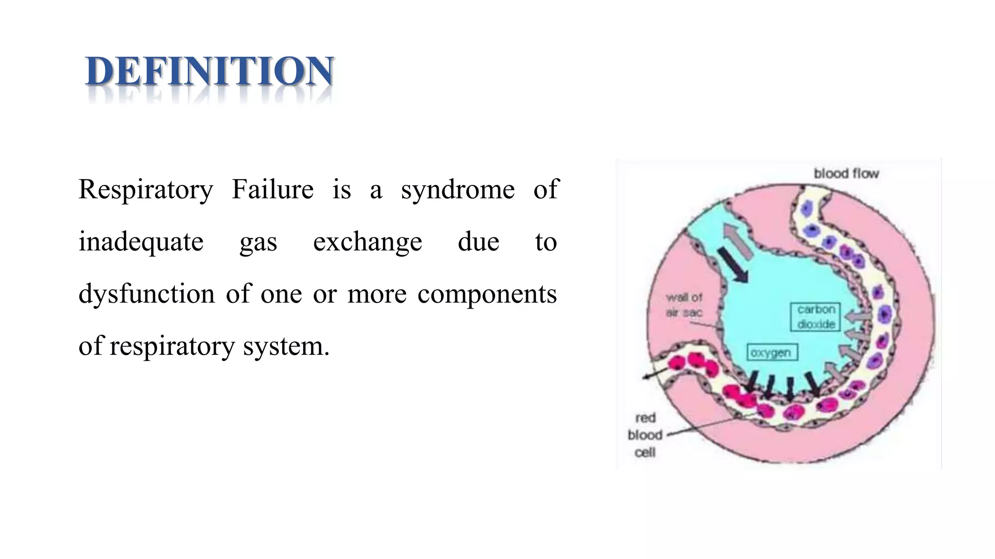
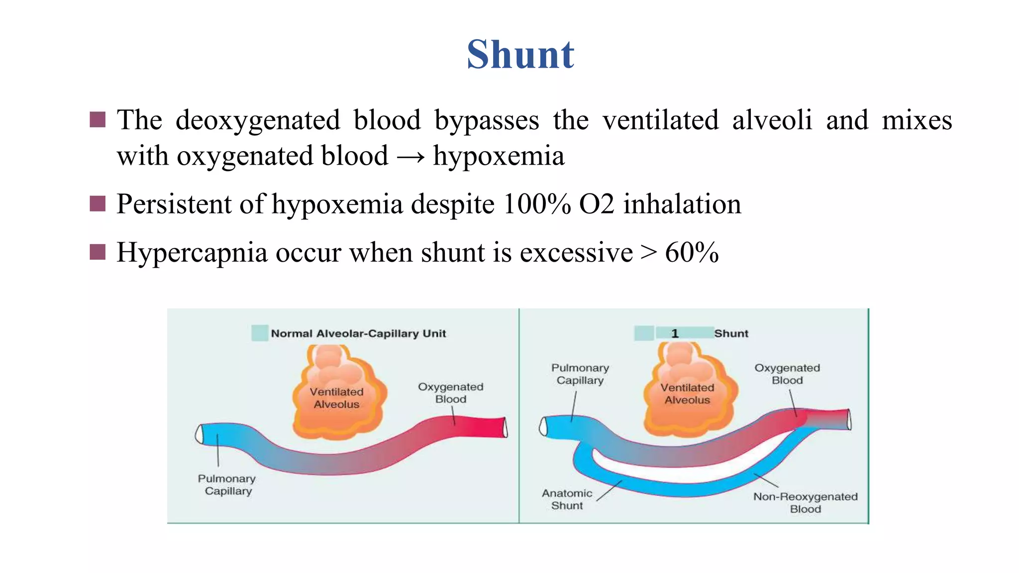
ACUTE RESPIRATORY DISTRESS
SYNDROME (ARDS)
INTRODUCTION
• Acute respiratory distress syndrome (ARDS) is a life-threatening lung
condition that prevents enough oxygen from getting into the blood.
• ARDS is also referred with variety of terms like
Stiff Lung
Shock lung
Wet lung
Post traumatic lung
Capillary leak syndrome &
Congestive atelectasis.
DEFINITION
Acute respiratory distress syndrome (ARDS) is a sudden and
progressive form of acute respiratory failure in which the alveolar
capillary membrane becomes damaged and more permeable to
intravascular fluid resulting in severe dyspnea, hypoxemia and diffuse
pulmonary infiltrates.
ETIOLOGY & RISK FACTORS
Direct Lung Injury
• Aspiration of gastric contents or other substances.
• Viral/bacterial pneumonia
• Chest trauma
• Embolism: fat, air, amniotic fluid
• Inhalation of toxic substances, massive smoking
• O2 toxicity: Prolonged exposure to high concentrations of oxygen
• Radiation pneumonitis
CONT..
Indirect Lung Injury
• Sepsis
• Multisystem trauma
• Acute pancreatitis
• Anaphylaxis
• Prolonged Cardiopulmonary bypass surgery
• Disseminated intravascular coagulation
• Narcotic drug overdose (e.g., heroin)
• Severe head injury
• Shock
• Massive blood transfusion.
PATHOPHYSIOLOGY
Surfactant production
Release of Vasoactive substances
(serotonin, histamine, bradykinin)
Damaged Type II alveolar cell
Bronchoconstriction
Alveolocapillary
membrane
permeability
Vascular
narrowing &
obstruction
Lung injury (direct and indirect)
Alveolar compliance
and recoil
Pulmonary edema
Outward migration
of blood cells &
fluids from capillaries
Atelectasis
Impairment in gas exchange Pulmonary
hypertension
ARDS
PHASES
1. Exudative stage (0-6 Days): Accumulation of excessive fluid in the
lungs due to exudation (leaking of fluids) and acute injury.
2. Proliferative stage (7-10 Days): Connective tissue and other
structural elements in the lungs proliferate in response to the initial
injury, including development of fibroblasts.
3. Fibrotic stage ( >10-14 Days): Thickening or scarring of the tissue.
Inflammation resolves, Oxygenation improves and extubation
becomes possible.
CLINICAL MANIFESTATIONS
Early signs/symptoms
– Restlessness
– Dyspnea
– Low blood pressure
– Confusion
– Extreme tiredness
– Change in patient’s behavior
• Mood swing
• Disorientation
• Change in LOC
– If pneumonia is causing ARDS then client may have
• Cough
• Fever
CONT..
Late signs & symptoms
– Severe difficulty in breathing i.e., labored, rapid breathing.
– Shortness of breath.
– Tachycardia
– Cyanosis (blue skin, lips and nails)
– Think frothy sputum
– Metabolic acidosis
– Abnormal breath sounds, like crackles
– PaCo2 with respiratory alkalosis.
DIAGNOSTIC EVALUATION
• Complete history
• On physical examination
– Auscultation reveals abnormal breath sounds: wheezing, crackles.
• The first tests done are :
– Arterial blood gas analysis
– Blood tests
– Chest x-ray
– Bronchoscopy
– Sputum cultures and analysis
• Other tests are :
– Chest CT Scan
– Echocardiogram
MEDICAL MANAGEMENT
• Persons with ARDS are hospitalized and require treatment in an
intensive care unit.
• No specific therapy for ARDS exists.
• Supportive measures :
Supplemental oxygen (lowest concentration)
Mechanical ventilation
• Positioning strategies
Turn the patient from supine to prone.
Another position is lateral rotation therapy
• Fluid therapy
CONT..
• Medications :
– Antibiotics
– Anti-inflammatory drugs; such as corticosteroids
– Diuretics
– Drugs to raise blood pressure
– Anti-anxiety
– Muscle relaxers
–Bronchodilators
Prone Positioning
• About two-thirds of patients with ARDS improve their oxygenation
after being placed in a prone position.
• Mechanisms include:
increased functional residual capacity
change in regional diaphragmatic motion
perfusion redistribution
improved clearance of secretions
Pao2 level in prone is more than supine position
Lateral rotation therapy
• To stimulate postural drainage
and help mobilized the secretion.
• The lateral movement of bed is
done for 18-24 hours slowly.
NURSING MANAGEMENT
1. Ineffective breathing pattern related to decreased lung compliance, decreased
energy as characterized by dyspnea, use of accessory muscles.
2. Impaired gas exchange related to diffusion defect as characterized by hypoxia,
restlessness, hypercapnia, cyanosis.
3. Risk for infection related to compromised pulmonary function, retained secretions
and positive pressure ventilation.
4. Impaired physical mobility related to monitoring devices, mechanical ventilation
& medications as evidenced by imposed restrictions of movement, decreased
muscle strength & limited range of motion.
5. Risk for impaired skin integrity related to prolonged bed rest, prolonged
intubation & immobility
6. Knowledge deficit related to health condition, new equipment as evidenced by
increased frequency of questions posed by patient and significant others.
COMPLICATIONS
• Common complications are:
– Nosocomial pneumonia
– Barotrauma
– Renal failure
• Other complications are:
– O2 toxicity
– Stress ulcers
– Tracheal ulceration,
– Blood clots leading to deep vein thrombosis & pulmonary embolism.
RESPIRATORY FAILURE
DEFINITION
Respiratory Failure is a syndrome of
inadequate gas exchange due to
dysfunction of one or more components
of respiratory system.
CLASSIFICATION
Type 1(Hypoxemic RF **)
 PaO2 < 60 mmHg with normal or ↓
PaCO2
 Associated with acute diseases of the
lung
 Pulmonary edema (Cardiogenic,
noncardiogenic (ARDS), pneumonia,
pulmonary hemorrhage and collapse
Type 2 (Hypercapnic RF)
 PaCO2 > 50 mmHg
 Hypoxemia is common
 Polycythemia
 Drug overdose,
neuromuscular disease, chest
wall deformity, COPD, and
Bronchial asthma
CONT..
Acute RF
 Develops over minutes to
hours
 ↓ pH quickly to <7.2
 Example: Pneumonia
Chronic RF
 Develops over days
 ↑ in HCO3
 ↓ pH slightly
 Example: COPD, Cor-pulmonale
CAUSES
Respiratory system
•ARDS
•COPD
•Pneumonia
•Toxic inhalation
•Massive pulmonary embolism
•Pulmonary artery laceration
•Hemorrhage
•Inflammatory state & related alveolar injury
CONT..
Cardiac System
• Anatomic shunt
• Shock
• High cardiac output state
CNS
•Sedative and opioid overdose
•Spinal cord injury
•Severe head injury
CONT..
Neuromuscular system
•Myasthenia Gravis
•Critical illness polyneuropathy
•Phrenic nerve injury
•Guillain Barre Syndrome
•Poliomyelitis
•Muscular dystrophy
•Multiple sclerosis
PATHOPHYSIOLOGICAL CHANGES
Hypoventilation
Ventilatory perfusion mismatch
 Shunt
 Diffusion Limitation
Hypoventilation
 Occurs when ventilation ↓ 4-6 /min
 Causes
 Depression of CNS from drugs
 Neuromuscular disease of respiratory system
 ↑PaCO2 and ↓PaO2
V/Q mismatch
 Most common cause of hypoxemia
 Low V/Q ratio, may occur either from
 Decrease of ventilation 2ry to airway or interstitial lung disease
 Under perfusion in the presence of normal ventilation e.g. PE
 Admin. of 100% O2 eliminate hypoxemia
Shunt
 The deoxygenated blood bypasses the ventilated alveoli and mixes
with oxygenated blood → hypoxemia
 Persistent of hypoxemia despite 100% O2 inhalation
 Hypercapnia occur when shunt is excessive > 60%
CONT..
Causes of Shunt
 Intracardiac
 Right to left shunt
 Fallot’s tetralogy
 Pulmonary
 A/V malformation
 Pneumonia
 Pulmonary edema
 Atelectasis/collapse
 Pulmonary contusion
Diffusion abnormality
 Less common
 Due to
• abnormality of the alveolar membrane
• ↓ the number of the alveoli
 Causes
• ARDS
• Fibrotic lung disease
CLINICAL MANIFESTATIONS
Hypoxemia
 Dyspnea, Cyanosis
 Confusion, somnolence, fits
 Tachycardia, arrhythmia
 Tachypnea (good sign)
 Use of accessory muscles
 Nasal flaring
 Recession of intercostal muscle
 Polycythemia
 Pulmonary HTN, Corpulmonale.
Hypercapnia
 ↑Cerebral blood flow, and CSF
Pressure
 Headache
 Asterixis
 Papilloedema
 Warm extremities, collapsing pulse
 Acidosis (respiratory and metabolic)
 ↓pH, ↑ lactic acid
INVESTIGATIONS
 ABG
 CBC, Hb
 Anemia → tissue hypoxemia
 Polycythemia → chronic RF
 Urea, Creatinine
 LFT → clues to RF or its complications
 Electrolytes (K, Mg, Ph) → Aggravate RF
 ↑ CPK, ↑ Troponin I → MI
 TSH → Hypothyroidism.
Arterial Blood Gases (ABG)
Normal values
 pH: 7.35-7.45
 PaO2: >70 mmHg
 PaCO2: 35-45 mmHg
 HCO3: 22-28 mmol/l
 ↓pH: Acidosis
 ↑pH: Alkalosis
 ↓ PaO2: Hypoxemia
 ↑PaCO2: Hypercapnia
 ↓pH+↑PaCO2: R. Acidosis
 ↑HCO3
 ↑pH+↓PaCO2: R. Alkalosis
 ↓HCO3
CONT..
 Chest x ray → Pulmonary edema, ARDS
 Echocardiography → Cardiogenic pulmonary edema
→ Rt ventricular hypertrophy
 PFT → Airflow obstruction
→ Restrictive lung disease
 ECG → cardiac cause of RF
→ Arrhythmia due to hypoxemia
MEDICAL MANAGEMENT
OXYGEN THERAPY
•Primary goal is to correct hypoxemia
•Supplemental oxygen administration at 1-3 L/minute by nasal canula
or 24%-32% by face mask should be provided.
•If hypoxemia is secondary to intrapulmonary shunt, patient requires
PPV
•Patient with severe hypercapnia (COPD) O2 therapy should be
provided through nasal cannula at 1-2 L /min
CONT..
QUAD COUGHING
 It may be have benefit of patient with
neuromuscular weakness from a disease
 It performed by placing one or both hand
on the anterolateral base of the lungs
 This measure helps increase expiratory
flow and facilitates secretion clearance
CONT..
HUFF COUGH
 Begin in a sitting position with the chin slightly upward
 Diaphragm should be used to breath in slowly
 Hold the breath for 2-3 seconds
 Force the breath out of the mouth in one quick brust of air
CONT..
HYDRATION & HUMIDIFICATION
 Patient should be encourage for adequate fluid intake (2-3 L /day)
 If the patient unable to take the fluid orally then IV hydration is used
 Aerosols of sterile normal saline should be administered to liquefy
secretions
 Mucolytic agents (Acetylcysteine) mixed with bronchodilator may be
used by nebulizer to thin secretions
CONT..
CHEST PHYSIOTHERAPY
 Indicates for patient produces more than 30ml of sputum per day
 Postural drainage, percussion & vibration to the affected lung should be
done to remove the secretion by coughing or suctioning
 Head down and lateral positioning should be avoided because of
extreme dyspnea or hypoxemia
CONT..
PHARMACOTHERAPY
 Relief of bronchospasm:
• Short acting bronchodilator (Metaproterenol, Albuterol) should be
provided
 Reduction of airway inflammation:
• Corticosteroids (Methylprednisolone) may be used with
bronchodialating agent
• It is given IV in acute Asthma
• Inhaled corticosteroids require 4-5 days for optimum therapeutic
effects
CONT..
PHARMACOTHERAPY
 Reduction of pulmonary congestion:
• IV diuretics (Furosemide), Morphine, Nitroglycerin are used to
decrease the pulmonary congestion caused by heart failure
• If atrial fibrillation is present calcium channel blockers (Diltiazem)
& b-adrenergic blockers (Metoprolol) are used to decrease heart rate
& to improve cardiac output
 Treatment of pulmonary infection:
• IV antibiotics (Azithromycin, Ceftriaxone) are often given to treat
infections
NURSING MANAGEMENT
Impaired gas exchange is related to alveolar hypoventilation, intrapulmonary
shunting, V/Q mismatch as evidenced by PaO2 <60mm Hg, PaCO2 >45mm Hg
Ineffective breathing pattern is related to neuromuscular impairment, pain,
decreased label of consciousness, respiratory muscle fatigue as evidenced by
tachypnea, dyspnea, rapid rate of shallow respiration
Ineffective airway clearance is related to presence of artificial airway, excessive
secretion, neuromuscular disfunction, pain as evidenced by crackles, rhonchi,
labored respiration
Risk for fluid volume imbalance related to sodium and water retention
CONT..
Self care deficit is related to pain, immobility as evidenced by inability to carry
out own activities, poor personal hygiene
Imbalance nutrition less than body requirement is related to inability to oral
intake, presence of artificial airway as evidenced by weight loss, fatigue,
weakness
Sleep pattern disturbances is related to progress of disease, hospitalization as
evidenced by sleeplessness, lathery
Anxiety is related to lack of knowledge about treatment outcome financial
needs as evidenced by frequent asking of question
ARDS , RESPIRATORY FAILURE_085830.pptx

ARDS , RESPIRATORY FAILURE_085830.pptx

  • 1.
  • 2.
    INTRODUCTION • Acute respiratorydistress syndrome (ARDS) is a life-threatening lung condition that prevents enough oxygen from getting into the blood. • ARDS is also referred with variety of terms like Stiff Lung Shock lung Wet lung Post traumatic lung Capillary leak syndrome & Congestive atelectasis.
  • 3.
    DEFINITION Acute respiratory distresssyndrome (ARDS) is a sudden and progressive form of acute respiratory failure in which the alveolar capillary membrane becomes damaged and more permeable to intravascular fluid resulting in severe dyspnea, hypoxemia and diffuse pulmonary infiltrates.
  • 5.
    ETIOLOGY & RISKFACTORS Direct Lung Injury • Aspiration of gastric contents or other substances. • Viral/bacterial pneumonia • Chest trauma • Embolism: fat, air, amniotic fluid • Inhalation of toxic substances, massive smoking • O2 toxicity: Prolonged exposure to high concentrations of oxygen • Radiation pneumonitis
  • 6.
    CONT.. Indirect Lung Injury •Sepsis • Multisystem trauma • Acute pancreatitis • Anaphylaxis • Prolonged Cardiopulmonary bypass surgery • Disseminated intravascular coagulation • Narcotic drug overdose (e.g., heroin) • Severe head injury • Shock • Massive blood transfusion.
  • 7.
    PATHOPHYSIOLOGY Surfactant production Release ofVasoactive substances (serotonin, histamine, bradykinin) Damaged Type II alveolar cell Bronchoconstriction Alveolocapillary membrane permeability Vascular narrowing & obstruction Lung injury (direct and indirect) Alveolar compliance and recoil Pulmonary edema Outward migration of blood cells & fluids from capillaries Atelectasis Impairment in gas exchange Pulmonary hypertension ARDS
  • 8.
    PHASES 1. Exudative stage(0-6 Days): Accumulation of excessive fluid in the lungs due to exudation (leaking of fluids) and acute injury. 2. Proliferative stage (7-10 Days): Connective tissue and other structural elements in the lungs proliferate in response to the initial injury, including development of fibroblasts. 3. Fibrotic stage ( >10-14 Days): Thickening or scarring of the tissue. Inflammation resolves, Oxygenation improves and extubation becomes possible.
  • 10.
    CLINICAL MANIFESTATIONS Early signs/symptoms –Restlessness – Dyspnea – Low blood pressure – Confusion – Extreme tiredness – Change in patient’s behavior • Mood swing • Disorientation • Change in LOC – If pneumonia is causing ARDS then client may have • Cough • Fever
  • 11.
    CONT.. Late signs &symptoms – Severe difficulty in breathing i.e., labored, rapid breathing. – Shortness of breath. – Tachycardia – Cyanosis (blue skin, lips and nails) – Think frothy sputum – Metabolic acidosis – Abnormal breath sounds, like crackles – PaCo2 with respiratory alkalosis.
  • 12.
    DIAGNOSTIC EVALUATION • Completehistory • On physical examination – Auscultation reveals abnormal breath sounds: wheezing, crackles. • The first tests done are : – Arterial blood gas analysis – Blood tests – Chest x-ray – Bronchoscopy – Sputum cultures and analysis • Other tests are : – Chest CT Scan – Echocardiogram
  • 13.
    MEDICAL MANAGEMENT • Personswith ARDS are hospitalized and require treatment in an intensive care unit. • No specific therapy for ARDS exists. • Supportive measures : Supplemental oxygen (lowest concentration) Mechanical ventilation • Positioning strategies Turn the patient from supine to prone. Another position is lateral rotation therapy • Fluid therapy
  • 14.
    CONT.. • Medications : –Antibiotics – Anti-inflammatory drugs; such as corticosteroids – Diuretics – Drugs to raise blood pressure – Anti-anxiety – Muscle relaxers –Bronchodilators
  • 15.
    Prone Positioning • Abouttwo-thirds of patients with ARDS improve their oxygenation after being placed in a prone position. • Mechanisms include: increased functional residual capacity change in regional diaphragmatic motion perfusion redistribution improved clearance of secretions Pao2 level in prone is more than supine position
  • 16.
    Lateral rotation therapy •To stimulate postural drainage and help mobilized the secretion. • The lateral movement of bed is done for 18-24 hours slowly.
  • 17.
    NURSING MANAGEMENT 1. Ineffectivebreathing pattern related to decreased lung compliance, decreased energy as characterized by dyspnea, use of accessory muscles. 2. Impaired gas exchange related to diffusion defect as characterized by hypoxia, restlessness, hypercapnia, cyanosis. 3. Risk for infection related to compromised pulmonary function, retained secretions and positive pressure ventilation. 4. Impaired physical mobility related to monitoring devices, mechanical ventilation & medications as evidenced by imposed restrictions of movement, decreased muscle strength & limited range of motion. 5. Risk for impaired skin integrity related to prolonged bed rest, prolonged intubation & immobility 6. Knowledge deficit related to health condition, new equipment as evidenced by increased frequency of questions posed by patient and significant others.
  • 18.
    COMPLICATIONS • Common complicationsare: – Nosocomial pneumonia – Barotrauma – Renal failure • Other complications are: – O2 toxicity – Stress ulcers – Tracheal ulceration, – Blood clots leading to deep vein thrombosis & pulmonary embolism.
  • 19.
  • 20.
    DEFINITION Respiratory Failure isa syndrome of inadequate gas exchange due to dysfunction of one or more components of respiratory system.
  • 21.
    CLASSIFICATION Type 1(Hypoxemic RF**)  PaO2 < 60 mmHg with normal or ↓ PaCO2  Associated with acute diseases of the lung  Pulmonary edema (Cardiogenic, noncardiogenic (ARDS), pneumonia, pulmonary hemorrhage and collapse Type 2 (Hypercapnic RF)  PaCO2 > 50 mmHg  Hypoxemia is common  Polycythemia  Drug overdose, neuromuscular disease, chest wall deformity, COPD, and Bronchial asthma
  • 22.
    CONT.. Acute RF  Developsover minutes to hours  ↓ pH quickly to <7.2  Example: Pneumonia Chronic RF  Develops over days  ↑ in HCO3  ↓ pH slightly  Example: COPD, Cor-pulmonale
  • 23.
    CAUSES Respiratory system •ARDS •COPD •Pneumonia •Toxic inhalation •Massivepulmonary embolism •Pulmonary artery laceration •Hemorrhage •Inflammatory state & related alveolar injury
  • 24.
    CONT.. Cardiac System • Anatomicshunt • Shock • High cardiac output state CNS •Sedative and opioid overdose •Spinal cord injury •Severe head injury
  • 25.
    CONT.. Neuromuscular system •Myasthenia Gravis •Criticalillness polyneuropathy •Phrenic nerve injury •Guillain Barre Syndrome •Poliomyelitis •Muscular dystrophy •Multiple sclerosis
  • 26.
  • 27.
    Hypoventilation  Occurs whenventilation ↓ 4-6 /min  Causes  Depression of CNS from drugs  Neuromuscular disease of respiratory system  ↑PaCO2 and ↓PaO2
  • 28.
    V/Q mismatch  Mostcommon cause of hypoxemia  Low V/Q ratio, may occur either from  Decrease of ventilation 2ry to airway or interstitial lung disease  Under perfusion in the presence of normal ventilation e.g. PE  Admin. of 100% O2 eliminate hypoxemia
  • 30.
    Shunt  The deoxygenatedblood bypasses the ventilated alveoli and mixes with oxygenated blood → hypoxemia  Persistent of hypoxemia despite 100% O2 inhalation  Hypercapnia occur when shunt is excessive > 60%
  • 31.
    CONT.. Causes of Shunt Intracardiac  Right to left shunt  Fallot’s tetralogy  Pulmonary  A/V malformation  Pneumonia  Pulmonary edema  Atelectasis/collapse  Pulmonary contusion
  • 32.
    Diffusion abnormality  Lesscommon  Due to • abnormality of the alveolar membrane • ↓ the number of the alveoli  Causes • ARDS • Fibrotic lung disease
  • 33.
    CLINICAL MANIFESTATIONS Hypoxemia  Dyspnea,Cyanosis  Confusion, somnolence, fits  Tachycardia, arrhythmia  Tachypnea (good sign)  Use of accessory muscles  Nasal flaring  Recession of intercostal muscle  Polycythemia  Pulmonary HTN, Corpulmonale. Hypercapnia  ↑Cerebral blood flow, and CSF Pressure  Headache  Asterixis  Papilloedema  Warm extremities, collapsing pulse  Acidosis (respiratory and metabolic)  ↓pH, ↑ lactic acid
  • 34.
    INVESTIGATIONS  ABG  CBC,Hb  Anemia → tissue hypoxemia  Polycythemia → chronic RF  Urea, Creatinine  LFT → clues to RF or its complications  Electrolytes (K, Mg, Ph) → Aggravate RF  ↑ CPK, ↑ Troponin I → MI  TSH → Hypothyroidism.
  • 35.
    Arterial Blood Gases(ABG) Normal values  pH: 7.35-7.45  PaO2: >70 mmHg  PaCO2: 35-45 mmHg  HCO3: 22-28 mmol/l  ↓pH: Acidosis  ↑pH: Alkalosis  ↓ PaO2: Hypoxemia  ↑PaCO2: Hypercapnia  ↓pH+↑PaCO2: R. Acidosis  ↑HCO3  ↑pH+↓PaCO2: R. Alkalosis  ↓HCO3
  • 36.
    CONT..  Chest xray → Pulmonary edema, ARDS  Echocardiography → Cardiogenic pulmonary edema → Rt ventricular hypertrophy  PFT → Airflow obstruction → Restrictive lung disease  ECG → cardiac cause of RF → Arrhythmia due to hypoxemia
  • 37.
    MEDICAL MANAGEMENT OXYGEN THERAPY •Primarygoal is to correct hypoxemia •Supplemental oxygen administration at 1-3 L/minute by nasal canula or 24%-32% by face mask should be provided. •If hypoxemia is secondary to intrapulmonary shunt, patient requires PPV •Patient with severe hypercapnia (COPD) O2 therapy should be provided through nasal cannula at 1-2 L /min
  • 38.
    CONT.. QUAD COUGHING  Itmay be have benefit of patient with neuromuscular weakness from a disease  It performed by placing one or both hand on the anterolateral base of the lungs  This measure helps increase expiratory flow and facilitates secretion clearance
  • 39.
    CONT.. HUFF COUGH  Beginin a sitting position with the chin slightly upward  Diaphragm should be used to breath in slowly  Hold the breath for 2-3 seconds  Force the breath out of the mouth in one quick brust of air
  • 40.
    CONT.. HYDRATION & HUMIDIFICATION Patient should be encourage for adequate fluid intake (2-3 L /day)  If the patient unable to take the fluid orally then IV hydration is used  Aerosols of sterile normal saline should be administered to liquefy secretions  Mucolytic agents (Acetylcysteine) mixed with bronchodilator may be used by nebulizer to thin secretions
  • 41.
    CONT.. CHEST PHYSIOTHERAPY  Indicatesfor patient produces more than 30ml of sputum per day  Postural drainage, percussion & vibration to the affected lung should be done to remove the secretion by coughing or suctioning  Head down and lateral positioning should be avoided because of extreme dyspnea or hypoxemia
  • 42.
    CONT.. PHARMACOTHERAPY  Relief ofbronchospasm: • Short acting bronchodilator (Metaproterenol, Albuterol) should be provided  Reduction of airway inflammation: • Corticosteroids (Methylprednisolone) may be used with bronchodialating agent • It is given IV in acute Asthma • Inhaled corticosteroids require 4-5 days for optimum therapeutic effects
  • 43.
    CONT.. PHARMACOTHERAPY  Reduction ofpulmonary congestion: • IV diuretics (Furosemide), Morphine, Nitroglycerin are used to decrease the pulmonary congestion caused by heart failure • If atrial fibrillation is present calcium channel blockers (Diltiazem) & b-adrenergic blockers (Metoprolol) are used to decrease heart rate & to improve cardiac output  Treatment of pulmonary infection: • IV antibiotics (Azithromycin, Ceftriaxone) are often given to treat infections
  • 44.
    NURSING MANAGEMENT Impaired gasexchange is related to alveolar hypoventilation, intrapulmonary shunting, V/Q mismatch as evidenced by PaO2 <60mm Hg, PaCO2 >45mm Hg Ineffective breathing pattern is related to neuromuscular impairment, pain, decreased label of consciousness, respiratory muscle fatigue as evidenced by tachypnea, dyspnea, rapid rate of shallow respiration Ineffective airway clearance is related to presence of artificial airway, excessive secretion, neuromuscular disfunction, pain as evidenced by crackles, rhonchi, labored respiration Risk for fluid volume imbalance related to sodium and water retention
  • 45.
    CONT.. Self care deficitis related to pain, immobility as evidenced by inability to carry out own activities, poor personal hygiene Imbalance nutrition less than body requirement is related to inability to oral intake, presence of artificial airway as evidenced by weight loss, fatigue, weakness Sleep pattern disturbances is related to progress of disease, hospitalization as evidenced by sleeplessness, lathery Anxiety is related to lack of knowledge about treatment outcome financial needs as evidenced by frequent asking of question