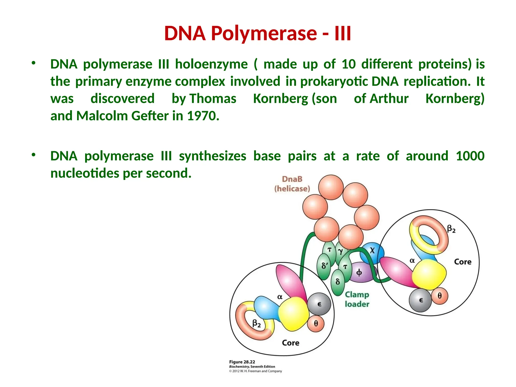
IMPORTANT ENZYMES AND
PROTEINS INVOLVED IN THE
PROKARYOTIC
DNA REPLICATION
Dr. S. SATHISH, ASST. PROFESSOR,
DEPT. OF BIOCHEMISTRY, APCAS,
KALAVAI.
Mail id: sathishapcasbio@gmail.com
PROKARYOTIC & EUKARYOTIC
DNA
Prokaryotic DNA:
• It is found freely in the cytoplasm (within a
region called the nucleoid)
• It is naked (i.e. not bound with proteins and
therefore doesn’t form chromatin)
• Genomes are compact (contain little
repetitive DNA and no introns)
• Contains extra-chromosomal plasmids
• It is circular in shape
Eukaryotic DNA:
• It is contained within a nucleus
• It is bound to histone proteins
• Genomes contain large amounts of non-
coding and repetitive DNA (including introns)
• Do not contain plasmids (but organelles such
as the mitochondria may contain their own
chromosomes)
• They are linear in shape
Prokaryotic vs Eukaryotic DNA
HUMAN CHROMOSOMES- 23
PAIRS
FEMALE MALE
SIZE OF DNA
• The human genome is comprised of 23 pairs of linear
chromosomes, and approximately 3000 megabases
(Mb) of DNA, But the genome of the bacterium
Escherichia coli consists of a single 4.6 Mb circular
chromosome.
• A base pair (bp) is a fundamental unit of double-
stranded nucleic acids consisting of two nucleobases
bound to each other by hydrogen bonds.
• 1 kb (= kbp) = kilo base pairs = 1,000 bp
• 1 Mb (Mbp) = mega base pairs = 1,000,000 bp (1million)
• 1 Gb = giga base pairs = 1,000,000,000 bp (one billion,
or one thousand million)
UNIT OF REPLICATION
Replisome complex/Replisome
Apparatus
• The replisome is a complex molecular machine that carries
out replication of DNA.
• All the enzymes and proteins involved in DNA replication
(DNA duplication or DNA synthesis) are collectively known
as Replisome.
• In terms of structure, the replisome is composed of two
replicative polymerase complexes, one of which
synthesizes the leading strand, while the other synthesizes
the lagging strand.
IMPORTANT ENZYMES AND PROTEINS INVOLVED IN
THE PROKARYOTIC DNA REPLICATION
The replisome is composed of a number of proteins/Enzymes
including
1. DNA Helicase (DNaB protein)
2. DNA Primase (DNaG protein)
3. DNA topoisomerase (DNA Gyrase)
4. DNA Polymerase
5. DNA Ligase (Polynucleotide Ligase)
6. Nucleases - RNase H
7. Single-Strand Binding (SSB) Protein
8. DNaA protein
9. DNaC protein
DNA Helicase/ DNA unwinding enzyme
• It is an enzyme that unwinds the dsDNA (double helix to
single strands) by breaking hydrogen bonds between the
complementary bases. It uses one ATP molecule for each
base that is separated.
• They are motor proteins that move directionally along
a nucleic acid phosphodiester backbone, separating
two nucleic acid strands (i.e., DNA, RNA, or RNA-DNA
hybrid) using energy derived from ATP hydrolysis.
• In E. coli, DNaB protein functions as helicase; this protein is a
hexamer and it moves with the replication fork.
• Approximately 1% of eukaryotic genes code for helicases.
The human genome codes for 95 helicases: 64 RNA helicases
and 31 DNA helicases.
DNA Helicase
• DNA helicases were discovered in E. coli in 1976.
• Helicases adopt different structures and oligomerization states.
Whereas DnaB-like helicases unwind DNA as ring-
shaped hexamers, other enzymes have been shown to be active
as monomers or dimers.
• Many cellular processes, such as DNA
replication, transcription, translation, recombination, DNA
repair, ribosome biogenesis, RNA splicing, RNA transport, RNA
editing, and RNA degradation involve the separation of nucleic
acid strands that are facilitated by helicases.
DNA Primase
Primase is an enzyme that synthesizes short RNA
sequences called primers complementary to
a ssDNA template to initiate DNA replication.
In E. coli, DnaG functions as primase. But in
eukaryotes, DNA polymerase α provides this function.
Since primase produces RNA molecules, the enzyme is
a type of RNA polymerase.
• In bacteria, primase binds to the DNA
helicase forming a complex called the primosome.
• It synthesizes a short RNA primer approximately
11±1 nucleotides long, to which
new nucleotides can be added by DNA polymerase.
DNA Primase
• Primases in organisms such as E. coli, synthesize
around 2000 to 3000 primers at the rate of one
primer per second.
• The crystal structure of primase in E. coli with a core
containing the DnaG protein was determined in the
year 2000. The DnaG and primase complex is cashew
shaped and contains three subdomains.
• Eukaryotic primases belong to the archaea-
eukaryotic primase (AEP) superfamily.
Human PrimPol serves both primase and polymerase
functions.
DNA Primase
• Topoisomerases are enzymes that participate in the
overwinding or underwinding of DNA (It removes
negative supercoiling).
• They do this by making an incision that breaks the
DNA backbone, so they can then pass the DNA
strands through one another, swivelling and
relaxing/coiling the DNA before resealing the breaks
DNA topoisomerases
DNA topoisomerases
• DNA topoisomerases can be divided into two
groups based on the number of strands that they
break.
TYPE I TOPOISOMERASE
• A type I topoisomerase cuts one strand of a DNA
double helix, relaxation occurs, and then the cut
strand is re-ligated. Cutting one strand allows the
part of the molecule on one side of the cut to rotate
around the uncut strand, thereby reducing stress.
Type I topoisomerases do not require ATP hydrolysis
(ATP independent).
Replication Causes DNA to Supercoil
TYPE II TOPOISOMERASE
• A type II topoisomerase (DNA gyrase) cuts
both strands of one DNA double helix, passes
another unbroken DNA helix through it, and
then re-ligates the cut strands. Type II
topoisomerases utilize ATP hydrolysis for its
activity (ATP dependent).
TOPOISOMERASE PREVENTS SUPERCOILING
SSBP - SINGLE-STRANDED DNA-BINDING PROTEIN
 Binds to single-stranded DNA and prevents annealing of
single-stranded DNA into double-stranded DNA, also
prevents single strand DNA from degradation by nucleases.
 177 amino acids and has a molecular weight of 18,873Da.
 It helps to keep the DNA template in an extended, single-
strand conformation, with the purine and pyrimidine bases
exposed so that they can base-pair readily with incoming
nucleotides.
 In eukaryotic cells, a heterotrimeric protein called
replication factor A (RFA) serves the role of SSB in DNA
replication.
SSBP - Single-stranded DNA-binding protein
DNA ligase
• DNA ligase is a specific type of enzyme, that facilitates the
joining of DNA strands together by catalyzing the formation
of a phosphodiester bond.
•
• DNA ligase is used in both DNA repair and DNA replication.
• DNA ligase in E. coli, as well as most prokaryotes, uses
energy gained by cleaving nicotinamide adenine
dinucleotide (NAD) to create the phosphodiester bond.
Mammalian DNA ligases
 DNA ligase I: ligates the nascent DNA of the lagging strand after
the DNA polymerase I has removed the RNA primer from the
Okazaki fragments.
 DNA ligase II: alternatively spliced form of DNA ligase III found
in non-dividing cells.
 DNA ligase III: complexes with DNA repair protein XRCC1 to aid
in sealing base excision mutations and recombinant fragments.
 DNA ligase IV: It catalyzes the final step in the non-homologous
end joining DNA double-strand break repair pathway.
 Energy gained from ATP hydrolysis.
H
H2O +
DNA Polymerase
• First discovered in 1956 by Kornberg
• E.coli have 3 types - DNA Pol I, II, and III
• DNA Pol III involved in replication of DNA
• DNA Pol II involved in repair
• DNA Pol I involved in repair
• Humans have 5 types
• DNA Pol alpha, beta, gamma, delta and epsilon
A DNA polymerase is a member of a family of enzymes that catalyze the
synthesis of DNA molecules from deoxynucleotides, the molecular
precursors of DNA. These enzymes are essential for DNA replication and
usually work in groups to create two identical DNA duplexes from a single
original DNA duplex.
• In 1956, Arthur Kornberg and colleagues
discovered DNA polymerase I (Pol I), in Escherichia coli.
They described that the DNA polymerase copies the
base sequence of a template DNA strand during DNA
replication process.
• Kornberg was later awarded the Nobel Prize in
Physiology or Medicine in 1959 for this work.
• DNA polymerase II & III were discovered by Thomas
Kornberg (the son of Arthur Kornberg) and Malcolm E.
DNA Polymerase
DNA Polymerase…
• ALL DNA Pol’s have 2 properties
–Only synthesize DNA in one direction
5’ to 3’
–Only add to the end of existing double
stranded DNA
Therefore they CANNOT start synthesis
of DNA without primer.
• This repair polymerase is involved in excision repair with
both 3'–5' and 5'–3' exonuclease activity and processing
of Okazaki fragments generated during lagging strand
synthesis.
• Pol I is the most abundant polymerase, accounting for >95%
of polymerase activity in E. coli. Pol I adds ~15-20
nucleotides per second, thus showing poor processivity.
DNA Polymerase-I (Kornberg enzyme)
Proofreading Activity
a. Insertion of the wrong nucleotide causes the DNA polymerase to stall,
b. And then the 3’-to-5’ exonuclease activity removes the mispaired A nt.
c. The polymerase then continues adding nts to the primer.
DNA polymerase
DNA Polymerase - II
• Pol II have 5'–3' polymerase & 3'–5' exonuclease activities
and participates in DNA repair, replication restart.
• Pol II is also thought to be a backup to Pol III as it can
interact with holoenzyme proteins and assume a high level
of processivity.
• The main role of Pol II is thought to be the ability to direct
polymerase activity at the replication fork and helped
stalled
DNA Polymerase - III
• DNA polymerase III holoenzyme ( made up of 10 different proteins) is
the primary enzyme complex involved in prokaryotic DNA replication. It
was discovered by Thomas Kornberg (son of Arthur Kornberg)
and Malcolm Gefter in 1970.
• DNA polymerase III synthesizes base pairs at a rate of around 1000
nucleotides per second.
The replisome is composed of the following:
2 DNA Pol III enzymes, each comprising core enyzme complex (α, ε and θ subunits)
and clamp loader.
 the α subunit has the polymerase activity.
 the ε subunit has 3'→5' exonuclease activity.
 the θ subunit stimulates the ε subunit's proofreading.
 2 β subunits which act as sliding DNA clamps, they keep the polymerase bound
to the DNA.
DNA Polymerase - III
 2 β subunits which act as sliding DNA clamps, they keep the polymerase
bound to the DNA.
 2 τ units which acts to dimerize two of the core enzymes (α, ε, and θ
subunits).
 1 γ unit which acts as a clamp loader for the lagging strand Okazaki fragments,
helping the two β subunits to form a unit and bind to DNA.
 The δ and δ' is involved in copying of the lagging strand.
 Χ and Ψ which form a 1:1 complex and bind to γ or τ.
 X can also mediate the switch from RNA primer to DNA.
DNA Polymerase – III - CLAMP LOADER
PROKARYOTIC DNA POLYMERASE
ORIGIN OF REPLICATION (OriC)
Replication of the bacterial DNA initiates at a single origin of
replication that is called oriC. This occurs via the intensive
action of numerous proteins, including DnaA, which acts as an
initiator.
ORIGIN OF REPLICATION (OriC)
• DnaA is a protein that activates initiation of DNA replication
in bacteria
• It is a replication initiation factor which promotes the
unwinding of DNA at oriC.
• Replication begins with active DnaA (The active form DnaA is
bound to ATP) binding to 9-mer (9-bp) repeats upstream of
oriC.
• Binding of DnaA leads to strand separation at the 13-mer
repeats.
• This binding causes the DNA to loop in preparation for
melting open by the helicase-DnaB.
DnaA protein
• Around 10 DnaA molecules bind to the 9 bp regions, which
wrap around the proteins causing the DNA at the AT-rich
region to unwind.
• There are 8 DnaA binding sites within oriC, to which DnaA
binds with differential affinity.
• The denatured AT-rich region allows for the recruitment of
DnaB (helicase), which complexes with DnaC (helicase
loader).
• DnaC helps the helicase to bind to DNA and to properly
accommodate the ssDNA at the 13 bp region; this is
accomplished by ATP hydrolysis, after which DnaC is released.
DnaA protein
DnaA protein interaction to OriC
DnaC (245 amino acids, 40kDa) is a loading factor for helicase
dnaB. dnaB and dnaC associate with the dnaA bound origin.
After loading dnaB at the origin, dnaC is released.
This interaction of dnaC with dnaB requires the hydrolysis of
ATP.
DnaC protein
/Rnase H
THANK YOU
DnaA is made up of four domains: the first is the N-terminal
that associates with regulatory proteins, the second is a helical
linker region, the third domain is a AAA+ region that binds to
ATP, and the fourth domain is the C-terminal DNA binding
region.
DnaA contains two conserved regions: the first is located in the
central part of the protein and corresponds to the ATP-binding
domain, the second is located in the C-terminal half and is
involved in DNA-binding
DnaA protein

Proteins and enzymes involved in prokaryotic DNA replication.pptx

  • 1.
    IMPORTANT ENZYMES AND PROTEINSINVOLVED IN THE PROKARYOTIC DNA REPLICATION Dr. S. SATHISH, ASST. PROFESSOR, DEPT. OF BIOCHEMISTRY, APCAS, KALAVAI. Mail id: sathishapcasbio@gmail.com PROKARYOTIC & EUKARYOTIC DNA
  • 2.
    Prokaryotic DNA: • Itis found freely in the cytoplasm (within a region called the nucleoid) • It is naked (i.e. not bound with proteins and therefore doesn’t form chromatin) • Genomes are compact (contain little repetitive DNA and no introns) • Contains extra-chromosomal plasmids • It is circular in shape
  • 4.
    Eukaryotic DNA: • Itis contained within a nucleus • It is bound to histone proteins • Genomes contain large amounts of non- coding and repetitive DNA (including introns) • Do not contain plasmids (but organelles such as the mitochondria may contain their own chromosomes) • They are linear in shape
  • 6.
  • 7.
  • 8.
    SIZE OF DNA •The human genome is comprised of 23 pairs of linear chromosomes, and approximately 3000 megabases (Mb) of DNA, But the genome of the bacterium Escherichia coli consists of a single 4.6 Mb circular chromosome. • A base pair (bp) is a fundamental unit of double- stranded nucleic acids consisting of two nucleobases bound to each other by hydrogen bonds. • 1 kb (= kbp) = kilo base pairs = 1,000 bp • 1 Mb (Mbp) = mega base pairs = 1,000,000 bp (1million) • 1 Gb = giga base pairs = 1,000,000,000 bp (one billion, or one thousand million)
  • 9.
  • 10.
    Replisome complex/Replisome Apparatus • Thereplisome is a complex molecular machine that carries out replication of DNA. • All the enzymes and proteins involved in DNA replication (DNA duplication or DNA synthesis) are collectively known as Replisome. • In terms of structure, the replisome is composed of two replicative polymerase complexes, one of which synthesizes the leading strand, while the other synthesizes the lagging strand.
  • 11.
    IMPORTANT ENZYMES ANDPROTEINS INVOLVED IN THE PROKARYOTIC DNA REPLICATION The replisome is composed of a number of proteins/Enzymes including 1. DNA Helicase (DNaB protein) 2. DNA Primase (DNaG protein) 3. DNA topoisomerase (DNA Gyrase) 4. DNA Polymerase 5. DNA Ligase (Polynucleotide Ligase) 6. Nucleases - RNase H 7. Single-Strand Binding (SSB) Protein 8. DNaA protein 9. DNaC protein
  • 12.
    DNA Helicase/ DNAunwinding enzyme • It is an enzyme that unwinds the dsDNA (double helix to single strands) by breaking hydrogen bonds between the complementary bases. It uses one ATP molecule for each base that is separated. • They are motor proteins that move directionally along a nucleic acid phosphodiester backbone, separating two nucleic acid strands (i.e., DNA, RNA, or RNA-DNA hybrid) using energy derived from ATP hydrolysis. • In E. coli, DNaB protein functions as helicase; this protein is a hexamer and it moves with the replication fork. • Approximately 1% of eukaryotic genes code for helicases. The human genome codes for 95 helicases: 64 RNA helicases and 31 DNA helicases.
  • 13.
    DNA Helicase • DNAhelicases were discovered in E. coli in 1976. • Helicases adopt different structures and oligomerization states. Whereas DnaB-like helicases unwind DNA as ring- shaped hexamers, other enzymes have been shown to be active as monomers or dimers. • Many cellular processes, such as DNA replication, transcription, translation, recombination, DNA repair, ribosome biogenesis, RNA splicing, RNA transport, RNA editing, and RNA degradation involve the separation of nucleic acid strands that are facilitated by helicases.
  • 15.
    DNA Primase Primase isan enzyme that synthesizes short RNA sequences called primers complementary to a ssDNA template to initiate DNA replication. In E. coli, DnaG functions as primase. But in eukaryotes, DNA polymerase α provides this function. Since primase produces RNA molecules, the enzyme is a type of RNA polymerase.
  • 16.
    • In bacteria,primase binds to the DNA helicase forming a complex called the primosome. • It synthesizes a short RNA primer approximately 11±1 nucleotides long, to which new nucleotides can be added by DNA polymerase. DNA Primase
  • 17.
    • Primases inorganisms such as E. coli, synthesize around 2000 to 3000 primers at the rate of one primer per second. • The crystal structure of primase in E. coli with a core containing the DnaG protein was determined in the year 2000. The DnaG and primase complex is cashew shaped and contains three subdomains. • Eukaryotic primases belong to the archaea- eukaryotic primase (AEP) superfamily. Human PrimPol serves both primase and polymerase functions. DNA Primase
  • 19.
    • Topoisomerases areenzymes that participate in the overwinding or underwinding of DNA (It removes negative supercoiling). • They do this by making an incision that breaks the DNA backbone, so they can then pass the DNA strands through one another, swivelling and relaxing/coiling the DNA before resealing the breaks DNA topoisomerases
  • 20.
    DNA topoisomerases • DNAtopoisomerases can be divided into two groups based on the number of strands that they break. TYPE I TOPOISOMERASE • A type I topoisomerase cuts one strand of a DNA double helix, relaxation occurs, and then the cut strand is re-ligated. Cutting one strand allows the part of the molecule on one side of the cut to rotate around the uncut strand, thereby reducing stress. Type I topoisomerases do not require ATP hydrolysis (ATP independent).
  • 21.
  • 22.
    TYPE II TOPOISOMERASE •A type II topoisomerase (DNA gyrase) cuts both strands of one DNA double helix, passes another unbroken DNA helix through it, and then re-ligates the cut strands. Type II topoisomerases utilize ATP hydrolysis for its activity (ATP dependent).
  • 23.
  • 25.
    SSBP - SINGLE-STRANDEDDNA-BINDING PROTEIN  Binds to single-stranded DNA and prevents annealing of single-stranded DNA into double-stranded DNA, also prevents single strand DNA from degradation by nucleases.  177 amino acids and has a molecular weight of 18,873Da.  It helps to keep the DNA template in an extended, single- strand conformation, with the purine and pyrimidine bases exposed so that they can base-pair readily with incoming nucleotides.  In eukaryotic cells, a heterotrimeric protein called replication factor A (RFA) serves the role of SSB in DNA replication.
  • 26.
    SSBP - Single-strandedDNA-binding protein
  • 27.
    DNA ligase • DNAligase is a specific type of enzyme, that facilitates the joining of DNA strands together by catalyzing the formation of a phosphodiester bond. • • DNA ligase is used in both DNA repair and DNA replication. • DNA ligase in E. coli, as well as most prokaryotes, uses energy gained by cleaving nicotinamide adenine dinucleotide (NAD) to create the phosphodiester bond.
  • 28.
    Mammalian DNA ligases DNA ligase I: ligates the nascent DNA of the lagging strand after the DNA polymerase I has removed the RNA primer from the Okazaki fragments.  DNA ligase II: alternatively spliced form of DNA ligase III found in non-dividing cells.  DNA ligase III: complexes with DNA repair protein XRCC1 to aid in sealing base excision mutations and recombinant fragments.  DNA ligase IV: It catalyzes the final step in the non-homologous end joining DNA double-strand break repair pathway.  Energy gained from ATP hydrolysis.
  • 29.
  • 30.
    DNA Polymerase • Firstdiscovered in 1956 by Kornberg • E.coli have 3 types - DNA Pol I, II, and III • DNA Pol III involved in replication of DNA • DNA Pol II involved in repair • DNA Pol I involved in repair • Humans have 5 types • DNA Pol alpha, beta, gamma, delta and epsilon A DNA polymerase is a member of a family of enzymes that catalyze the synthesis of DNA molecules from deoxynucleotides, the molecular precursors of DNA. These enzymes are essential for DNA replication and usually work in groups to create two identical DNA duplexes from a single original DNA duplex.
  • 31.
    • In 1956,Arthur Kornberg and colleagues discovered DNA polymerase I (Pol I), in Escherichia coli. They described that the DNA polymerase copies the base sequence of a template DNA strand during DNA replication process. • Kornberg was later awarded the Nobel Prize in Physiology or Medicine in 1959 for this work. • DNA polymerase II & III were discovered by Thomas Kornberg (the son of Arthur Kornberg) and Malcolm E. DNA Polymerase
  • 32.
    DNA Polymerase… • ALLDNA Pol’s have 2 properties –Only synthesize DNA in one direction 5’ to 3’ –Only add to the end of existing double stranded DNA Therefore they CANNOT start synthesis of DNA without primer.
  • 33.
    • This repairpolymerase is involved in excision repair with both 3'–5' and 5'–3' exonuclease activity and processing of Okazaki fragments generated during lagging strand synthesis. • Pol I is the most abundant polymerase, accounting for >95% of polymerase activity in E. coli. Pol I adds ~15-20 nucleotides per second, thus showing poor processivity. DNA Polymerase-I (Kornberg enzyme)
  • 34.
    Proofreading Activity a. Insertionof the wrong nucleotide causes the DNA polymerase to stall, b. And then the 3’-to-5’ exonuclease activity removes the mispaired A nt. c. The polymerase then continues adding nts to the primer. DNA polymerase
  • 35.
    DNA Polymerase -II • Pol II have 5'–3' polymerase & 3'–5' exonuclease activities and participates in DNA repair, replication restart. • Pol II is also thought to be a backup to Pol III as it can interact with holoenzyme proteins and assume a high level of processivity. • The main role of Pol II is thought to be the ability to direct polymerase activity at the replication fork and helped stalled
  • 36.
    DNA Polymerase -III • DNA polymerase III holoenzyme ( made up of 10 different proteins) is the primary enzyme complex involved in prokaryotic DNA replication. It was discovered by Thomas Kornberg (son of Arthur Kornberg) and Malcolm Gefter in 1970. • DNA polymerase III synthesizes base pairs at a rate of around 1000 nucleotides per second.
  • 37.
    The replisome iscomposed of the following: 2 DNA Pol III enzymes, each comprising core enyzme complex (α, ε and θ subunits) and clamp loader.  the α subunit has the polymerase activity.  the ε subunit has 3'→5' exonuclease activity.  the θ subunit stimulates the ε subunit's proofreading.  2 β subunits which act as sliding DNA clamps, they keep the polymerase bound to the DNA. DNA Polymerase - III
  • 38.
     2 βsubunits which act as sliding DNA clamps, they keep the polymerase bound to the DNA.  2 τ units which acts to dimerize two of the core enzymes (α, ε, and θ subunits).  1 γ unit which acts as a clamp loader for the lagging strand Okazaki fragments, helping the two β subunits to form a unit and bind to DNA.  The δ and δ' is involved in copying of the lagging strand.  Χ and Ψ which form a 1:1 complex and bind to γ or τ.  X can also mediate the switch from RNA primer to DNA. DNA Polymerase – III - CLAMP LOADER
  • 39.
  • 40.
    ORIGIN OF REPLICATION(OriC) Replication of the bacterial DNA initiates at a single origin of replication that is called oriC. This occurs via the intensive action of numerous proteins, including DnaA, which acts as an initiator.
  • 41.
  • 42.
    • DnaA isa protein that activates initiation of DNA replication in bacteria • It is a replication initiation factor which promotes the unwinding of DNA at oriC. • Replication begins with active DnaA (The active form DnaA is bound to ATP) binding to 9-mer (9-bp) repeats upstream of oriC. • Binding of DnaA leads to strand separation at the 13-mer repeats. • This binding causes the DNA to loop in preparation for melting open by the helicase-DnaB. DnaA protein
  • 43.
    • Around 10DnaA molecules bind to the 9 bp regions, which wrap around the proteins causing the DNA at the AT-rich region to unwind. • There are 8 DnaA binding sites within oriC, to which DnaA binds with differential affinity. • The denatured AT-rich region allows for the recruitment of DnaB (helicase), which complexes with DnaC (helicase loader). • DnaC helps the helicase to bind to DNA and to properly accommodate the ssDNA at the 13 bp region; this is accomplished by ATP hydrolysis, after which DnaC is released. DnaA protein
  • 44.
  • 45.
    DnaC (245 aminoacids, 40kDa) is a loading factor for helicase dnaB. dnaB and dnaC associate with the dnaA bound origin. After loading dnaB at the origin, dnaC is released. This interaction of dnaC with dnaB requires the hydrolysis of ATP. DnaC protein
  • 46.
  • 48.
  • 50.
    DnaA is madeup of four domains: the first is the N-terminal that associates with regulatory proteins, the second is a helical linker region, the third domain is a AAA+ region that binds to ATP, and the fourth domain is the C-terminal DNA binding region. DnaA contains two conserved regions: the first is located in the central part of the protein and corresponds to the ATP-binding domain, the second is located in the C-terminal half and is involved in DNA-binding DnaA protein