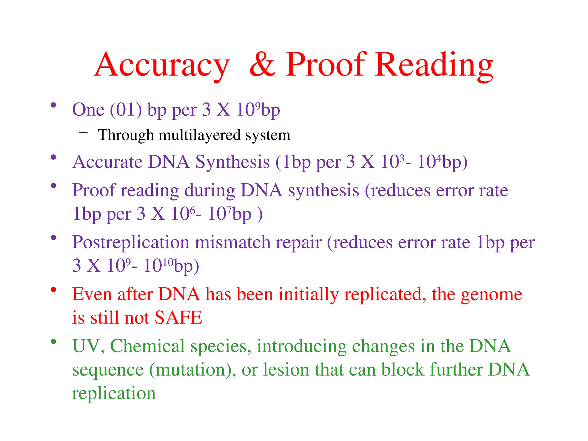
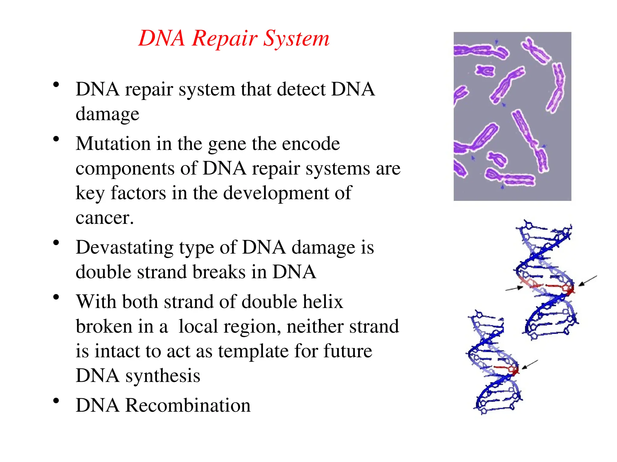
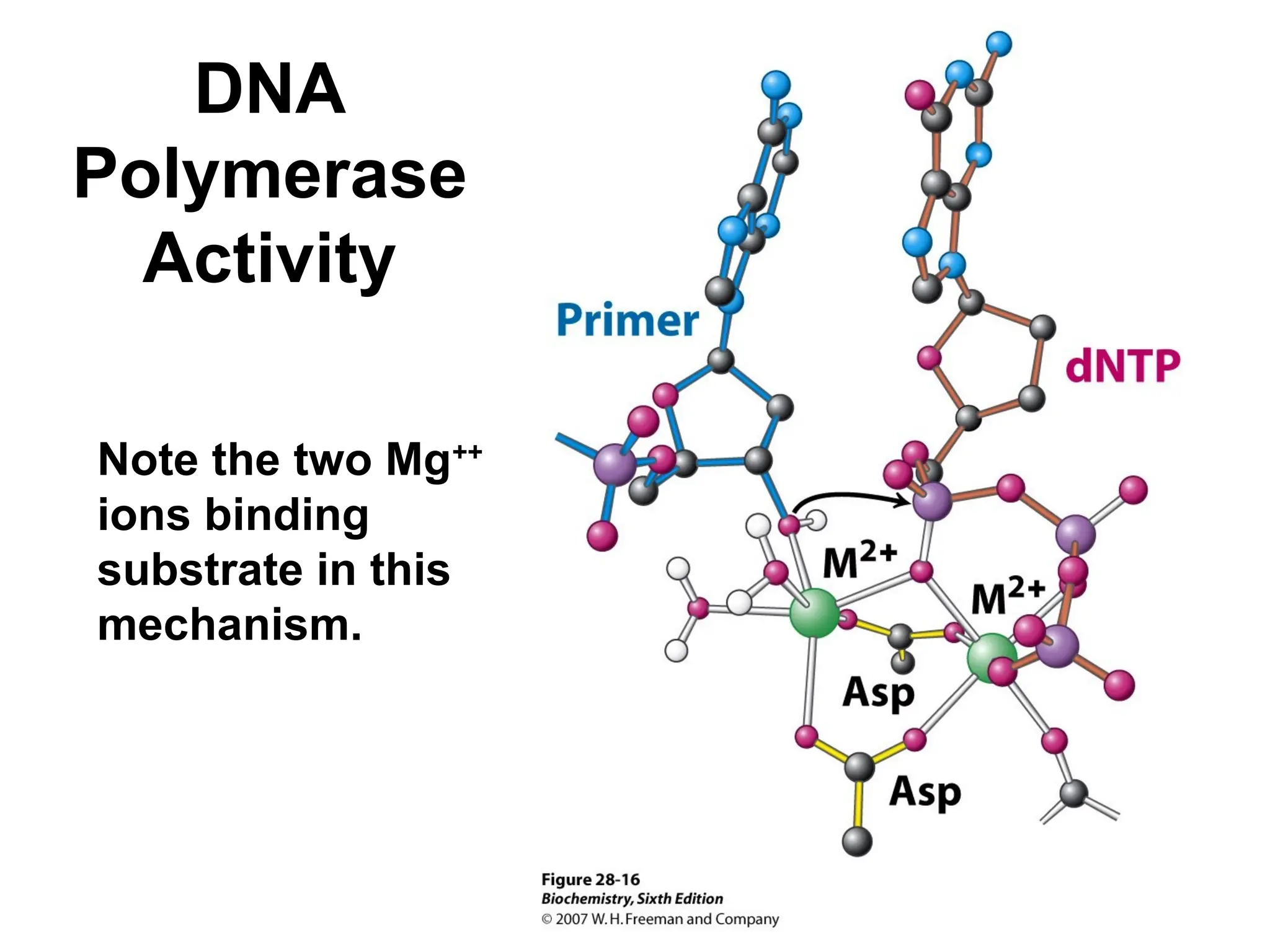
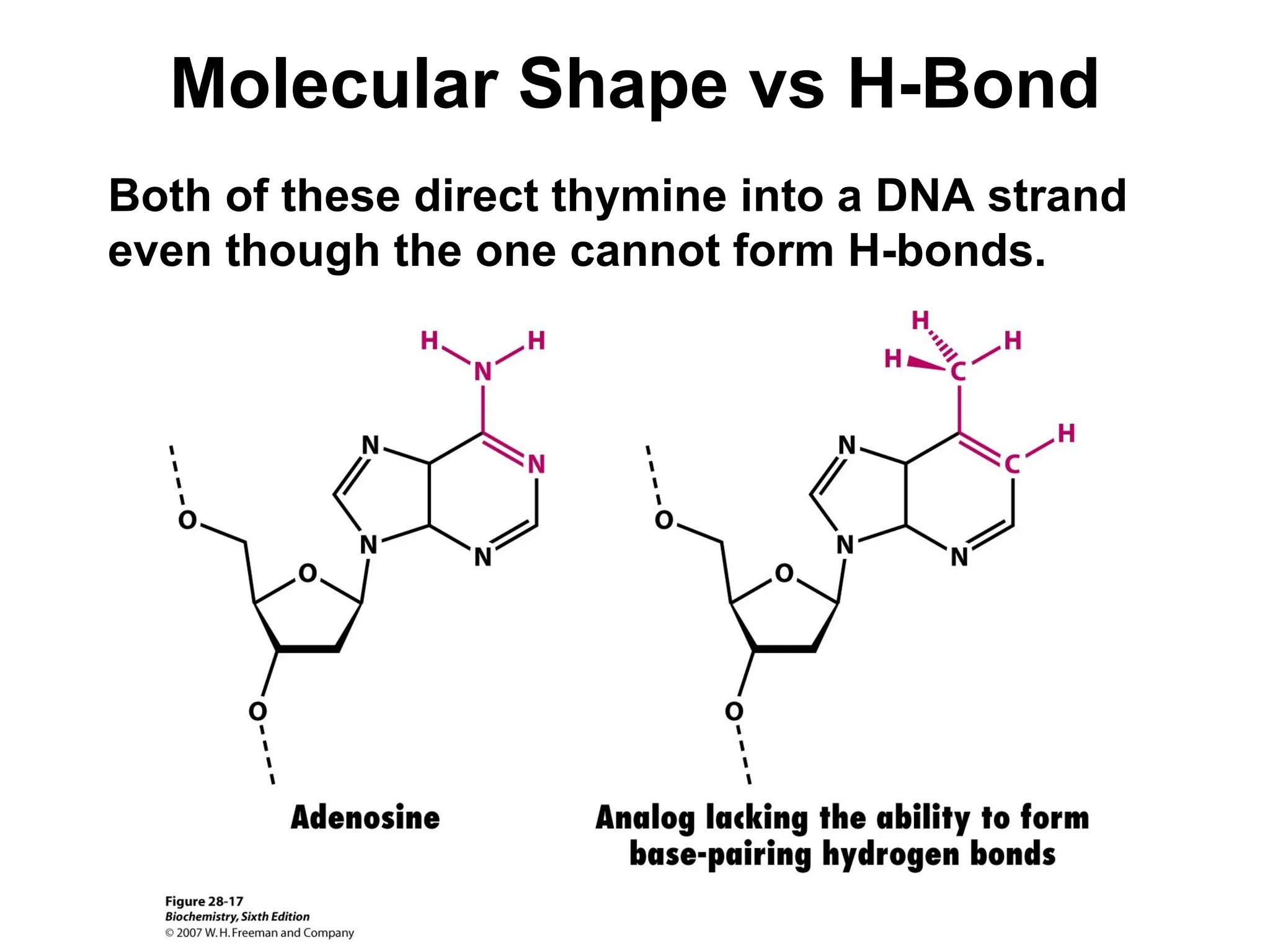
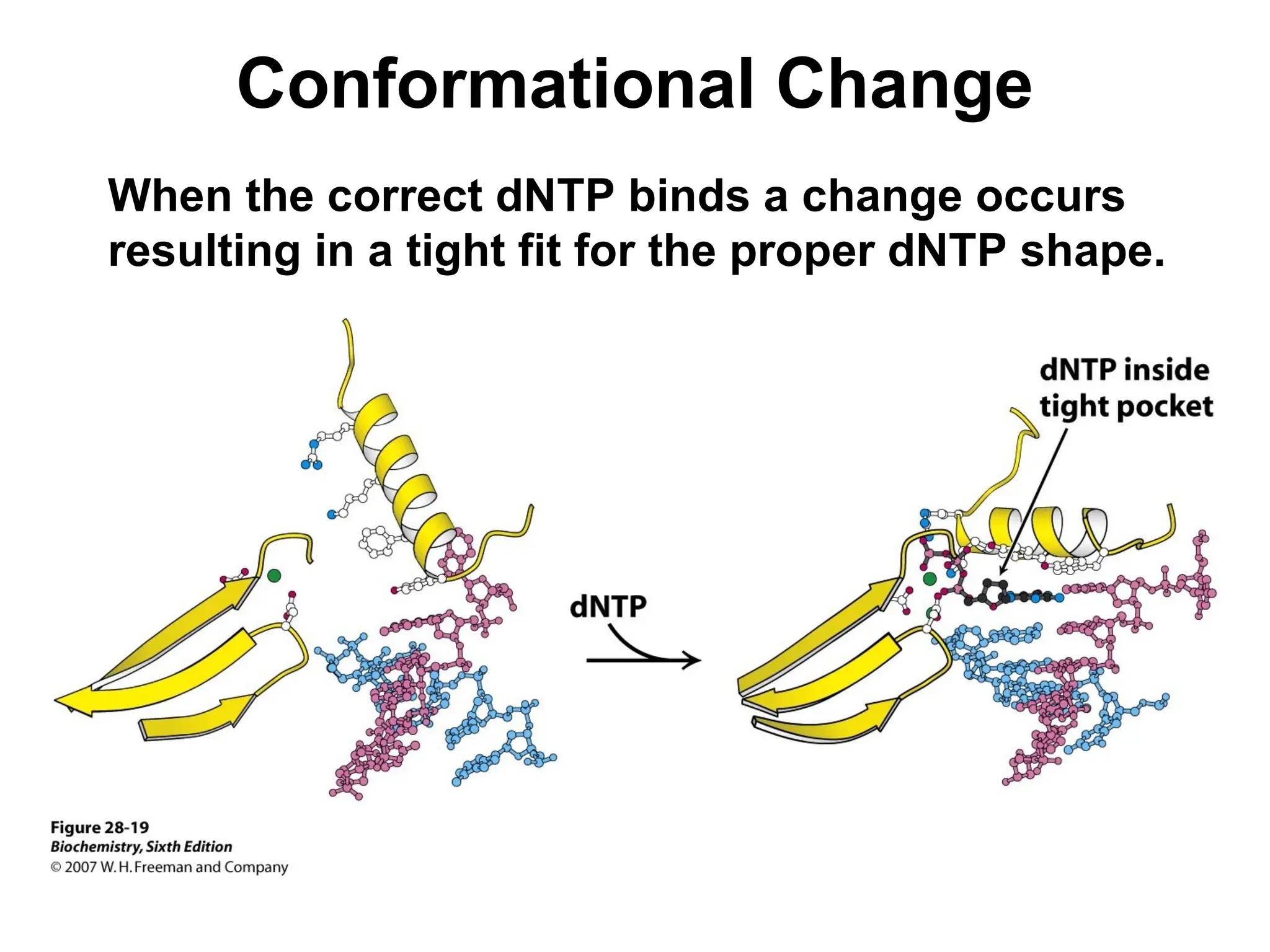
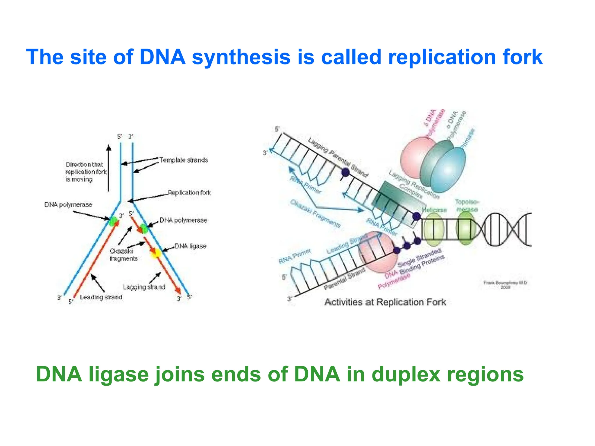
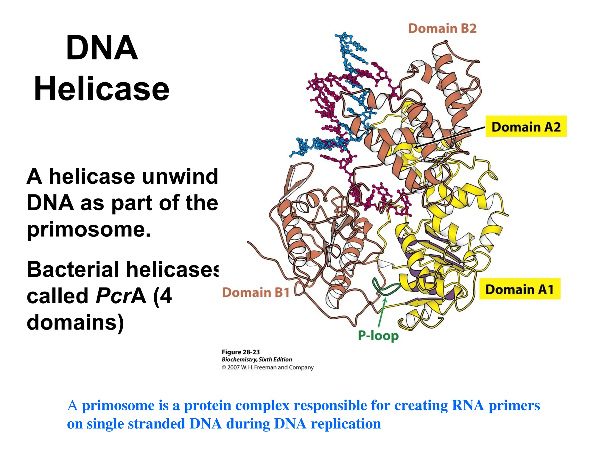
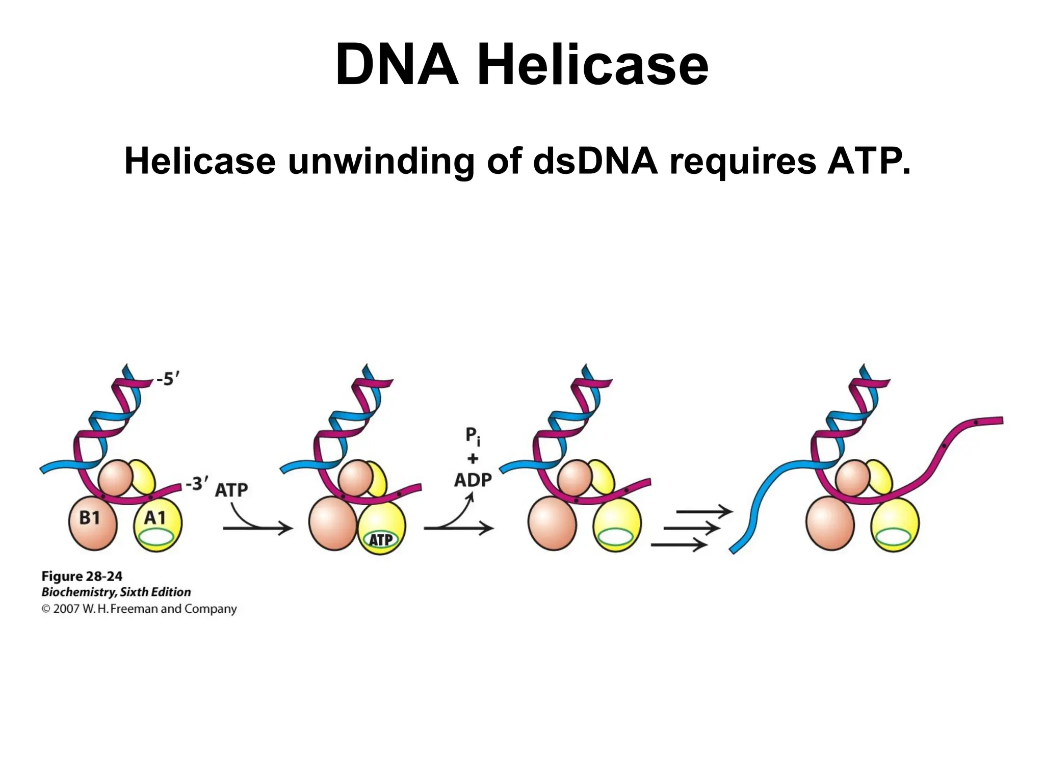
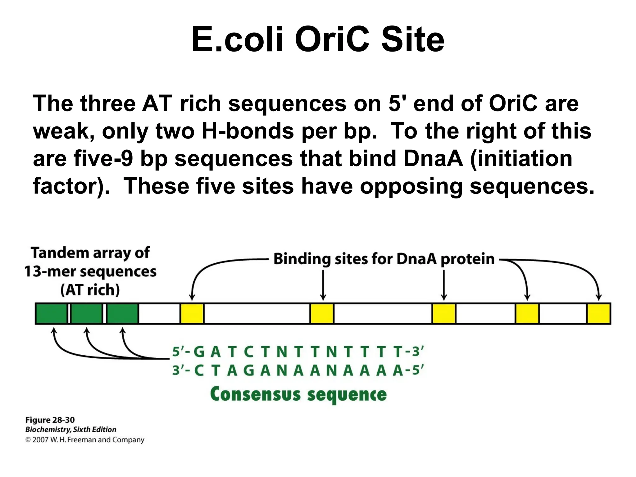
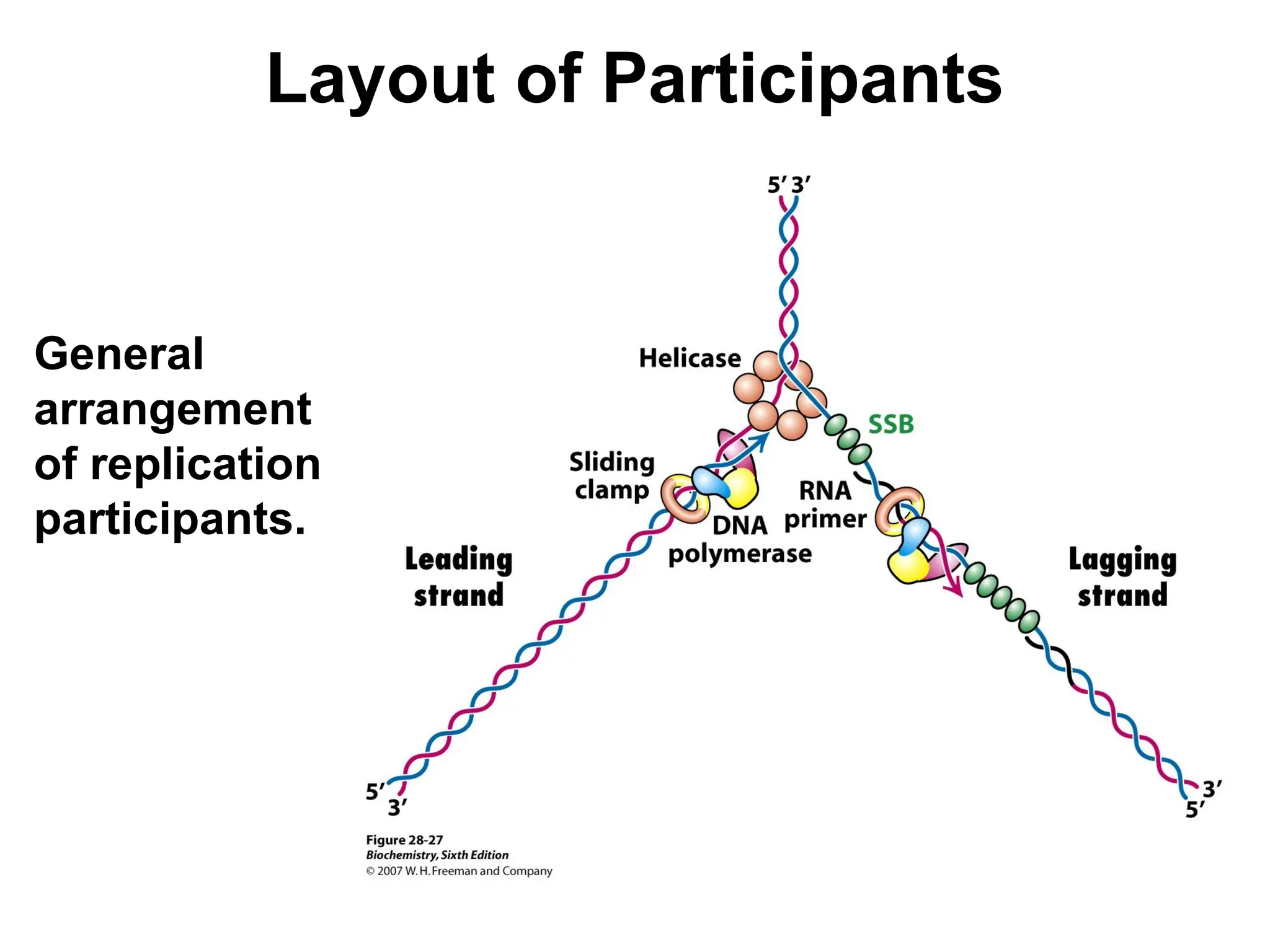
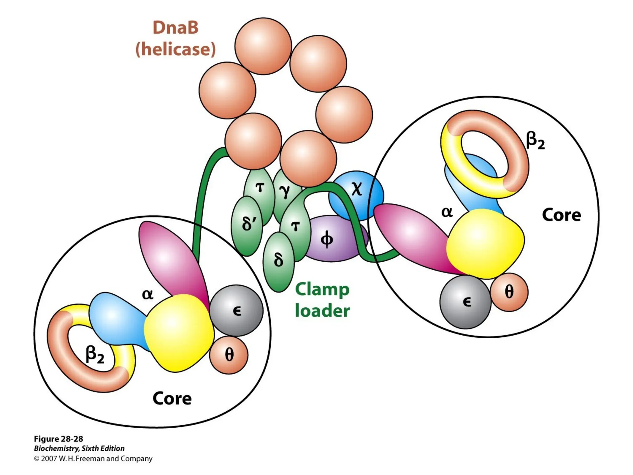
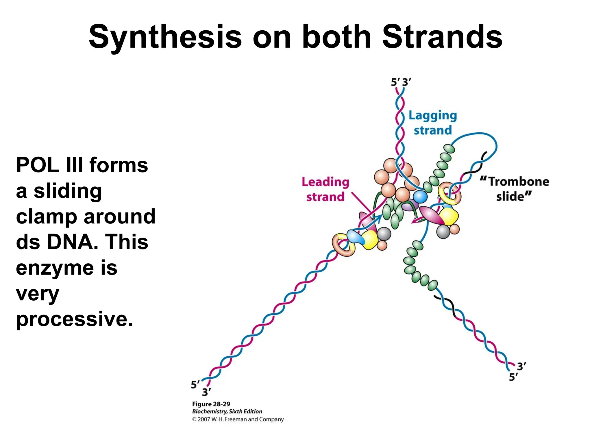
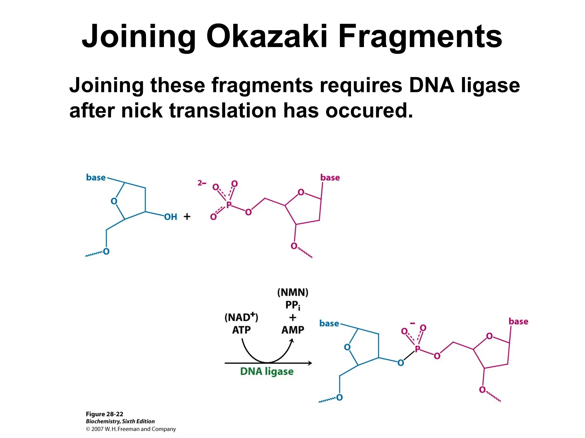
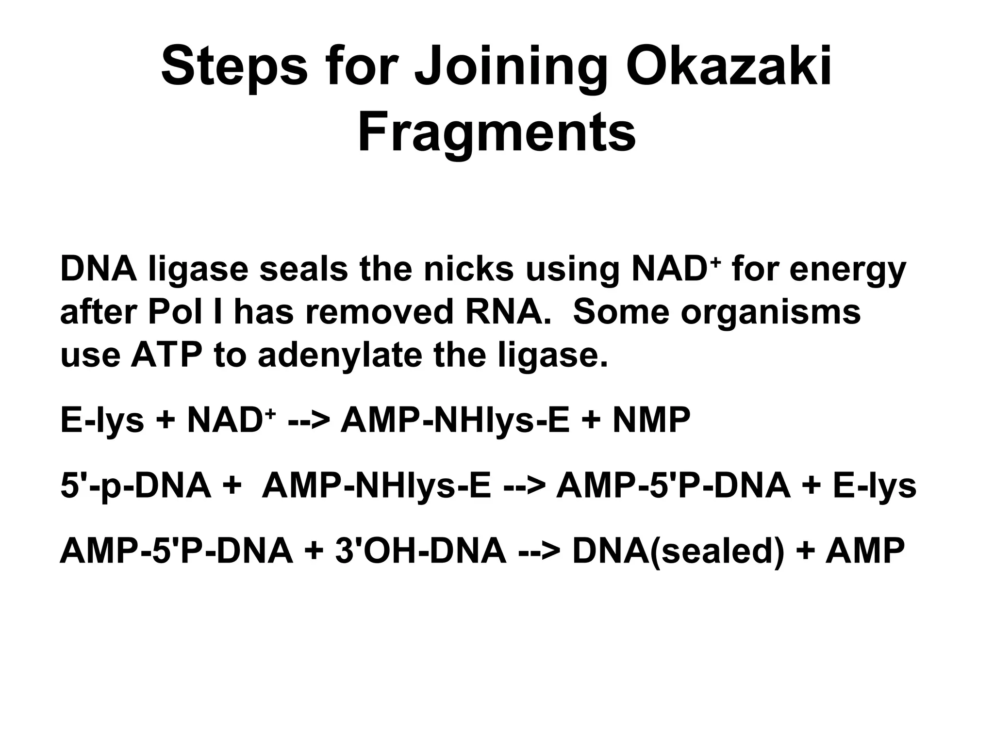
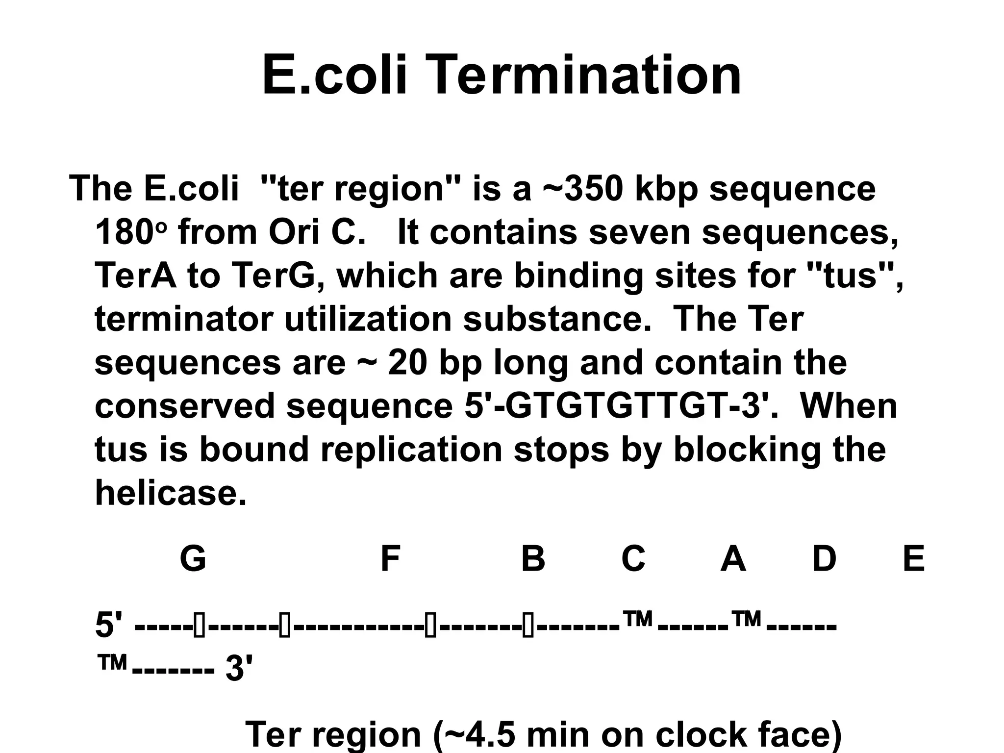
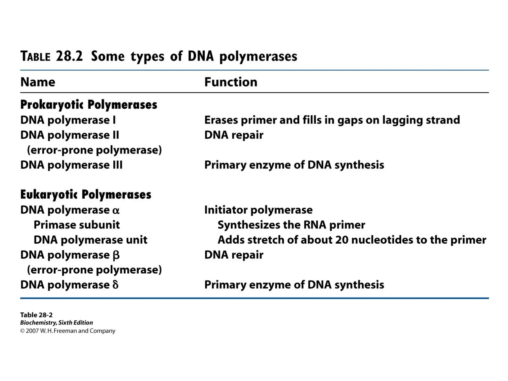
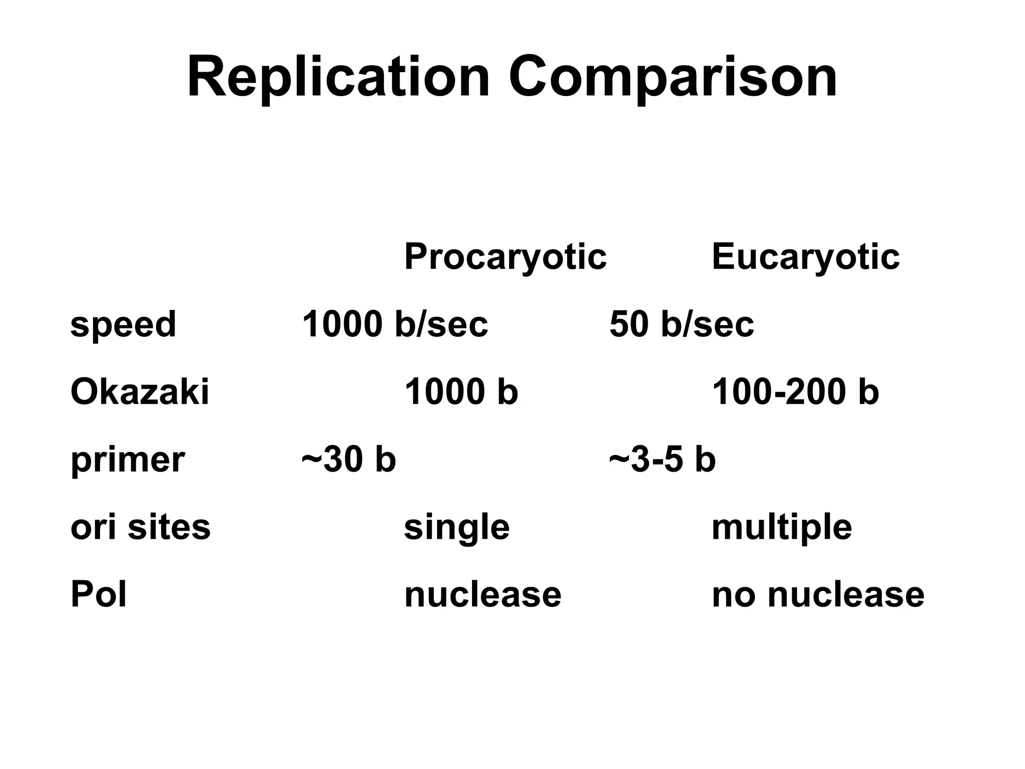
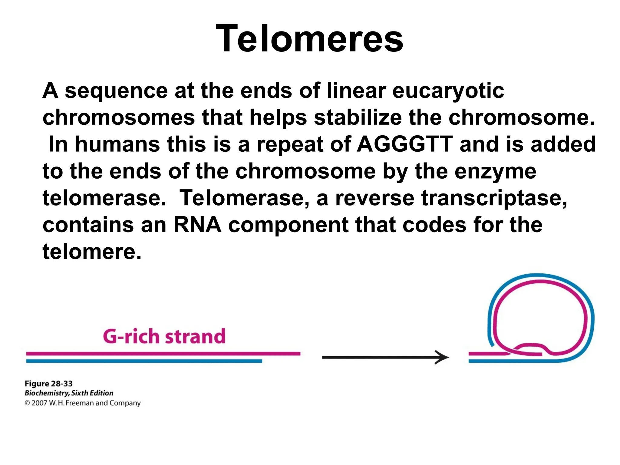
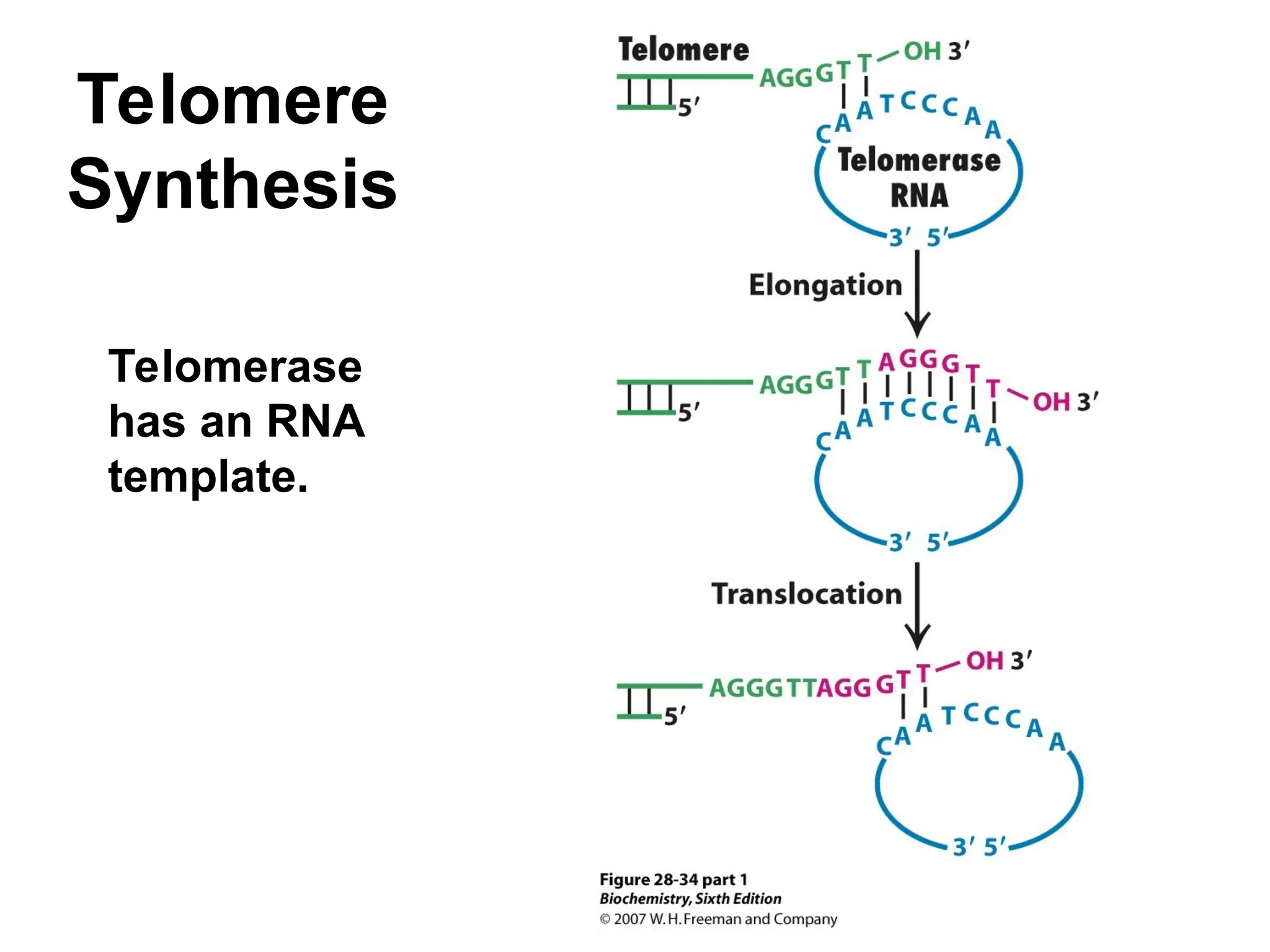
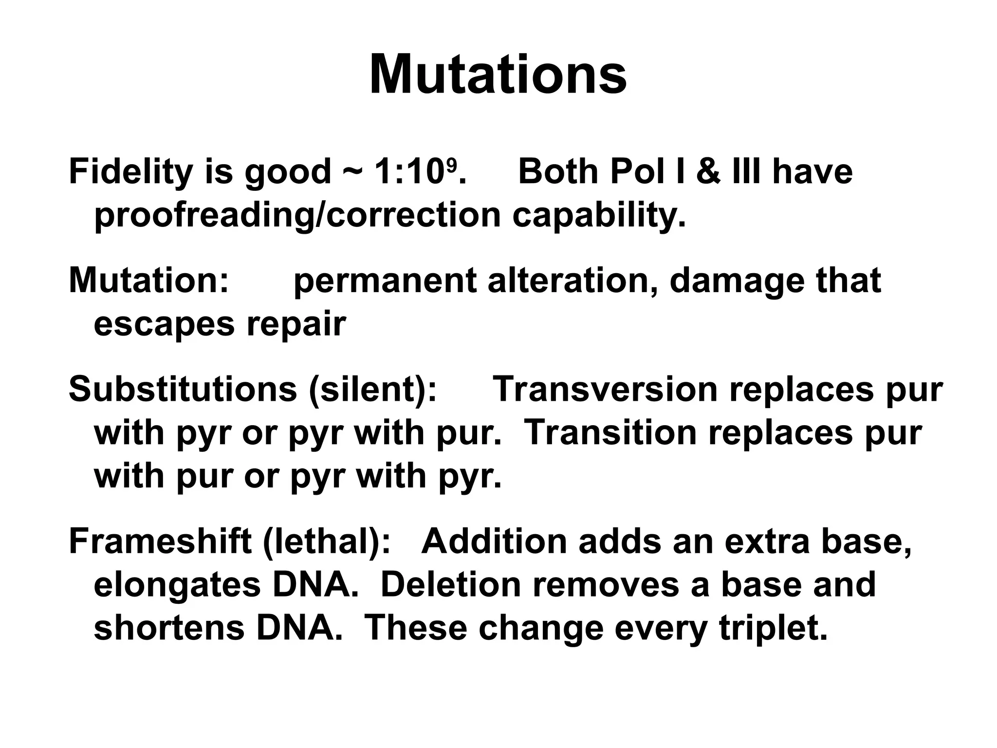
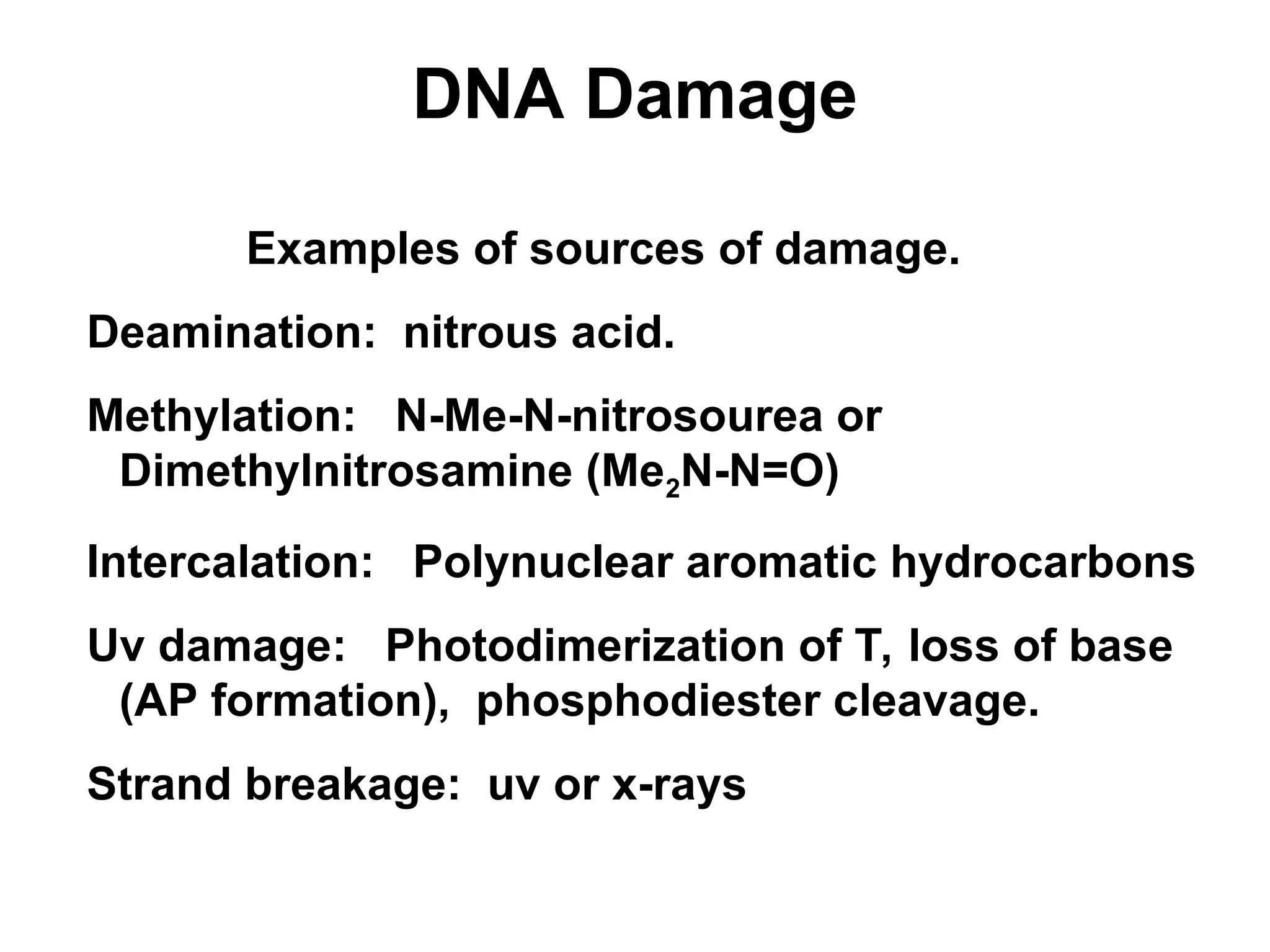
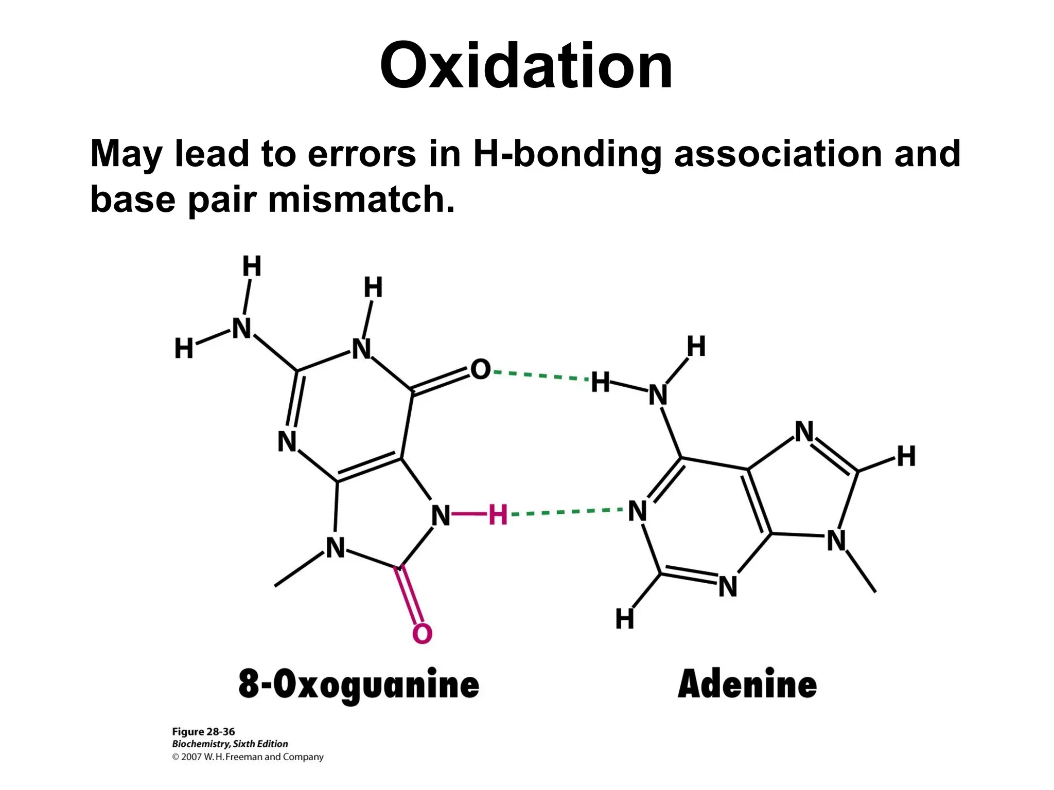
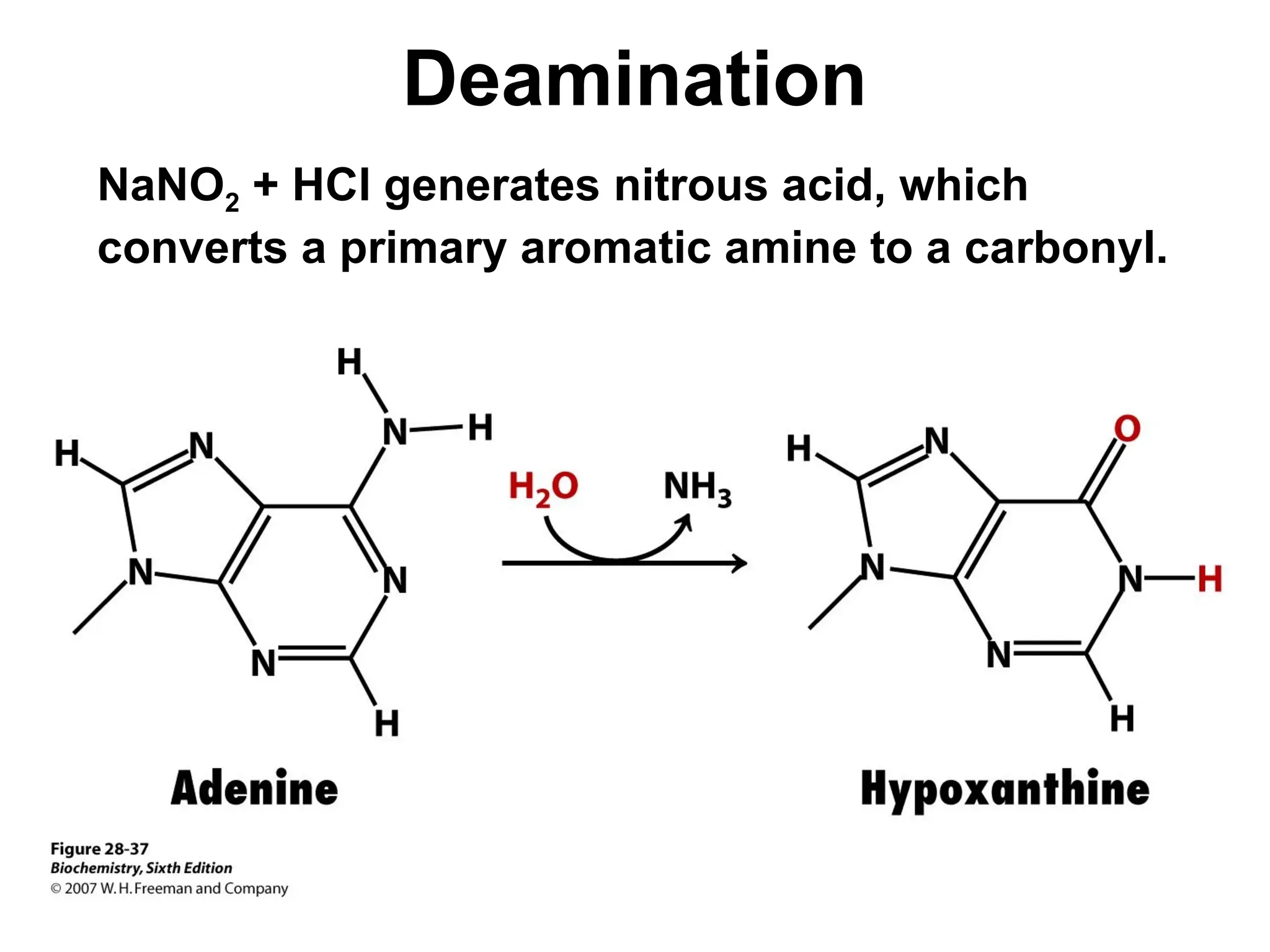
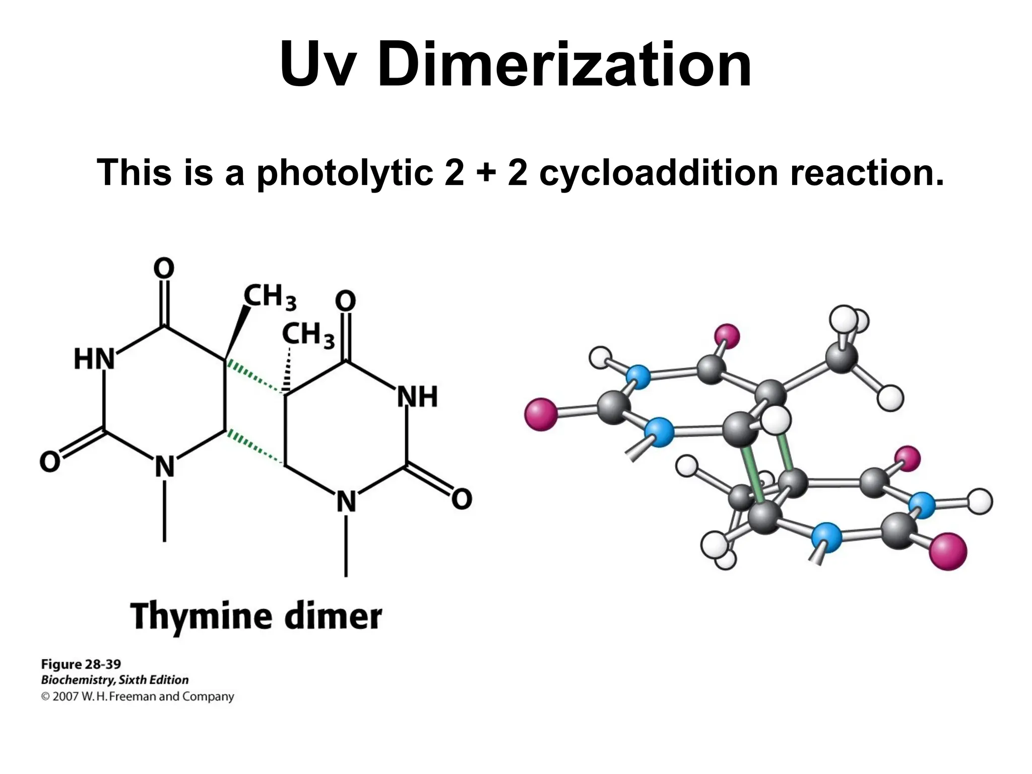
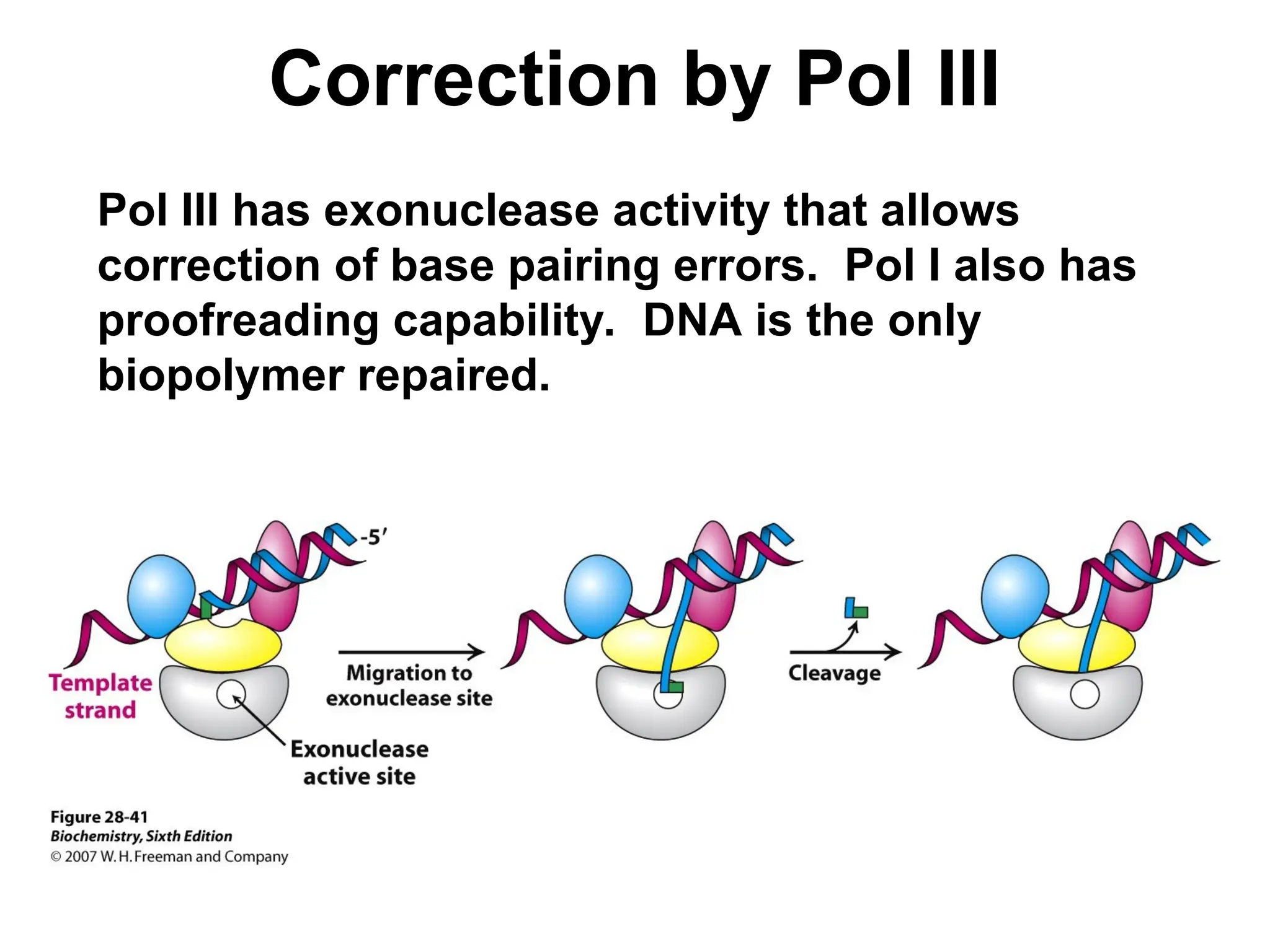
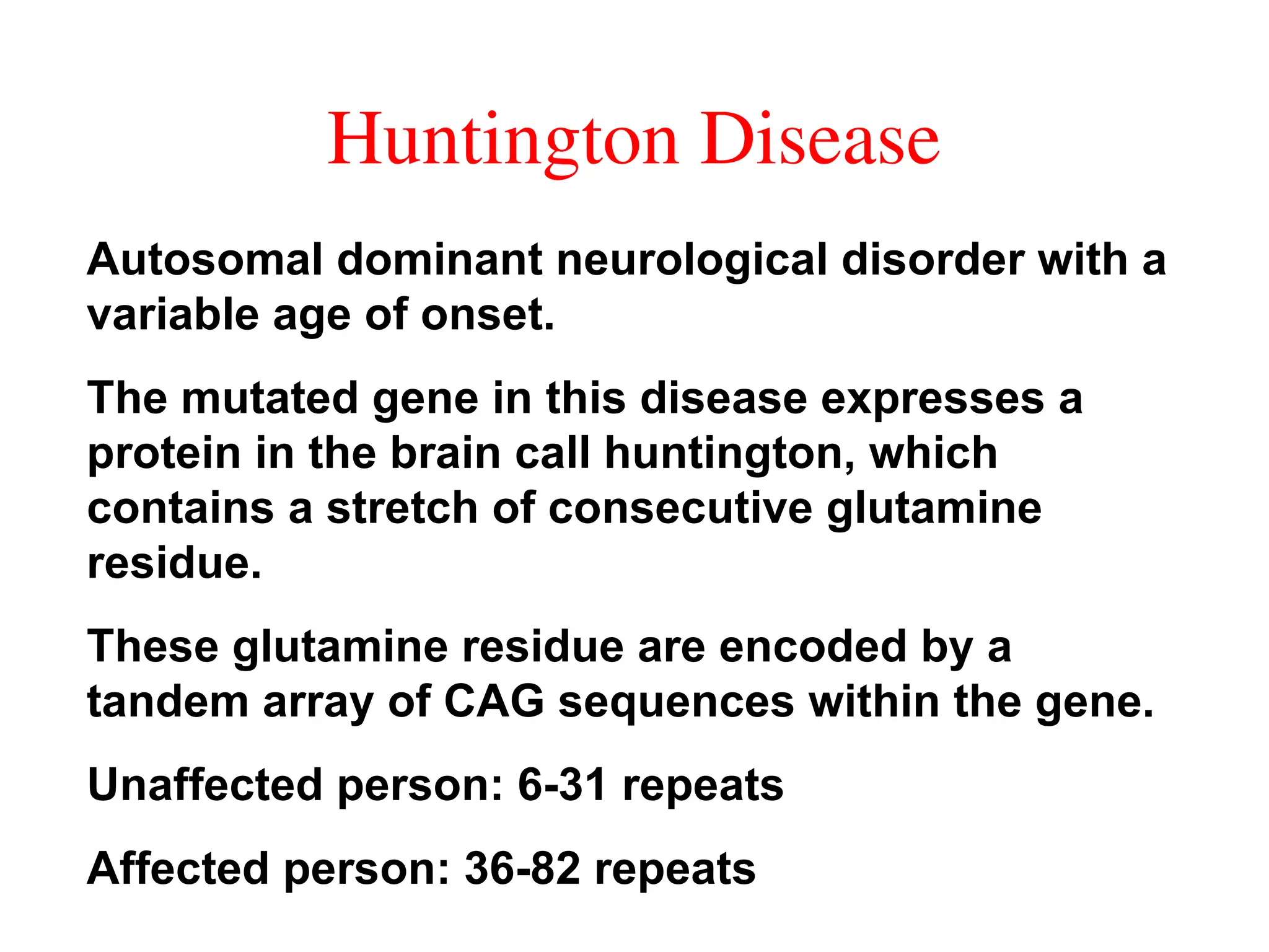
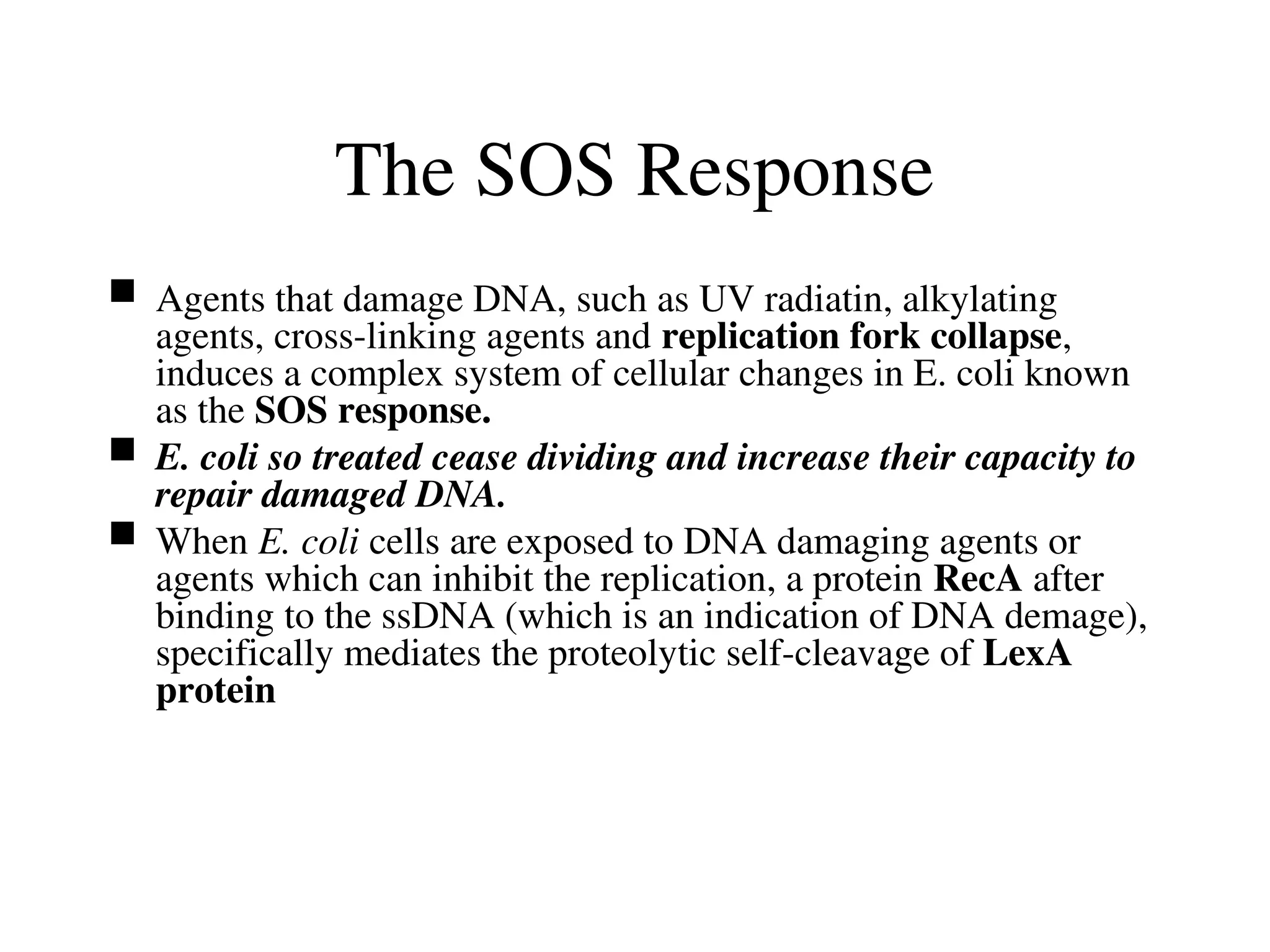
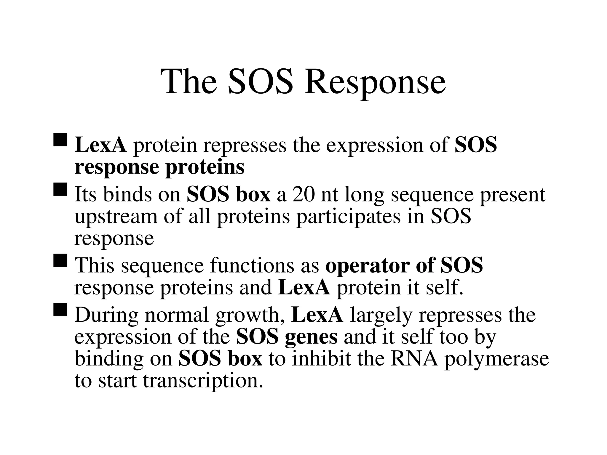
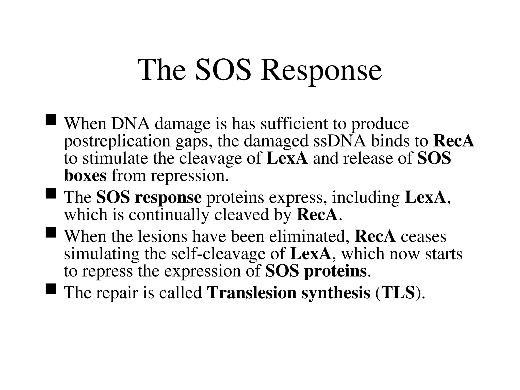
Chapter 28:
DNA Replication, Repair, and
Recombination
Copyright © 2007 by W. H. Freeman and Company
Berg • Tymoczko • Stryer
Biochemistry
Sixth Edition
Accuracy & Proof Reading
• One (01) bp per 3 X 10⁹
bp
– Through multilayered system
• Accurate DNA Synthesis (1bp per 3 X 10³
- 104
bp)
• Proof reading during DNA synthesis (reduces error rate
1bp per 3 X 10⁶
- 107
bp )
• Postreplication mismatch repair (reduces error rate 1bp per
3 X 10⁹
- 1010
bp)
• Even after DNA has been initially replicated, the genome
is still not SAFE
• UV, Chemical species, introducing changes in the DNA
sequence (mutation), or lesion that can block further DNA
replication
DNA Repair System
• DNA repair system that detect DNA
damage
• Mutation in the gene the encode
components of DNA repair systems are
key factors in the development of
cancer.
• Devastating type of DNA damage is
double strand breaks in DNA
• With both strand of double helix
broken in a local region, neither strand
is intact to act as template for future
DNA synthesis
• DNA Recombination
Basics of DNA Structure
• The two strands of double stranded DNA run in
opposite directions (anti-parallel).
• The purine and pyrimidine bases appended to
the deoxyribose-phosphate polymer strands are
on the inside of the double helix.
• In the Watson-Crick model Adenine (A) base
pairs with Thymine (T) through two H-bonds and
Guanine (G) always base pairs with Cytosine (C)
through three H-bonds.
Semiconservative
Replication
of DNA
Based on studies
by Meselson and
Stahl.
Replication always
occurs moving
5'-3'.
A and B
DNA
Both are right-
handed helices
B = common,
10.4 bp/turn
base tilt = 5-6o
A = low humidity,
11 bp/turn
base tilt = 11-20o
Ribose Conformations
In form A the 3' C is up and in form B the 2' C is up.
Bases are typically anti to the ribose ring so that
base pairing is facilitated.
Ribose Attachments Ribose in relationship to
the groves in ds DNA.
The Minor groove contains the pyrimidine O-2 and Purine N-3 of the base pair and
the major groove is on the opposite side of the pair.
Methyl group of thymine lies in the major groove
N-3 of A and G and o-2 of T or C can serve as H bond acceptor and NH2 group
attached to C-2 of G can be a H bond donor
Major groove– N-7 of G or A is a potential acceptor, as are O4 of T and O-6 of G
NH2 gp attached to C-6 of A and C-4 of C can serve as H bond doner
Propeller Twist
Study of a short segment of dsDNA (dodecamer)
shows non-coplanarity between some base pairs.
DNA, Z Form
A left handed helix with alternating GCGCGC and alternating
syn and anti purines.
Protein domain has been discovered that bind nucleic acids
specifically in the Z form.
This observation strongly suggests that such structures are
present in cells and perform specific function
Three Forms of DNA
DNA Supercoiling
Relaxed DNA in the B form has 10.4 base pairs
per turn of the helix. The linear structure below
shows the number of turns about the helix axis.
Supercoiling Parameters
The Linking number (L) is the number of times
one strand of circular dsDNA passes over the
other. L is constant unless the strands break
and reform.
The Twist (T) is the number of turns that DNA
makes about the duplex axis.
The Writhe (W) is the number of supercoils (turns
about the superhelix axis). A clockwise turn = a
(+) supercoil and ccw = a (-) supercoil.
So: L = T + W
Circular
DNA
Closing the linear
DNA seen
previously gives
relaxed, circular
ds DNA with no
supercoils so:
L = T + W
25 = 25 + 0
Changing L
Open the circular DNA and unravel two turns.
This decreases L by two.
Circular
DNA
Closing the DNA
makes gives
relaxed, circular
ds DNA with no
supercoils, now:
L = T + W
23 = 23 + 0
Introducing Supercoils
Making two right-
handed coils in
the previous
helix without
breaking the
strands is equal
to a W of –2 and
T changes:
23 = 25 - 2
Electron Micrograph
E.Coli has about 5 supercoils per 1000 bp.
Topoisomerase Prepare the Double helix for Unwinding
• Most naturally occuring DNA molecules are negatively supercoiled.
• Negatively supercoiling prepares DNA for processes requiring
separation of the DNA strands, like replication
• Positive super coiling condenses DNA as effectively, but it makes
strand separation more difficult
• Specific enzymes called topoisomerases that introduce or eliminate
supercoils
• Type 1 topoisomerases catalyze the relaxation of supercoiled DNA, a
thermodynamically favorable process
• Type 2 topoisomerases utilize free enerty from ATP hydrolysis to add
negetive supercoils to DNA
Topoisomerases alter the Lk number
• Three step process
1. The cleavage of one or both strands of DNA
2. The passage of a segment of DNA through this break
3. The resealing of the DNA break
• Type 1 topoisomerases cleave just one strand of DNA,
whereas, type II enzyme cleave both strands
Topoisomerase I
• Human type 1 comprises
four domains having central
cavity (20Ao
)
• This cavity also includes a
tyrosine residue (Tyr 723),
which acts as a nuclephile to
cleave the DNA backbone in
the course of catalysis
Tyr of Topoisomerase I
Tyr of Topo I cleaves one strand of DNA to permit
unwinding and a change the linking number (L).
Topoisomerase II
Topo II breaks
both strands of
DNA and
requires ATP.
Topo I breaks
only one strand.
• The bacterial topoisomerases II (often called DNA gyrase)
is the target of several antibiotics that inhibit the
prokaryotic enzyme much more than the eukaryotic one
• Novobiocin blocks the binding of ATP to gyrase.
• Nalidixic acid and ciprofloxacin, in contrast interfere with
the breakage and rejoining of DNA chains.
• These two gyrase inhibitors are widely used to treat UTI
including those due to Bacillus antracis (anthrax).
• Campthethecin, an antitumor agent, inhibits human
topoisomerase I by stablizing the form of enzyme
covalentaly
Complication of DNA Replication
• Two strands run in opposite direction
– Replication 5` to 3` direction, so must have special mechanism to
accommodate the oppositely directed strand
• The two strands of double helix interact with one another
in such a way that the edges of the bases on which the
newly synthesized DNA is to be assembled and occupied.
– Thus two strand must be separted
• The two strands of double helix wrap around each other
– Thus strand seperation also entails the unwinding of the double
helix. This unwinding creates supercoils that must themselves be
resolved as replition continues.
DNA Polymerases Requires a Template and a
Primer
• DNA polymerases are template directed enzymes.
• Primer: The initial segment of a polymer that is to be
extended on which elongation depends.
• Template: A sequence of DNA or RNA that directs the
sysnthesis of a complementary sequence
DNA Polymerase I
DNA Pol I was the first polymerase isolated. This
was obtained from E.coli by Arthur Kornberg
and is known as the Kornberg enzyme.
This enzyme has a 5'-3' exonuclease activity that
cleaves RNA primers, a 5'-3' polymerase activity
that makes DNA and a 3'-5' exonuclease activity
that repairs DNA.
The Klenow fragment is the large portion after
cleaving off the 5'-3' exonuclease and has been
used as a polymerase in lab work.
DNA
Polymerase
This is the
Klenow fragment
of the E.coli
enzyme.
DNA
Polymerase
Activity
Note the two Mg++
ions binding
substrate in this
mechanism.
Molecular Shape vs H-Bond
Both of these direct thymine into a DNA strand
even though the one cannot form H-bonds.
Conformational Change
When the correct dNTP binds a change occurs
resulting in a tight fit for the proper dNTP shape.
RNA primer synthesized by Primase
enables DNA synthesis to Begin
• DNA Polymerase cannot initiate DNA synthesis without a
primer.
• RNA primes the synthesis of DNA .
• RNA polymerase called primase synthesizes a short
stretch of RNA (5nt).
• Primase, like other RNA polymerases can initiate synthesis
without a primer.
• After DNA synthesis has been initiated, the short stretch of
RNA is removed by hydrolysis and replaced by DNA
The site of DNA synthesis is called replication fork
DNA ligase joins ends of DNA in duplex regions
DNA
Helicase
A helicase unwinds
DNA as part of the
primosome.
Bacterial helicases
called PcrA (4
domains)
A primosome is a protein complex responsible for creating RNA primers
on single stranded DNA during DNA replication
DNA Helicase
Helicase unwinding of dsDNA requires ATP.
E.coli Replication
Three Steps:
1. Initiation: Ori site in E.coli = Ori C
This is a 245 bp highly conserved seq.
2. Polymerization: chain elongation in the 5’-
3’ direction.
3. Termination: “ter region” is ~350 kbp
sequence that is 180o
from Ori C.
E.coli OriC Site
The three AT rich sequences on 5' end of OriC are
weak, only two H-bonds per bp. To the right of this
are five-9 bp sequences that bind DnaA (initiation
factor). These five sites have opposing sequences.
Priming Events
DnaA binds to the 9 bp sequences along with ATP
and causes opening in the AT rich region. HU a
histone-like protein prevents DnaA from binding
at sites other than OriC.
When the loop is open DnaB, a helicase, binds at
each fork as a complex (DnaB6•DnaC6•ATP6).
DnaT assists and this is the pre-priming
complex.
DnaB continues to unwind increasing the bubble
size displacing DnaA as it moves. SSB binds to
ssDNA to prevent annealing. Topo II binds ahead
of the fork to relieve stress cause by opening.
Possible DnaA Binding
Proposals
suggest that
up to 20 or
more DnaA
are bound
and that a
nucleosome
type
structure is
formed.
Priming Events
PriA, PriB and PriC enter the bubble along with
DnaG (DNA primase). This completes the
primosome which makes RNA primers. Topo II
and SSB are not part of the primosome. DNA
primase does not need a primer to begin
synthesis of RNA primers.
The primers (10-30 bp) begin at the center base of
any GTT sequence and start a primer about every
1000 bp. Only one primer is needed on the
leading strand. After this is made, the
primosome moves to the lagging strand.
Replication Events
Two Pol III holoenzymes (DnaE) enter with a few
other proteins to complete the replisome. Pol III
is an asymmetric dimer that synthesizes DNA
from both template strands simultaneously.
Pol III needs a primer to begin synthesis. The
leading strand is synthesized continuously and
the lagging strand discontinously. Both are
synthesized in the 5'-3' direction.
Bidirectional synthesis occurs at both replication
forks.
DNA Pol III, the Replicase
Pol III forms a
sliding clamp
around ds
DNA.
This enzyme is
the workhorse
of replication
and is very
processive.
Priming DNA Synthesis
A primer is require to start DNA synthesis.
Leading and Lagging Strand
Synthesis
Okazaki fragments are made on the lagging strand.
Layout of Participants
General
arrangement
of replication
participants.
Synthesis on both Strands
POL III forms
a sliding
clamp around
ds DNA. This
enzyme is
very
processive.
Lagging Strand Synthesis
The lagging strand loops to enable both Pol III
core units to move in the direction of the
replication fork.
The lagging strand begins replication at a primer
and proceeds until it runs into another Okazaki
fragment. At this point the core unit dissociates,
the chain shifts to position another primer, the
core rebinds and makes another Okazaki piece.
The lagging strand will always be a little behind
the leading strand.
Joining Okazaki Fragments
Joining these fragments requires DNA ligase
after nick translation has occured.
Steps for Joining Okazaki
Fragments
DNA ligase seals the nicks using NAD+
for energy
after Pol I has removed RNA. Some organisms
use ATP to adenylate the ligase.
E-lys + NAD+
--> AMP-NHlys-E + NMP
5'-p-DNA + AMP-NHlys-E --> AMP-5'P-DNA + E-lys
AMP-5'P-DNA + 3'OH-DNA --> DNA(sealed) + AMP
E.coli Termination
The E.coli ''ter region'' is a ~350 kbp sequence
180o
from Ori C. It contains seven sequences,
TerA to TerG, which are binding sites for ''tus'',
terminator utilization substance. The Ter
sequences are ~ 20 bp long and contain the
conserved sequence 5'-GTGTGTTGT-3'. When
tus is bound replication stops by blocking the
helicase.
G F B C A D E
5' ------------------------------------™------™------
™------- 3'
Ter region (~4.5 min on clock face)
E.coli Termination
The clockwise replication is stopped at ter B,C,F or
G and counterclockwise replication at ter A,D or
E. The process is complete when synthesis
from the opposite direction reaches the stopped
strand.
At this point, the two new DNAs are intertwined
and Topo II mediates unraveling these by
cleavage and reassembly.
Replication Comparison
Procaryotic Eucaryotic
speed 1000 b/sec 50 b/sec
Okazaki 1000 b 100-200 b
primer ~30 b ~3-5 b
ori sites single multiple
Pol nuclease no nuclease
Telomeres
A sequence at the ends of linear eucaryotic
chromosomes that helps stabilize the chromosome.
In humans this is a repeat of AGGGTT and is added
to the ends of the chromosome by the enzyme
telomerase. Telomerase, a reverse transcriptase,
contains an RNA component that codes for the
telomere.
Telomere
Synthesis
Telomerase
has an RNA
template.
Telomere
Synthesis
Mutations
Fidelity is good ~ 1:109
. Both Pol I & III have
proofreading/correction capability.
Mutation: permanent alteration, damage that
escapes repair
Substitutions (silent): Transversion replaces pur
with pyr or pyr with pur. Transition replaces pur
with pur or pyr with pyr.
Frameshift (lethal): Addition adds an extra base,
elongates DNA. Deletion removes a base and
shortens DNA. These change every triplet.
DNA Damage
Examples of sources of damage.
Deamination: nitrous acid.
Methylation: N-Me-N-nitrosourea or
Dimethylnitrosamine (Me2N-N=O)
Intercalation: Polynuclear aromatic hydrocarbons
Uv damage: Photodimerization of T, loss of base
(AP formation), phosphodiester cleavage.
Strand breakage: uv or x-rays
Oxidation
May lead to errors in H-bonding association and
base pair mismatch.
Deamination
NaNO2 + HCl generates nitrous acid, which
converts a primary aromatic amine to a carbonyl.
Uv Dimerization
This is a photolytic 2 + 2 cycloaddition reaction.
Correction by Pol III
Pol III has exonuclease activity that allows
correction of base pairing errors. Pol I also has
proofreading capability. DNA is the only
biopolymer repaired.
Direct Repair
Direct Repair:
Photolyase, a photoreactivating enzyme that
reverses a uv induced thymine dimer (needs vis).
Insertase, is an enzyme that can replace a
specific base at AP site.
O6
-methylguanine methyltransferase is a
suicide enzyme (TON = 1) that transfers methyl
from O6
-methylguanine to a Cys on the enzyme
and as a result loses activity.
Excision Repair
Excision Repair: (1. base and 2. nucleotide)
Base: Deamination or methylation may
modify a base. A glycosylase (AlkA) recognizes
and cleaves the modified base to produce an AP
site. AP endonuclease cleaves the strand, Pol I
fills and DNA ligase seals the gap. Note: thermal
effects spontaneously produce AP sites ~ 5 x 10³
per day.
Nucleotide or general: Exinuclease
(excision repair endonuclease) cleaves both sides
of the damaged site making a ssDNA gap, Pol I
fills and DNA ligase seals the gap.
Glycosylase
AlkA in E.coli.
About 20 of
these enzymes
are known.
There is one
specific for
uracil.
Base Excision
Repair
Uracil Glycosylase:
Deamination of
cytosine gives uracil.
This error is corrected
by base excision
repair. Leaving uracil
in place would
produce a C-G to U-T
transition.
Nucleotide
Excision
Repair
Mismatch Repair
Mismatch Repair:
Focus is primarily on correcting non-
Watson-Crick base pairs called Hoogsteen base
pairs. C-C is least responsive to repair and T-G
is the easiest. The newly made strand is
corrected to match the template. This involves
three proteins: MutS, Mut L and Mut H. MutS
binds to the mismatch, then Mut H binds to 6-MeA
in GATC of the parent near the mismatch and
cleaves. MutL links MutS and MutH. Exonuclease
I removes the mismatch segment 3’-5’. Pol III
comes in and fills in from the template and DNA
ligase seals the gap.
Mismatch
Repair
Diseases Associated with defective Repair system
• Defects in DNA-repair systems are expected to increase
the overall frequency of mutations and, hence, the
likelihood of a cancer-causing mutation
• There are many genetic disease associated with defective
DNA repair
Xeroderma pigmentosum
• Xeroderma pigmentosum, a rare human skin disease, is
genetically transmitted as an autosomal recessive trait.
• The skin in an affected homozygote is extremely
sensitive to sunlight or ultraviolet light. The skin
becomes dry, and there is a marked atrophy of the
dermis.
• Keratoses appear, the eyelids become scarred, and the
cornea ulcerates. Skin cancer usually develops at several
sites.
• Many patients die before age 30 from metastases of these
malignant skin tumors
• In normal cell, half the pyrimidine dimers produced by
ultraviolet radiation are excised in less than 24 hours.
• In contrast, almost no dimers are excised in this time interval
in cells derived from patients with xeroderma pigmentosum
Hereditary nonpolyposis colorectal cancer (HNPCC, or Lynch
syndrome)
• HNPCC results from defective DNA mismatch repair.
HNPCC is not rare as many as 1 in 200 people will develop
this form of cancer
• HNPCC results due to defects in mismatch repair system,
which leads to the accumulation of mutations throughout the
genome. In time, genes important in controlling cell
proliferation become altered, resulting in the onset of cancer.
Huntington Disease
Autosomal dominant neurological disorder with a
variable age of onset.
The mutated gene in this disease expresses a
protein in the brain call huntington, which
contains a stretch of consecutive glutamine
residue.
These glutamine residue are encoded by a
tandem array of CAG sequences within the gene.
Unaffected person: 6-31 repeats
Affected person: 36-82 repeats
The SOS Response
 Agents that damage DNA, such as UV radiatin, alkylating
agents, cross-linking agents and replication fork collapse,
induces a complex system of cellular changes in E. coli known
as the SOS response.
 E. coli so treated cease dividing and increase their capacity to
repair damaged DNA.
 When E. coli cells are exposed to DNA damaging agents or
agents which can inhibit the replication, a protein RecA after
binding to the ssDNA (which is an indication of DNA demage),
specifically mediates the proteolytic self-cleavage of LexA
protein
The SOS Response
 LexA protein represses the expression of SOS
response proteins
 Its binds on SOS box a 20 nt long sequence present
upstream of all proteins participates in SOS
response
 This sequence functions as operator of SOS
response proteins and LexA protein it self.
 During normal growth, LexA largely represses the
expression of the SOS genes and it self too by
binding on SOS box to inhibit the RNA polymerase
to start transcription.
The SOS Response
 When DNA damage is has sufficient to produce
postreplication gaps, the damaged ssDNA binds to RecA
to stimulate the cleavage of LexA and release of SOS
boxes from repression.
 The SOS response proteins express, including LexA,
which is continually cleaved by RecA.
 When the lesions have been eliminated, RecA ceases
simulating the self-cleavage of LexA, which now starts
to repress the expression of SOS proteins.
 The repair is called Translesion synthesis (TLS).
The SOS Response
 The repair is filled by error-rone DNA Pol IV or V.
 The DNA Pol V is also called Pol V mutasome tends to
incorporates G about half as often as A opposite thymine
dimmers and AP sites, with pyrimidines installed
infrequently.
 In TLS some time deletion and insertions also take place
resulting in Frameshift mutations.
 After synthesizing about 7 nt, the DNA Pol V
mutasome is replaced by Pol III holoenzyme, which
replicate DNA about 1000 fold more fidelity.
The SOS Response
• Similarly, Pol II with high fidelity also
participate in TLS and express before the
DNA Pol V, the role of DNA Pol II is to
mediate the error-free TLS, and only if this
process fails, it is replaced by DNA Pol V
to carry out error-prone TLS.
• The remaining nick after filling the gap is
filled by DNA ligase.
The SOS Response
 SOS repair is an error-prone and hence mutagenic
process.
 It is therefore a last resort that is only initiated about 50
minutes after SOS induction, if the DNA has not already
been repaired by other mechanisms.
 In E. coli, most mutations arise from the action of the
SOS repair system
 Under conditions of environmental stress, the SOS
system functions to increase the rate of mutation so as to
increase the rate at which the E. coli adapt to the new
conditions.
SOS Response
SOS Response:
Repair in this instance almost always results
in inaccurate or “error prone” production of a
daughter strand due to polymerization through an
unreadable template. As a result, a mutant is
produced. The error(s) produced in the daughter
strand may or may not be lethal, however this is
the only path available. The alternative is cell
death caused by incomplete replication.
SOS Response
SOS Response:
A protein, LexA, normally binds to the SOS
box to prevent expression. Major DNA damage
results in RecA protein binding to the ssDNA and
this in turn promotes autolysis of LexA. The SOS
box is now unprotected and is expressed. The
resulting SOS proteins are capable of repairing
large segments of DNA, even filling in gaps where
bases are missing.
Recombination
This is sometimes called post-replication repair.
End of Chapter 28
Copyright © 2007 by W. H. Freeman and Company
Berg • Tymoczko • Stryer
Biochemistry
Sixth Edition

Ch28web of genetics involving different genes.ppt

  • 1.
    Chapter 28: DNA Replication,Repair, and Recombination Copyright © 2007 by W. H. Freeman and Company Berg • Tymoczko • Stryer Biochemistry Sixth Edition
  • 2.
    Accuracy & ProofReading • One (01) bp per 3 X 10⁹ bp – Through multilayered system • Accurate DNA Synthesis (1bp per 3 X 10³ - 104 bp) • Proof reading during DNA synthesis (reduces error rate 1bp per 3 X 10⁶ - 107 bp ) • Postreplication mismatch repair (reduces error rate 1bp per 3 X 10⁹ - 1010 bp) • Even after DNA has been initially replicated, the genome is still not SAFE • UV, Chemical species, introducing changes in the DNA sequence (mutation), or lesion that can block further DNA replication
  • 3.
    DNA Repair System •DNA repair system that detect DNA damage • Mutation in the gene the encode components of DNA repair systems are key factors in the development of cancer. • Devastating type of DNA damage is double strand breaks in DNA • With both strand of double helix broken in a local region, neither strand is intact to act as template for future DNA synthesis • DNA Recombination
  • 4.
    Basics of DNAStructure • The two strands of double stranded DNA run in opposite directions (anti-parallel). • The purine and pyrimidine bases appended to the deoxyribose-phosphate polymer strands are on the inside of the double helix. • In the Watson-Crick model Adenine (A) base pairs with Thymine (T) through two H-bonds and Guanine (G) always base pairs with Cytosine (C) through three H-bonds.
  • 5.
    Semiconservative Replication of DNA Based onstudies by Meselson and Stahl. Replication always occurs moving 5'-3'.
  • 6.
    A and B DNA Bothare right- handed helices B = common, 10.4 bp/turn base tilt = 5-6o A = low humidity, 11 bp/turn base tilt = 11-20o
  • 7.
    Ribose Conformations In formA the 3' C is up and in form B the 2' C is up. Bases are typically anti to the ribose ring so that base pairing is facilitated.
  • 8.
    Ribose Attachments Ribosein relationship to the groves in ds DNA. The Minor groove contains the pyrimidine O-2 and Purine N-3 of the base pair and the major groove is on the opposite side of the pair. Methyl group of thymine lies in the major groove N-3 of A and G and o-2 of T or C can serve as H bond acceptor and NH2 group attached to C-2 of G can be a H bond donor Major groove– N-7 of G or A is a potential acceptor, as are O4 of T and O-6 of G NH2 gp attached to C-6 of A and C-4 of C can serve as H bond doner
  • 10.
    Propeller Twist Study ofa short segment of dsDNA (dodecamer) shows non-coplanarity between some base pairs.
  • 11.
    DNA, Z Form Aleft handed helix with alternating GCGCGC and alternating syn and anti purines. Protein domain has been discovered that bind nucleic acids specifically in the Z form. This observation strongly suggests that such structures are present in cells and perform specific function
  • 12.
  • 13.
    DNA Supercoiling Relaxed DNAin the B form has 10.4 base pairs per turn of the helix. The linear structure below shows the number of turns about the helix axis.
  • 14.
    Supercoiling Parameters The Linkingnumber (L) is the number of times one strand of circular dsDNA passes over the other. L is constant unless the strands break and reform. The Twist (T) is the number of turns that DNA makes about the duplex axis. The Writhe (W) is the number of supercoils (turns about the superhelix axis). A clockwise turn = a (+) supercoil and ccw = a (-) supercoil. So: L = T + W
  • 15.
    Circular DNA Closing the linear DNAseen previously gives relaxed, circular ds DNA with no supercoils so: L = T + W 25 = 25 + 0
  • 16.
    Changing L Open thecircular DNA and unravel two turns. This decreases L by two.
  • 17.
    Circular DNA Closing the DNA makesgives relaxed, circular ds DNA with no supercoils, now: L = T + W 23 = 23 + 0
  • 18.
    Introducing Supercoils Making tworight- handed coils in the previous helix without breaking the strands is equal to a W of –2 and T changes: 23 = 25 - 2
  • 19.
    Electron Micrograph E.Coli hasabout 5 supercoils per 1000 bp.
  • 20.
    Topoisomerase Prepare theDouble helix for Unwinding • Most naturally occuring DNA molecules are negatively supercoiled. • Negatively supercoiling prepares DNA for processes requiring separation of the DNA strands, like replication • Positive super coiling condenses DNA as effectively, but it makes strand separation more difficult • Specific enzymes called topoisomerases that introduce or eliminate supercoils • Type 1 topoisomerases catalyze the relaxation of supercoiled DNA, a thermodynamically favorable process • Type 2 topoisomerases utilize free enerty from ATP hydrolysis to add negetive supercoils to DNA
  • 21.
    Topoisomerases alter theLk number • Three step process 1. The cleavage of one or both strands of DNA 2. The passage of a segment of DNA through this break 3. The resealing of the DNA break • Type 1 topoisomerases cleave just one strand of DNA, whereas, type II enzyme cleave both strands
  • 22.
    Topoisomerase I • Humantype 1 comprises four domains having central cavity (20Ao ) • This cavity also includes a tyrosine residue (Tyr 723), which acts as a nuclephile to cleave the DNA backbone in the course of catalysis
  • 23.
    Tyr of TopoisomeraseI Tyr of Topo I cleaves one strand of DNA to permit unwinding and a change the linking number (L).
  • 24.
    Topoisomerase II Topo IIbreaks both strands of DNA and requires ATP. Topo I breaks only one strand.
  • 25.
    • The bacterialtopoisomerases II (often called DNA gyrase) is the target of several antibiotics that inhibit the prokaryotic enzyme much more than the eukaryotic one • Novobiocin blocks the binding of ATP to gyrase. • Nalidixic acid and ciprofloxacin, in contrast interfere with the breakage and rejoining of DNA chains. • These two gyrase inhibitors are widely used to treat UTI including those due to Bacillus antracis (anthrax). • Campthethecin, an antitumor agent, inhibits human topoisomerase I by stablizing the form of enzyme covalentaly
  • 26.
    Complication of DNAReplication • Two strands run in opposite direction – Replication 5` to 3` direction, so must have special mechanism to accommodate the oppositely directed strand • The two strands of double helix interact with one another in such a way that the edges of the bases on which the newly synthesized DNA is to be assembled and occupied. – Thus two strand must be separted • The two strands of double helix wrap around each other – Thus strand seperation also entails the unwinding of the double helix. This unwinding creates supercoils that must themselves be resolved as replition continues.
  • 27.
    DNA Polymerases Requiresa Template and a Primer • DNA polymerases are template directed enzymes. • Primer: The initial segment of a polymer that is to be extended on which elongation depends. • Template: A sequence of DNA or RNA that directs the sysnthesis of a complementary sequence
  • 28.
    DNA Polymerase I DNAPol I was the first polymerase isolated. This was obtained from E.coli by Arthur Kornberg and is known as the Kornberg enzyme. This enzyme has a 5'-3' exonuclease activity that cleaves RNA primers, a 5'-3' polymerase activity that makes DNA and a 3'-5' exonuclease activity that repairs DNA. The Klenow fragment is the large portion after cleaving off the 5'-3' exonuclease and has been used as a polymerase in lab work.
  • 29.
    DNA Polymerase This is the Klenowfragment of the E.coli enzyme.
  • 30.
    DNA Polymerase Activity Note the twoMg++ ions binding substrate in this mechanism.
  • 31.
    Molecular Shape vsH-Bond Both of these direct thymine into a DNA strand even though the one cannot form H-bonds.
  • 32.
    Conformational Change When thecorrect dNTP binds a change occurs resulting in a tight fit for the proper dNTP shape.
  • 33.
    RNA primer synthesizedby Primase enables DNA synthesis to Begin • DNA Polymerase cannot initiate DNA synthesis without a primer. • RNA primes the synthesis of DNA . • RNA polymerase called primase synthesizes a short stretch of RNA (5nt). • Primase, like other RNA polymerases can initiate synthesis without a primer. • After DNA synthesis has been initiated, the short stretch of RNA is removed by hydrolysis and replaced by DNA
  • 34.
    The site ofDNA synthesis is called replication fork DNA ligase joins ends of DNA in duplex regions
  • 35.
    DNA Helicase A helicase unwinds DNAas part of the primosome. Bacterial helicases called PcrA (4 domains) A primosome is a protein complex responsible for creating RNA primers on single stranded DNA during DNA replication
  • 36.
    DNA Helicase Helicase unwindingof dsDNA requires ATP.
  • 37.
    E.coli Replication Three Steps: 1.Initiation: Ori site in E.coli = Ori C This is a 245 bp highly conserved seq. 2. Polymerization: chain elongation in the 5’- 3’ direction. 3. Termination: “ter region” is ~350 kbp sequence that is 180o from Ori C.
  • 38.
    E.coli OriC Site Thethree AT rich sequences on 5' end of OriC are weak, only two H-bonds per bp. To the right of this are five-9 bp sequences that bind DnaA (initiation factor). These five sites have opposing sequences.
  • 39.
    Priming Events DnaA bindsto the 9 bp sequences along with ATP and causes opening in the AT rich region. HU a histone-like protein prevents DnaA from binding at sites other than OriC. When the loop is open DnaB, a helicase, binds at each fork as a complex (DnaB6•DnaC6•ATP6). DnaT assists and this is the pre-priming complex. DnaB continues to unwind increasing the bubble size displacing DnaA as it moves. SSB binds to ssDNA to prevent annealing. Topo II binds ahead of the fork to relieve stress cause by opening.
  • 40.
    Possible DnaA Binding Proposals suggestthat up to 20 or more DnaA are bound and that a nucleosome type structure is formed.
  • 41.
    Priming Events PriA, PriBand PriC enter the bubble along with DnaG (DNA primase). This completes the primosome which makes RNA primers. Topo II and SSB are not part of the primosome. DNA primase does not need a primer to begin synthesis of RNA primers. The primers (10-30 bp) begin at the center base of any GTT sequence and start a primer about every 1000 bp. Only one primer is needed on the leading strand. After this is made, the primosome moves to the lagging strand.
  • 42.
    Replication Events Two PolIII holoenzymes (DnaE) enter with a few other proteins to complete the replisome. Pol III is an asymmetric dimer that synthesizes DNA from both template strands simultaneously. Pol III needs a primer to begin synthesis. The leading strand is synthesized continuously and the lagging strand discontinously. Both are synthesized in the 5'-3' direction. Bidirectional synthesis occurs at both replication forks.
  • 43.
    DNA Pol III,the Replicase Pol III forms a sliding clamp around ds DNA. This enzyme is the workhorse of replication and is very processive.
  • 44.
    Priming DNA Synthesis Aprimer is require to start DNA synthesis.
  • 45.
    Leading and LaggingStrand Synthesis Okazaki fragments are made on the lagging strand.
  • 46.
  • 48.
    Synthesis on bothStrands POL III forms a sliding clamp around ds DNA. This enzyme is very processive.
  • 49.
    Lagging Strand Synthesis Thelagging strand loops to enable both Pol III core units to move in the direction of the replication fork. The lagging strand begins replication at a primer and proceeds until it runs into another Okazaki fragment. At this point the core unit dissociates, the chain shifts to position another primer, the core rebinds and makes another Okazaki piece. The lagging strand will always be a little behind the leading strand.
  • 50.
    Joining Okazaki Fragments Joiningthese fragments requires DNA ligase after nick translation has occured.
  • 51.
    Steps for JoiningOkazaki Fragments DNA ligase seals the nicks using NAD+ for energy after Pol I has removed RNA. Some organisms use ATP to adenylate the ligase. E-lys + NAD+ --> AMP-NHlys-E + NMP 5'-p-DNA + AMP-NHlys-E --> AMP-5'P-DNA + E-lys AMP-5'P-DNA + 3'OH-DNA --> DNA(sealed) + AMP
  • 52.
    E.coli Termination The E.coli''ter region'' is a ~350 kbp sequence 180o from Ori C. It contains seven sequences, TerA to TerG, which are binding sites for ''tus'', terminator utilization substance. The Ter sequences are ~ 20 bp long and contain the conserved sequence 5'-GTGTGTTGT-3'. When tus is bound replication stops by blocking the helicase. G F B C A D E 5' ------------------------------------™------™------ ™------- 3' Ter region (~4.5 min on clock face)
  • 53.
    E.coli Termination The clockwisereplication is stopped at ter B,C,F or G and counterclockwise replication at ter A,D or E. The process is complete when synthesis from the opposite direction reaches the stopped strand. At this point, the two new DNAs are intertwined and Topo II mediates unraveling these by cleavage and reassembly.
  • 55.
    Replication Comparison Procaryotic Eucaryotic speed1000 b/sec 50 b/sec Okazaki 1000 b 100-200 b primer ~30 b ~3-5 b ori sites single multiple Pol nuclease no nuclease
  • 56.
    Telomeres A sequence atthe ends of linear eucaryotic chromosomes that helps stabilize the chromosome. In humans this is a repeat of AGGGTT and is added to the ends of the chromosome by the enzyme telomerase. Telomerase, a reverse transcriptase, contains an RNA component that codes for the telomere.
  • 57.
  • 58.
  • 59.
    Mutations Fidelity is good~ 1:109 . Both Pol I & III have proofreading/correction capability. Mutation: permanent alteration, damage that escapes repair Substitutions (silent): Transversion replaces pur with pyr or pyr with pur. Transition replaces pur with pur or pyr with pyr. Frameshift (lethal): Addition adds an extra base, elongates DNA. Deletion removes a base and shortens DNA. These change every triplet.
  • 60.
    DNA Damage Examples ofsources of damage. Deamination: nitrous acid. Methylation: N-Me-N-nitrosourea or Dimethylnitrosamine (Me2N-N=O) Intercalation: Polynuclear aromatic hydrocarbons Uv damage: Photodimerization of T, loss of base (AP formation), phosphodiester cleavage. Strand breakage: uv or x-rays
  • 61.
    Oxidation May lead toerrors in H-bonding association and base pair mismatch.
  • 62.
    Deamination NaNO2 + HClgenerates nitrous acid, which converts a primary aromatic amine to a carbonyl.
  • 63.
    Uv Dimerization This isa photolytic 2 + 2 cycloaddition reaction.
  • 64.
    Correction by PolIII Pol III has exonuclease activity that allows correction of base pairing errors. Pol I also has proofreading capability. DNA is the only biopolymer repaired.
  • 65.
    Direct Repair Direct Repair: Photolyase,a photoreactivating enzyme that reverses a uv induced thymine dimer (needs vis). Insertase, is an enzyme that can replace a specific base at AP site. O6 -methylguanine methyltransferase is a suicide enzyme (TON = 1) that transfers methyl from O6 -methylguanine to a Cys on the enzyme and as a result loses activity.
  • 66.
    Excision Repair Excision Repair:(1. base and 2. nucleotide) Base: Deamination or methylation may modify a base. A glycosylase (AlkA) recognizes and cleaves the modified base to produce an AP site. AP endonuclease cleaves the strand, Pol I fills and DNA ligase seals the gap. Note: thermal effects spontaneously produce AP sites ~ 5 x 10³ per day. Nucleotide or general: Exinuclease (excision repair endonuclease) cleaves both sides of the damaged site making a ssDNA gap, Pol I fills and DNA ligase seals the gap.
  • 67.
    Glycosylase AlkA in E.coli. About20 of these enzymes are known. There is one specific for uracil.
  • 68.
    Base Excision Repair Uracil Glycosylase: Deaminationof cytosine gives uracil. This error is corrected by base excision repair. Leaving uracil in place would produce a C-G to U-T transition.
  • 69.
  • 70.
    Mismatch Repair Mismatch Repair: Focusis primarily on correcting non- Watson-Crick base pairs called Hoogsteen base pairs. C-C is least responsive to repair and T-G is the easiest. The newly made strand is corrected to match the template. This involves three proteins: MutS, Mut L and Mut H. MutS binds to the mismatch, then Mut H binds to 6-MeA in GATC of the parent near the mismatch and cleaves. MutL links MutS and MutH. Exonuclease I removes the mismatch segment 3’-5’. Pol III comes in and fills in from the template and DNA ligase seals the gap.
  • 71.
  • 72.
    Diseases Associated withdefective Repair system • Defects in DNA-repair systems are expected to increase the overall frequency of mutations and, hence, the likelihood of a cancer-causing mutation • There are many genetic disease associated with defective DNA repair Xeroderma pigmentosum • Xeroderma pigmentosum, a rare human skin disease, is genetically transmitted as an autosomal recessive trait. • The skin in an affected homozygote is extremely sensitive to sunlight or ultraviolet light. The skin becomes dry, and there is a marked atrophy of the dermis. • Keratoses appear, the eyelids become scarred, and the cornea ulcerates. Skin cancer usually develops at several sites.
  • 73.
    • Many patientsdie before age 30 from metastases of these malignant skin tumors • In normal cell, half the pyrimidine dimers produced by ultraviolet radiation are excised in less than 24 hours. • In contrast, almost no dimers are excised in this time interval in cells derived from patients with xeroderma pigmentosum Hereditary nonpolyposis colorectal cancer (HNPCC, or Lynch syndrome) • HNPCC results from defective DNA mismatch repair. HNPCC is not rare as many as 1 in 200 people will develop this form of cancer • HNPCC results due to defects in mismatch repair system, which leads to the accumulation of mutations throughout the genome. In time, genes important in controlling cell proliferation become altered, resulting in the onset of cancer.
  • 74.
    Huntington Disease Autosomal dominantneurological disorder with a variable age of onset. The mutated gene in this disease expresses a protein in the brain call huntington, which contains a stretch of consecutive glutamine residue. These glutamine residue are encoded by a tandem array of CAG sequences within the gene. Unaffected person: 6-31 repeats Affected person: 36-82 repeats
  • 75.
    The SOS Response Agents that damage DNA, such as UV radiatin, alkylating agents, cross-linking agents and replication fork collapse, induces a complex system of cellular changes in E. coli known as the SOS response.  E. coli so treated cease dividing and increase their capacity to repair damaged DNA.  When E. coli cells are exposed to DNA damaging agents or agents which can inhibit the replication, a protein RecA after binding to the ssDNA (which is an indication of DNA demage), specifically mediates the proteolytic self-cleavage of LexA protein
  • 76.
    The SOS Response LexA protein represses the expression of SOS response proteins  Its binds on SOS box a 20 nt long sequence present upstream of all proteins participates in SOS response  This sequence functions as operator of SOS response proteins and LexA protein it self.  During normal growth, LexA largely represses the expression of the SOS genes and it self too by binding on SOS box to inhibit the RNA polymerase to start transcription.
  • 77.
    The SOS Response When DNA damage is has sufficient to produce postreplication gaps, the damaged ssDNA binds to RecA to stimulate the cleavage of LexA and release of SOS boxes from repression.  The SOS response proteins express, including LexA, which is continually cleaved by RecA.  When the lesions have been eliminated, RecA ceases simulating the self-cleavage of LexA, which now starts to repress the expression of SOS proteins.  The repair is called Translesion synthesis (TLS).
  • 78.
    The SOS Response The repair is filled by error-rone DNA Pol IV or V.  The DNA Pol V is also called Pol V mutasome tends to incorporates G about half as often as A opposite thymine dimmers and AP sites, with pyrimidines installed infrequently.  In TLS some time deletion and insertions also take place resulting in Frameshift mutations.  After synthesizing about 7 nt, the DNA Pol V mutasome is replaced by Pol III holoenzyme, which replicate DNA about 1000 fold more fidelity.
  • 79.
    The SOS Response •Similarly, Pol II with high fidelity also participate in TLS and express before the DNA Pol V, the role of DNA Pol II is to mediate the error-free TLS, and only if this process fails, it is replaced by DNA Pol V to carry out error-prone TLS. • The remaining nick after filling the gap is filled by DNA ligase.
  • 80.
    The SOS Response SOS repair is an error-prone and hence mutagenic process.  It is therefore a last resort that is only initiated about 50 minutes after SOS induction, if the DNA has not already been repaired by other mechanisms.  In E. coli, most mutations arise from the action of the SOS repair system  Under conditions of environmental stress, the SOS system functions to increase the rate of mutation so as to increase the rate at which the E. coli adapt to the new conditions.
  • 81.
    SOS Response SOS Response: Repairin this instance almost always results in inaccurate or “error prone” production of a daughter strand due to polymerization through an unreadable template. As a result, a mutant is produced. The error(s) produced in the daughter strand may or may not be lethal, however this is the only path available. The alternative is cell death caused by incomplete replication.
  • 82.
    SOS Response SOS Response: Aprotein, LexA, normally binds to the SOS box to prevent expression. Major DNA damage results in RecA protein binding to the ssDNA and this in turn promotes autolysis of LexA. The SOS box is now unprotected and is expressed. The resulting SOS proteins are capable of repairing large segments of DNA, even filling in gaps where bases are missing.
  • 83.
    Recombination This is sometimescalled post-replication repair.
  • 86.
    End of Chapter28 Copyright © 2007 by W. H. Freeman and Company Berg • Tymoczko • Stryer Biochemistry Sixth Edition