Download to read offline
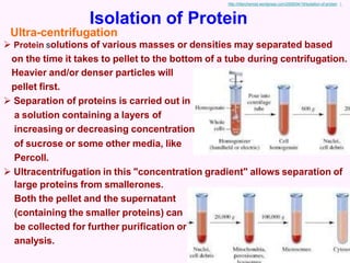
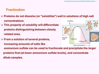
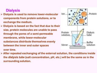
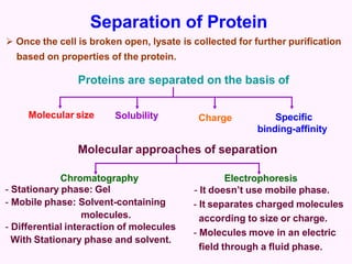
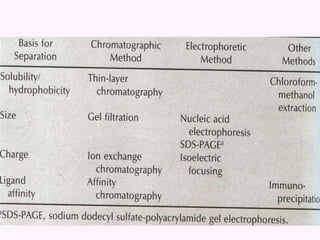
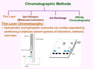
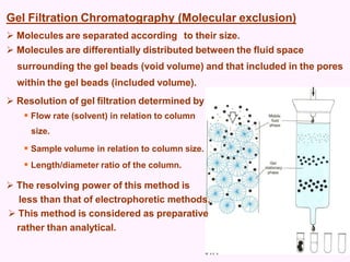
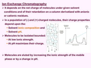
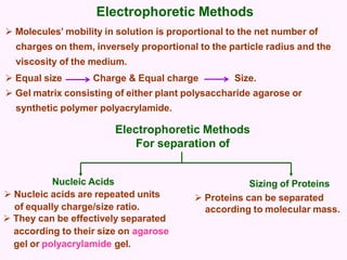
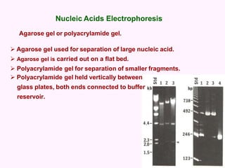
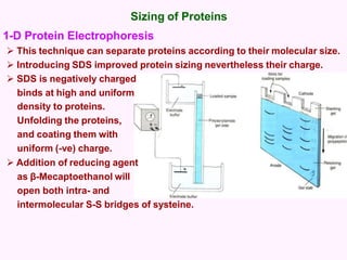
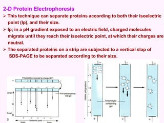

This document discusses several methods for isolating and separating proteins, including: - Ultracentrifugation, which separates proteins based on mass and density over time in a centrifuge. - Dialysis, which removes smaller molecules from a protein solution through a semi-permeable membrane. - Chromatography techniques like gel filtration, ion exchange, and affinity chromatography separate proteins based on size, charge, and binding affinity to separate mixtures. - Electrophoresis techniques like agarose/polyacrylamide gel electrophoresis and 2D electrophoresis separate proteins based on size and isoelectric point to further purify samples.


