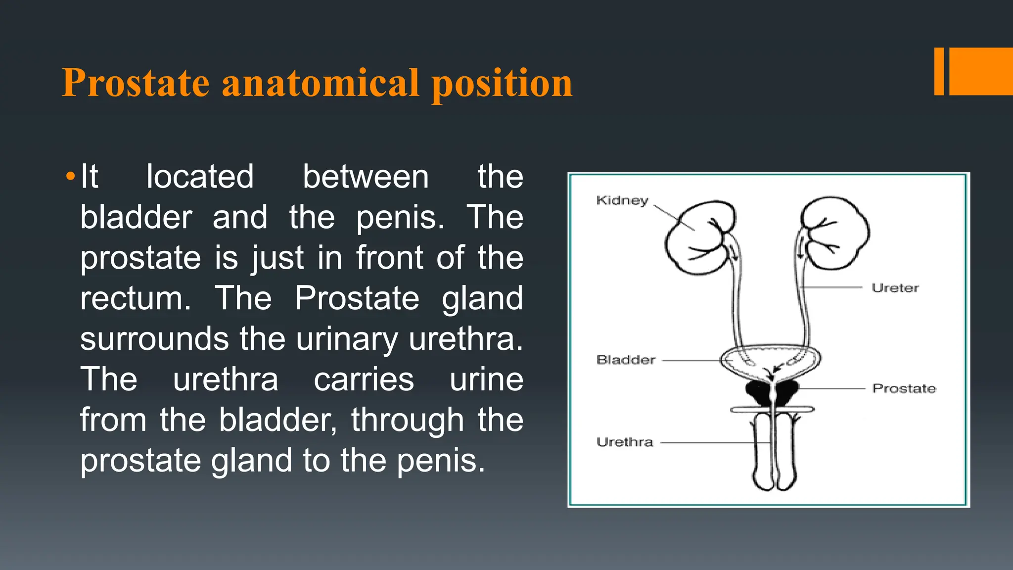
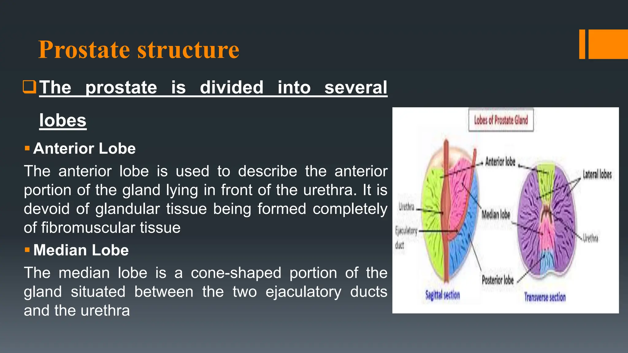
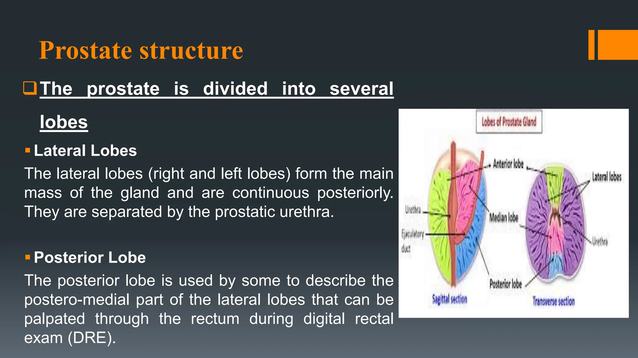
2. The prostate is the largest
accessory gland in the
male reproductive system
The prostate is a
approximately 4 cm wide
and 3 cm thick. The actual
size of the prostate varies
from man to man. It can
range from the size of a
walnut to a small apple.
3. Prostate anatomical position
•It located between the
bladder and the penis. The
prostate is just in front of the
rectum. The Prostate gland
surrounds the urinary urethra.
The urethra carries urine
from the bladder, through the
prostate gland to the penis.
5. Prostate structure
The prostate gland is divided histologically into four
zones
Central zone
is the area that surrounds the ejaculatory ducts. The ducts of
the glands from the central zone are obliquely emptying in
the prostatic urethra, thus being rather immune to urine reflux
Only a very small percentage of prostate cancers begin here
(less than 5%) and are thought to be more aggressive and
more likely to invade the seminal vesicles.
6. Prostate structure
The prostate gland is divided histologically into
four zones
The transition zone
is surrounds the urethra as it enters the prostate gland. It
is small in young adults, but it grows throughout life,
taking up a bigger percentage of the gland, and is
responsible for benign prostatic hyperplasia (BPH, or
normal gland enlargement that occurs with aging but can
cause urinary problems). Roughly 20% of prostate
cancers begin in this zone.
7. Prostate structure
The prostate gland is divided histologically into
four zones
The peripheral zone
It contains the majority of prostatic glandular tissue. The
largest area of the peripheral zone is at the back of the
gland, closest to the rectal wall. When a doctor performs
a digital rectal exam (DRE) it is the back surface of the
gland he is feeling. This is important because about 70-
80% of prostate cancers originate in the peripheral zone
8. Prostate structure
The prostate gland is divided histologically into
four zones
anterior fibromuscular stroma
is a thickened area of tissue that surrounds the apex of
the prostate. It is made of muscle fibers and fibrous
connective tissue. This area of the prostate doesn’t
contain any glands. Prostate cancer is rarely found in this
part of the prostate
9. Prostate structure
The prostate is divided into several
lobes
Anterior Lobe
The anterior lobe is used to describe the anterior
portion of the gland lying in front of the urethra. It is
devoid of glandular tissue being formed completely
of fibromuscular tissue
Median Lobe
The median lobe is a cone-shaped portion of the
gland situated between the two ejaculatory ducts
and the urethra
10. Prostate structure
The prostate is divided into several
lobes
Lateral Lobes
The lateral lobes (right and left lobes) form the main
mass of the gland and are continuous posteriorly.
They are separated by the prostatic urethra.
Posterior Lobe
The posterior lobe is used by some to describe the
postero-medial part of the lateral lobes that can be
palpated through the rectum during digital rectal
exam (DRE).
11. Physiology of prostate
Secretion
The prostate produces a fluid that makes up a large portion of
semen volume, the function of this secretion is nutrition for
sperm as they pass into the female body to fertilize ova.
It secretes proteolytic enzymes into the semen, which act to break
down clotting factors in the ejaculate. This allows the semen to
remain in a fluid state, moving throughout the female
reproductive tract for potential fertilization
prostate secrete alkaline chemicals which neutralize acidic
vaginal secretions to promote the survival of sperm in the female
body.
12. Physiology of prostate
Ejaculation
The vas deferens bring sperm from
the testes to the seminal vesicles.
The seminal vesicles contribute
fluid to semen during ejaculation.
The prostate contains the
ejaculatory duct that releases
sperm during ejaculation. During
orgasm, smooth muscle tissue in
the prostate contracts in order to
push semen through the urethra.
13. Physiology of prostate
Control urine flow
The prostate also plays a part in controlling the
flow of urine. The urethra runs from the bladder,
through the prostate, and out through the penis.
The muscle fibers of the prostate are wrapped
around the urethra. These fibers contract to slow
and stop the flow of urine
14. Prostate Tests
Digital rectal examination
(DRE):
A doctor inserts a lubricated,
gloved finger into the rectum
and feels the prostate. A DRE
can sometimes detect an
enlarged prostate, lumps or
nodules of prostate cancer, or
tenderness from prostatitis.
15. Prostate Tests
Prostate-specific antigen
(PSA):
The prostate makes a protein
called PSA, which can be
measured by a blood test. If
PSA is high than 4, prostate
cancer is more likely, but an
enlarged prostate can also
cause a high PSA.
16. Prostate Tests
Prostate ultrasound (transrectal ultrasound):
An ultrasound probe is inserted into the rectum, bringing it
close to the prostate. Ultrasound is often done with a biopsy
to test for prostate cancer.
Prostate biopsy:
A needle is inserted into the prostate to take tissue out to
check for prostate cancer. This is usually done through the
rectum.
18. Prostate Conditions
Benign prostatic hypertrophy or BPH
BPH, also known as an enlarged prostate, is growth of
the prostate gland to an unhealthy size. A man's
chances of having BPH go up with age:
Age 31-40: one in 12
Age 51-60: about one in two
Over age 80: more than eight in 10
However, only about half of men ever have BPH
symptoms that need treatment. BPH does not lead to
prostate cancer, although both are common in older
men.
19. Prostate Conditions
prostatitis
Prostatitis is an inflammation of the prostate gland,
that is caused by infectious agents.
Unlike most prostate problems, prostatitis --
inflammation or an infection of the prostate -- occurs
more often in young and middle-aged men.
It is the most common urologic problem in men younger
than 50 years of age.
20. Prostate Conditions
prostatitis
Types Of Prostatitis
oAcute bacterial prostatitis:
Is caused by an infection, is least common but easiest to diagnose and treat.
Symptoms come on strong and suddenly. This type of prostatitis can be quite
serious if the infection spreads to the blood or other parts of the body, treated with
antibiotics for about two to four weeks
oChronic bacterial prostatitis:
Is a recurrent infection and inflammation of the prostate and urinary tract.
Symptoms are less severe than those associated with acute bacterial prostatitis.
Chronic bacterial prostatitis is also a common cause of frequent urinary tract
infections in men, treated with alpha-blockers together with antibiotics for at least
four to six weeks and may need pain relieve
21. Prostate Conditions
prostatitis
Types Of Prostatitis
oChronic prostatitis/chronic pelvic pain syndrome:
Most cases of prostatitis fall into this category, however it is the least understood. It can
be further characterized as inflammatory or non-inflammatory, depending upon the
presence or absence of infection-fighting cells in the urine, semen, and prostatic fluid.
Often no specific cause can be identified. The symptoms can come and go or remain
chronically, treated with Anti-inflammatory, pain medications, heat, diet, lifestyle
changes and prostatic massage
oAsymptomatic inflammatory prostatitis:
This form of prostatitis doesn’t have any symptoms. This condition is often diagnosed
incidentally during the work-up for infertility or prostate cancer. Individuals with this form
of prostatitis will have the presence of infection-fighting cells present in semen/prostatic
fluid.
22. Prostate Conditions
prostatitis
signs and symptoms of prostatitis
oPainful, difficult and/or frequent urinating
oBlood in the urine
oGroin pain, rectal pain, abdominal pain and/or low back pain
oFever
oMalaise and body aches
oPainful ejaculation or sexual dysfunction
23. Prostate Conditions
prostatitis
the complications of prostatitis
oacute prostatitis becoming chronic prostatitis
obladder outlet obstruction or urinary retention
oRenal damage
oInfertility due to scarring of the urethra or ejaculatory ducts
ospreading of the infection to the blood stream
odeath.
24. Prostate Conditions
Prostate cancer
Prostate cancer is the most common cancer in men
(besides skin cancer). prostate cancer is usually slow
growing, only about one in 35 men will die of prostate
cancer.
Like BPH, the risk for prostate cancer increases with age.
About six out of every ten men with prostate cancer are
over age 65.
25. Prostate Conditions
Prostate cancer
Early warning signs of prostate cancer
oBurning or pain during urination
oDifficulty urinating, or trouble starting and stopping while urinating
oMore frequent urges to urinate at night
oLoss of bladder control
oDecreased flow or velocity of urine stream
oBlood in urine (hematuria)
oBlood in semen
oDifficulty getting an erection (erectile dysfunction)
oPainful ejaculation
26. Prostate Conditions
Prostate cancer
Risk factors associated with prostate cancer
oAge:
Prostate cancer is rare in men younger than 40, but the chance of having
prostate cancer rises rapidly after age 50. About 6 in 10 cases of prostate
cancer are found in men older than 65
oRace
Prostate cancer occurs more often in African-American men and in Caribbean
men of African ancestry than in men of other races. African-American men are
also more than twice as likely to die of prostate cancer as white men.
27. Prostate Conditions
Prostate cancer
Risk factors associated with prostate cancer
oFamily history
Prostate cancer seems to run in some families, The risk is higher for men who
have a brother with the disease than for those who have a father with it.
oGene changes
Several inherited gene changes seem to raise prostate cancer risk, but they
probably account for only a small percentage of cases overall
28.
29. Prostatectomy
Prostatectomy includes a number of surgical
procedures to remove part or all of the prostate gland
Prostatectomy can be performed in several ways,
depending on the condition involved and recommended
treatment approach
30. Types of prostatectomy
Simple prostatectomy
Simple prostatectomy is recommended for men with
severe urinary symptoms and very enlarged prostate
glands (rather than prostate cancer), and can be
performed open or robotically. This enlargement of the
prostate is called benign prostatic hyperplasia, or BPH.
Simple prostatectomy doesn't remove the entire prostate,
as in a radical prostatectomy, but instead removes just
the obstructive part of the prostate that's blocking the
flow of urine.
31. Types of prostatectomy
Radical prostatectomy
Radical prostatectomy is surgery
to remove the entire prostate gland
and surrounding lymph nodes as
treatment for men with localized
prostate cancer. A surgeon can
perform a radical prostatectomy
using different techniques.
32. Techniques of radical prostatectomy
Open prostatectomy
In this traditional method of radical prostatectomy,
the surgeon makes a vertical 8- to 10-inch incision
below the belly button. Radical prostatectomy is
performed through this incision. In rare cases, the
incision is made in the perineum, the space
between the scrotum and anus.
Laparoscopic prostatectomy
In laparoscopic prostatectomy, surgeons make
several small incisions across the belly. Surgical
tools and a camera are inserted through the
incisions, and radical prostatectomy is performed
from outside the body. The surgeon views the entire
operation on a video screen.
33. Techniques of radical prostatectomy
Robot-assisted laparoscopic
prostatectomy
Small incisions are made in the
belly, as in regular laparoscopic
prostatectomy. A surgeon controls
an advanced robotic system of
surgical tools from outside the
body. A high-tech interface lets
the surgeon use natural wrist
movements and a 3-D screen
during radical prostatectomy.
34. Complications of radical prostatectomy
Urinary incontinence:
There are two sphincter control urination process one at the point
where the bladder and urethra join known as the bladder neck
sphincter, which is closed most of the time to prevent urine leaking out
but when it gets permission from the brain, it opens to allow urine to be
passed. Another sphincter that is part of the set of muscles below the
prostate gland called the pelvic floor muscles is also involved in
bladder control. If the bladder neck sphincter is damaged during
radical prostatectomy, the group of muscles called pelvic floor muscles
can assist in the control of the passage of urine. If the pelvic floor is
weak you may experience urinary incontinence.
36. Complications of radical prostatectomy
Erectile dysfunction (ED):
Erectile dysfunction is reported to occur in between 30-70% of post
prostatectomy patients. Less than 10% of patients return to their pre-operative
erectile capacity following prostate surgery. The younger the man, the higher the
chance of maintaining potency after prostatectomy.
there are two bundles of nerves, responsible for controlling erections and blood
flow to the penis according to the Urology Care Foundation (UCF). Due to their
proximity to the prostate gland, there is the potential for damage to occur during
the procedure, thus hindering a patient’s ability to maintain an erection. If a man’s
ability to do so is fully intact prior to the surgery, a nerve-sparing approach is
followed and the surgeon will do his best to avoid damaging the nerves. In some
cases, if the cancer has spread very close to these nerves, they may need to be
removed altogether.
37. Complications of radical prostatectomy
Sterility :
RP cuts the connection between the testicles and the urethra and causes
retrograde ejaculation. This results in a man being unable to provide sperm for a
biological child. A man may be able to have an orgasm, but there will be no
ejaculate. In other words, the orgasm is "dry."
Lymphedema:
Although this complication is rare, if lymph nodes are removed during
prostatectomy, fluid may accumulate in the legs or genital region over time. Pain
and swelling result. Physical therapy is usually helpful in treating the effects of
lymphedema.
38. Complications of radical prostatectomy
Other complications of radical prostatectomy include:
Bleeding after the operation
Infection
Narrowing of the urethra, blocking urine flow
Injury to the rectum (rare)
39. Types of urinary incontinence
Stress incontinence:
which occurs when urine leaks from the bladder when you cough, laugh,
sneeze or do any other activity that places stress on the abdomen, Stress
incontinence occurs when the urethral sphincter, the pelvic floor muscles,
or both these structures have been weakened or damaged and cannot
dependably hold in urine.
As a woman gets older, the muscles in her pelvic floor and urethra
weaken, and it takes less pressure for the urethra to open and allow
leakage. In men, the most frequent cause of stress incontinence is
urinary sphincter damage sustained through prostate surgery or a pelvic
fracture
40. Types of urinary incontinence
Urge incontinence:
With this type, you have an urgent need to go to the bathroom and
may not get there in time. Urgency is caused when the bladder
muscle, the detrusor, begins to contract and signals a need to
urinate, even when the bladder is not full . Conditions such
as multiple sclerosis, Parkinson's disease, diabetes, and stroke can
affect nerves, leading to urge incontinence
Overflow incontinence:
Overflow incontinence occurs when something blocks urine from
flowing normally out of the bladder, as in the case of prostate
enlargement that partially closes off the urethra.
41. Types of urinary incontinence
Reflex incontinence:
Reflex incontinence occurs when the bladder muscle contracts and
urine leaks (often in large amounts) without any warning or urge. This
can happen as a result of damage to the nerves that normally warn the
brain that the bladder is filling. Reflex incontinence usually appears in
people with serious neurological impairment from multiple sclerosis,
spinal cord injury, other injuries, or damage from surgery or radiation
treatment
43. Physical therapy interventions for urinary incontinence
Pelvic Muscle Re-Training
Electrical stimulation
Electrical Biofeedback
Bladder Training
Behavioral change
44. Physical therapy interventions for urinary incontinence
Pelvic Muscle Re-Training
Pelvic floor exercises, also called Kegel exercises and pelvic muscle
exercises, are performed to strengthen the voluntary muscles that
contribute to the closing force of the urethra and to the support of the
pelvic organs. Pelvic floor exercises are performed to control urination
or defecation with minimal contraction of abdominal, buttock, or inner
thigh muscles.
Most men find it difficult to remember to do the exercises. You can do
the exercises every 2 hours during the day or just before a meal, e.g.
before breakfast, morning tea, lunch, afternoon tea, dinner and supper
45. Physical therapy interventions for urinary incontinence
Pelvic Muscle Re-Training
CONTRACT-RELAX TECHNIQUE:
Instruct to tighten the pelvic floor muscles as if attempting to stop urine flow or hold back gas. Hold
for 3to 5 seconds and relax for at least the same length of time. Repeat up to 10 times. Watch the
patient to encourage normal breathing pattern.
QUICK CONTRACTIONS:
Repeat contractions of pelvic floor muscles while maintaining a normal breathing rate and keeping
accessory muscles relaxed. Try for 15 to 20 repetitions per set .
ELEVATOR EXERCISES:
Instruct the patient to imagine riding in an elevator. As the elevator goes up from one floor to next,
contracts the pelvic floor muscles a little more. As strength and awareness improves, add more
floors to the sequence of the contraction. Instruct the patient to relax the muscles as if descending
one floor at a time.
47. Physical therapy interventions for urinary incontinence
Pelvic Muscle Re-Training
Effective especially in activities that increase bladder pressure
from change of position (sitting to standing, etc)
4 to 6 weeks before surgery is a good time to practice pelvic floor
exercises. The advantage of training before surgery is that you
learn how to control your muscles when you are pain –free and
sensation is normal. These exercises will prepare you in advance
to begin using the muscles immediately after catheter removal Try
doing them in different circumstances such as while walking,
sitting, standing and walking
48. Physical therapy interventions for urinary incontinence
Pelvic Muscle Re-Training
After surgery, while the catheter is still in place, doing pelvic floor
exercises can irritate the catheter and cause discomfort. Don’t do
too many deliberate pelvic floor contractions. Once the catheter is
out start Pelvic Floor Muscles Exercises straight away as doing
the exercises gets easier, do them in different positions: from
sitting to standing and on to walking.
Exercises should be performed until muscle fatigue occurs.
49. Physical therapy interventions for urinary incontinence
Electrical stimulation
Brief doses of electrical stimulation can strengthen muscles
in the lower pelvis in a way similar to exercising the
muscles. Electrodes are temporarily placed in the vagina or
rectum to stimulate nearby muscles. This can stabilize
overactive muscles and stimulate contraction of urethral
muscles.
50. Physical therapy interventions for urinary incontinence
Electrical Biofeedback Application Technique
Patient will be asked to evacuate their bladder before starting the
treatment sessions to ensure that patients relaxed and comfortable
during the session.
Patient will be placed on a plinth in fowler lying position with
exposed the premium area.
The perineum area will be shaved and the perineum skin will be
cleaned with alcohol before the placement of electrodes to reduce
skin impedance.
The earth electrode will connected to the thigh after soaked in
normal saline solution.
51. Physical therapy interventions for urinary incontinence
Electrical Biofeedback Application Technique
Positive electrode (red) will be base of the penis, and the site
of negative electrode (black) will be at the bulk of pelvic-floor
muscles.
The program of EMG bio feedback will be selected.
The patient will be instructed to make forceful perineal
contraction and then the EMG activities during maximal
contraction will be record and the device will give a visual and
auditory feedback for the patient
53. Physical therapy interventions for urinary incontinence
Bladder Training
Bladder training is an important form of behavior therapy that can
be effective in treating urinary incontinence.
The goals are to increase the amount of time between emptying
your bladder and the amount of fluids your bladder can hold. It
also can diminish leakage and the sense of urgency associated
with the problem.
Bladder training requires following a fixed voiding schedule,
whether or not you feel the urge to urinate.
54. Physical therapy interventions for urinary incontinence
Bladder Training
Bladder Retraining Instructions
oEmpty your bladder as soon as you get up in the morning. This act starts your
retraining schedule.
oGo to the bathroom at the specific times you and your health care provider
have discussed. Wait until your next scheduled time before you urinate again.
Be sure to empty your bladder even if you feel no urge to urinate. Follow the
schedule during waking hours only. At night, go to the bathroom only if you
awaken and find it necessary.
oWhen you feel the urge to urinate before the next designated time, use "urge
suppression" techniques or try relaxation techniques like deep breathing.
Focus on relaxing all other muscles. If possible, sit down until the sensation
passes.
55. Physical therapy interventions for urinary incontinence
Bladder Training
Bladder Retraining Instructions
oIf the urge is suppressed, adhere to the schedule. If you cannot suppress
the urge, wait five minutes then slowly make your way to the bathroom.
After urinating, re-establish the schedule. Repeat this process every time
an urge is felt.
oWhen you have accomplished your initial goal, gradually increase the
time between emptying your bladder by 15-minute intervals. Try to
increase your interval each week. Increase the time between each
urination until you reach a three- to four-hour voiding interval.
56. Physical therapy interventions for urinary incontinence
Bladder Training
Bladder Retraining Instructions
oIt should take between six to 12 weeks to accomplish your ultimate goal.
Don't be discouraged by setbacks. You may find you have good days and
bad days. As you continue bladder retraining, you will start to notice more
and more good days, so keep practicing.
oYou will hasten your success by doing your pelvic muscles
exercises faithfully every day..
57. Physical therapy interventions for urinary incontinence
BEHAVIORAL CHANGE
Decreasing any excessive fluid intake (you should not decrease your
fluid intake if you drink normal amounts of fluid).
Changing physical activities to avoid jumping or running movements,
which can cause more urine leakage.
Regulating bowel movements with dietary fiber or laxatives to avoid
constipation (which can worsen incontinence).
Quitting smoking to reduce coughing and bladder irritation (and your risk
of bladder cancer).
58. Physical therapy interventions for urinary incontinence
BEHAVIORAL CHANGE
Avoiding alcohol and caffeine, which can overstimulate the
bladder.
Losing weight if you are overweight.
Avoiding food and drinks that irritate the bladder, such as spicy
foods.
Keeping blood sugar under control if you have diabetes.