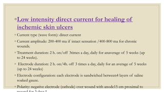
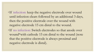
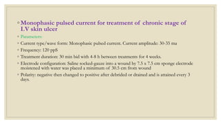
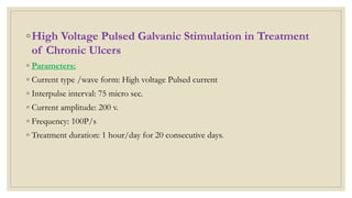
MANAGEMENT OF ULCERS
FOR MSC &POSTGRADUATE STUDENTS
SUPERVISED BY DR. MOSTAFA AHMED
Lecturer of physiotherapy at deraya
university .
◦Function
◦ At the very premise of gaining a thorough understanding of
wounds is acquiring solid foundational knowledge in skin
anatomy, physiology, and the biological cascade of normal
wound healing.
◦ Skin layers:
◦ Epidermis (superficial to deep): stratum corneum, stratum lucidum,
stratum granulosum, stratum spinosum, stratum basalis
◦ Primary cell: keratinocyte
◦ Dermis: 2 layers - papillary (superficial) and reticular (deep) dermis
◦ It contains hair follicles, sebaceous glands, sweat glands, blood
vessels, and nerve fibers.
◦ Skin offers protection from UV light and serves as a mechanical, chemical,
and thermal barrier. Moreover, the skin is involved in the Vitamin D
synthesis process.
◦
The series of events associated with wound healing instantaneously begins at the
moment of injury. Each of the potential underlying causes must be addressed and
optimized for the wound to progress successfully through a complex biological
healing procession of hemostasis, inflammation, proliferation, and remodeling. All
four highly integrated and overlapping physiological phases must occur in the
proper sequence and time frame for wound healing success.
◦
◦ Hemostasis begins shortly after wound infliction.
◦ Platelets are the first cells to arrive at the injury site and are
critical in clot formation to establish hemostasis.
◦ Platelets release platelet-derived growth factor (PDGF) and
transform growth factor-beta (TGF-B) to stimulate chemotaxis
and proliferation of inflammatory cells
◦ (Hemostasis is sometimes included within the inflammatory
phase, depending on the source)
◦ Inflammation is initiated at the moment of tissue disruption
(Days 1 to 6)
◦ Polymorphonuclear cells (PMNs) first appear during this phase, with a primary role
of clearing devitalized tissue, blood clot, foreign material, and bacteria from the
wound
◦ Macrophages appear within 48 hours and complete the clean-up activities.
Macrophages are the most critical cell in wound healing. They secrete cytokines
and growth factors to induce activation and recruitment of additional macrophages
while stimulating angiogenesis, the proliferation of fibroblasts, and extracellular
matrix (ECM) production, thus prompting the proliferative phase
◦ The defining role of the fibroproliferation/proliferative phase is ECM
production
(Day 4 - Week 3)
◦ While the inflammatory phase is most active, proliferation lays the
foundation of a transitional ECM to stabilize the wound edges and provide a
healing framework.
◦ Fibroblasts replace macrophages to generate new tissue and stimulate
collagen production for enhanced structural integrity.
◦ Tensile strength begins to increase on days 4 to 5
◦ Angiogenesis occurs simultaneously
◦
◦ Maturation/remodeling is characterized by continued synthesis and degradation of the ECM
components in efforts to establish a new equilibrium and organized scar formation (Week 3 -
Years)
◦ As the scar matures, late remodeling occurs
◦ Type I collagen replaces type III collagen
◦ Peak tensile strength at 60 days is approximately 80% of pre-injury strength.
◦ The scar eventually contracts and thins out
Clinicians perform wound assessments to determine the appropriate treatment for extremely
diverse disease processes.
◦ How do professionals then approach wound assessment when the causes are so diverse?
Below are some basic questions for the interprofessional team during initial and subsequent
wound assessments to best classify and treat a wound to optimize outcomes.
◦ The initial assessment is crucial. It should begin with the following:
◦ How: How was the wound created? If chronic, why is it still open? What is the underlying
etiology
◦ Where: Where on the body is it located? Is it in an area that is difficult to offload or to keep
clean? Is it in a region of high skin tension? Is it near any vital structures such as a major
artery?
◦ When: How long has this wound been present? (e.g., acute or chronic)
◦ What: What anatomy does it involve (e.g., epidermis, dermis, subcutaneous tissue, fascia,
muscle, tendon, bone, arteries, nerves)?
◦ What: What comorbidities or social factors does the patient have that might negatively
impact their healing ability?
◦ Is it life-threatening?
◦Issues of Concern
◦ While some wounds are simple, the majority of encountered wounds are caused by or become
complicated by confounding patient-related issues.
◦Examples
◦ A chronic wound will have a different structure and percentage of components than an acute
wound, requiring conversion for healing.
◦ An underlying infection will impede wound healing even if the infection is subacute.
◦ Damaged or constricted arterial supply will prevent appropriate blood flow to the wound.
◦ Impaired venous drainage may cause venous stasis.
◦ Physical pressure on chronic ulceration will cause repeated damage, preventing healing.
◦Universal Principles of Management
◦ Thorough evaluation
◦ Obtain source control
◦ Eliminate confounding/contributing factors
◦ Optimize the wound’s healing ability/environment
◦ Close the wound
◦
Considering this rational algorithm, in total, is the core of successful wound healing.
Similarly, there is no one-to-one correlation between a given wound and specific
treatment. A spectrum of options must be considered for every wound encountered;
selecting the best option for each patient remains challenging.
◦Clinical Significance
◦ Before determining the underlying cause, it is essential to establish what type of wound the
patient has. These subclassifications can be acute or chronic.
ACUTE
WOUND
CHRONIC
WOUND
Typesof Wounds
◦Types of Wounds
◦ Acute
◦ Clinicians assess acute wounds by the method of injury and resultant damage to soft
tissues and bony structures.
◦ For example, in crush or high impact injuries, there is an area of demarcation which
is not fully recognized until sometimes as much as a week or two later. For this
reason, it is important to determine the method of injury and to keep in mind that the
wound seen is not necessarily the entirety of the wound which will be present in a
week. In these cases, the patient and their family should be educated on this
progression to prevent frustration and misunderstanding.
◦ For all acute types of wounds, it is important to determine the length of time since injury
(days or hours), the involvement of neurovascular supply, muscle, tendon, ligament, and
bony structures, and the likelihood of contaminants in the wound.
◦ Also important is when the patient had their last tetanus vaccine/booster dose.
Clinicians should start antibiotics if the wound is severely contaminated or if it has
been longer than 3 hours since the injury. All underlying tissue should be repaired, and
the wound should be irrigated to remove contaminants and bacteria.
◦ In cases of traumatic/open fracture, the most used classification system is Gustillo-
Anderson:
◦ Type 1: Clean wound, low energy puncture wound of less than 1 cm size, minimal
contamination, minimal soft tissue damage, adequate soft tissue coverage of bone, without
periosteal stripping, minimal fracture comminution.
◦ Type 2: Moderate soft tissue damage and crushing, moderate contamination, laceration of
greater than 1cm in size, adequate soft tissue coverage of bone, without periosteal stripping,
minimal fracture comminution.
◦ Type 3A: High-energy open injury, massively contaminated wound, extensive/significant soft
tissue damage and crushing, adequate soft tissue coverage of bone, severely comminuted
and/or segmental fracture, periosteal stripping is present.
◦ Type 3B: Significantly contaminated wound, extensive soft tissue damage, unable to cover
bone with soft tissue (requiring graft), severely comminuted and/or segmental fracture,
periosteal stripping, and bone exposure.
◦ Type 3C: Similar to type A or B, however, associated with arterial injuries requiring repair.
◦Chronic
◦ If a wound becomes arrested during normal stages of inflammation and healing, this
classifies a chronic wound; deemed such at an untimely three months of non-progression.
They often stall in the inflammatory phase . Numerous factors and disease conditions
impair the wound healing process, resulting in chronic, non-healing wounds.
◦ The most important concept is a persistent pro-inflammatory condition that propagates an
unstable wound environment, recalcitrant to healing. Persistent hyper-inflammation is the
ubiquitous pathophysiological characteristic of chronic wounds, and macrophage
malfunction significantly contributes to the altered normal course.
◦ In the chronic setting, the main goal is to identify why the wound is not healing and
alleviate these obstacles. Chronic wound causalities can be categorized as follows, and the
related questions should be responded to accordingly.
◦ Arterial
◦ (Atherosclerosis, Raynaud Disease, Scleroderma)Is there enough blood flow?
◦ Generally, an ankle-brachial index (ABI) of less than 0.9 is diagnostic of peripheral arterial
disease (PAD) and limits blood supply to the tissues while impairing antibiotic delivery to
infected wounds.
◦ Venous
◦ Pressure-induced changes in blood vessel wall permeability lead to leakage of fibrin and other
plasma components into the perivascular space
◦ Accumulation of fibrin has direct and negative effects on wound healing as it down-regulates
collagen synthesis
◦
◦ Infection
◦ Underlying infectious processes, including cellulitis and osteomyelitis, will inhibit wound
healing
◦ Culturing for aerobic, anaerobic, and fungal pathogens is recommended
◦ Pressure
◦ Increased pressure to the area of concern will destroy new tissue growth and prevent proper
perfusion of blood to the wound site.
◦ These areas need to be offloaded to avoid undue pressure
◦ Radiation Therapy
◦ Causes stasis/occlusion of small vessels and damages fibroblasts and nuclei.
◦ Oncologic
◦ Always biopsy areas of concern in non-healing wounds, as this can be an atypical presentation
in various types of malignancies.
◦ Age and Hormones
◦ It is commonly recognized that, in healthy older adults, the effect of aging causes a temporal
delay in wound healing but not an actual impairment in terms of healing quality
◦ A review of age-related changes in healing capacity demonstrated that every phase of healing
undergoes characteristic age-related changes. Compared with females, aged males have been
shown to exhibit delayed healing of acute wounds. Studies indicate that estrogen can improve
the age-related impairment in healing in both men and women, while androgens negatively
regulate cutaneous wound healing.
◦ Systemic
◦ Multiple systemic diseases inhibit wound healing, with diabetes being the most common
culprit It has been determined that uncontrolled blood glucose levels suppress the body's
normal inflammatory response while inducing microvascular disease, which limits healing.
◦ Obesity is also a significant risk factor that impairs wound healing, perhaps secondary to
relative hypoperfusion and ischemia of subcutaneous adipose tissues.
◦ Immunodeficiencies/immunosuppression and renal failure are also implicated .
◦ Nutrition
◦ Although serum albumin is not a good predictor of wound healing, there is some evidence that
protein malnutrition, as well as insufficient levels of certain vitamins and minerals, will
hinder the body's ability to heal chronic wounds
◦ High glucose levels interfere with nutrient absorption, causing depletion of several nutrients
◦ Protein and energy requirements of chronic wound patients may rise by 250% and 50%,
respectively
◦ Pharmacological
◦ Hydroxyurea has been reported in multiple instances to cause non-healing ulcerations
◦ Chemotherapeutic drugs delay cell migration, decrease wound matrix formation, lower
collagen production, impair fibroblast proliferation, and inhibit wound contraction;
furthermore, they weaken immune functions, thereby impeding the natural inflammatory
phase of healing, and increasing the risk of infection
◦ Self-inflicted/Psychosocial
◦ There are instances where a patient causes the ulceration, either on purpose or as a result of
non-compliance.
◦ This is often the hardest factor to spot and overcome but must always be a consideration
◦ In addition, psychological stress impairs cell-mediated immunity, compromising normal
healing processes
◦ Genetic
◦ Predisposition to hypertrophic or keloid scarring, hereditary conditions, and various skin types
(pigmentation, elasticity, thickness, sebaceous quality, and location)
◦ Smoking
◦ Cigarette smoke contains more than 4000 constituents
◦ Nicotine constricts blood vessels and increases platelet adhesiveness, carbon monoxide binds
to hemoglobin and reduces oxygen delivery, and hydrogen cyanide inhibits oxygen transport
◦ Necrotizing Soft Tissue Infections
◦ Necrotizing soft tissue infections (NSTIs)can induce and instigate widespread necrosis
subsequent to their aggressive nature. Despite increased understanding of their disease
processes, NSTI's rarity, complexity, and occasional ambiguity of symptomatology continue to
pose diagnostic dilemmas.
◦ A clear approach allows rapid identification, early administration of antimicrobial agents, and
immediate surgical intervention, as they are limb- and life-threatening. Failure to recognize the
underlying disease process and intervene expeditiously results in extremely high mortality.
◦ Categorized based on the layer(s) of soft tissue association, these infections include skin
level necrotizing cellulitis, necrotizing fasciitis within the superficial or deep soft tissue, and
myonecrosis/necrotizing myositis indicating muscle involvement. Establishing the diagnosis
of these infections is often the main challenge. This differentiation is critical as necrotizing
infections warrant prompt, aggressive surgical debridement.
◦ It should be of no surprise after reading this review that susceptible patients at most significant
risk for these types of infections include elderly adults, patients with obesity,
immunosuppressed individuals, patients with renal failure or diabetes, those who suffer from
peripheral vascular disease, or patients with a combination of these factors.
◦ When the "hard signs" of bullae, crepitus, gas/emphysema on X-ray, hypotension with systolic
blood pressure less than 90 mmHg, or skin necrosis are present, establishing the diagnosis of
NSTI is not challenging. However, these indicators are often absent on presentation, thus
potentially delaying diagnosis and surgical intervention. Most commonly, the diagnosis is
clinical.
skin necrosis
hypotension emphysema
◦
Patients may clinically present or progress to develop sepsis or septic shock without obvious
causation. The extremities, perineum, trunk, and torso are commonly implicated. A detailed
examination should be undertaken for expression of turbid, grayish, semi-purulent material
("dishwater fluid") from an identifiable entry site, in addition to assessing the presence of skin
changes, blebs, or crepitus. At the site of infection, patients often develop pain out of proportion to
observed physical manifestations. Any of these findings mandate immediate surgical intervention,
which should consist of exposure and direct visualization of potentially infected tissue (including
deep soft tissue, fascia, and underlying muscle) and radical resection of affected areas.
◦
Patients may clinically present or progress to develop sepsis or septic shock without
obvious causation. The extremities, perineum, trunk, and torso are commonly
implicated. A detailed examination should be undertaken for expression of turbid,
grayish, semi-purulent material ("dishwater fluid") from an identifiable entry site, in
addition to assessing the presence of skin changes, blebs, or crepitus. At the site of
infection, patients often develop pain out of proportion to observed physical
manifestations. Any of these findings mandate immediate surgical intervention, which
should consist of exposure and direct visualization of potentially infected tissue
(including deep soft tissue, fascia, and underlying muscle) and radical resection of
affected areas.
◦ The Laboratory Risk Indicator for Necrotizing Fasciitis (LRINEC) score is also helpful as a
laboratory aid in distinguishing necrotizing from non-necrotizing skin and soft tissue infections
(SSTIs). Wong et al. originally published a score of >/= 6, yielding a positive predictive value
of 92% and a negative predictive value of 96%. Developed in 2004,
◦ LRINEC displayed promise for predicting the severity of skin and soft tissue infections.
◦ NSTIs necessitate prompt surgical debridement for satisfactory treatment following the
primary initiation of antimicrobial therapy. Because of the rapidly progressive and
potentially fatal outcome of this condition, if imaging cannot be performed expeditiously,
delaying treatment is not justified. Radiologic studies should be considered only in patients
in whom the diagnosis is not seriously considered. Plain film findings may
demonstrate extensive soft tissue gas
◦ CT examination can reveal asymmetric thickening of deep fascia in association with gas, or
the presence of abscesses
◦ MR imaging effectively differentiates between necrotizing and non-necrotizing infections;
although it can be overly sensitive as well as time-consuming, thus should not delay prompt
surgical intervention in the management of NSTIs.
◦ Classified into two types, NSTIs are either
◦ polymicrobial (type I) or monomicrobial (type II)
◦ dependent upon the offending organism(s). Antimicrobial therapy is an essential
component of their management. As in all serious life-threatening infections, it is important
to initiate early, empiric antimicrobial therapy. Prompt and appropriate treatment improves
outcomes for SSTIs, and furthermore, it is well established that aggressive and timely
antibiotic therapy reduces mortality.
◦ Polymicrobial infections are more common, occur secondary to a combination of aerobic
and anaerobic organisms, and primarily involve the trunk and perineum.
◦ antimicrobial agents directed against gram-positive and gram-negative aerobes and anaerobes
should be administered. Due to its inherent anti-toxin properties, Clindamycin is
recommended as an addition to any empiric regimen.
◦ Any chosen antibiotic regimen can be later refined based on culture and sensitivity,
particularly in the case of monomicrobial (type II) infections.
◦ The aforementioned findings constitute a surgical emergency, the mainstay of which remains
wide debridement of necrotic tissue until bleeding, viable tissue is reached. A grey serous fluid
at the level of the necrotic tissue is usually noted, and as the infection spreads, thrombosed
blood vessels can be visualized along the involved tissue planes.
◦ Typically, patients require serial trips to the operating room for wide debridement until
adequate infectious source control is obtained.
◦
In summary, successful treatment requires early initiation of broad-spectrum systemic
antibiotic therapy, aggressive surgical debridement of all necrotic tissue, and supportive care
(fluid resuscitation, organ, and critical care support) to maintain oxygenation and tissue
perfusion. Delay of definitive debridement is the most critical risk factor for death. Early
operative debridement is a significant determinant of success in NSTI treatment and an
independent predictor of improved outcomes
broad-spectrum
systemic antibiotic
therapy
surgical
debridement
supportive
care
◦ Other Issues
◦ A wound ensues following a breach in epithelial integrity and may be accompanied by disruption of
structure and/or function of underlying normal tissue. To restore the structure of the injured tissue, a
complex signaling network of biomolecular interactions must occur in a precisely programmed fashion.
Along each individual wound’s course of healing, repeated thorough assessments are necessary. Once a
wound has been fully evaluated, treatment should be designed to address any modifiable cause for the
wound and then to achieve specific targeted goals of
◦ (a) preventing complications resulting from the wound.
◦ (b) preserving or restoring critical functions.
◦ (c) achieving wound closure.
◦ (d) restoration of aesthetics.
◦ Enhancing Healthcare Team Outcomes
◦ The current review provides a basic understanding of the common types of wounds and the
underlying concerns. The takeaway is the need for appropriate assessment. Too often,
wounds are not treated properly because of a lack of understanding of the underlying
disease process. Most chronic wounds are complex and best managed by an
interprofessional team coordinating care that includes a wound care nurse, general and/or
vascular surgeon, podiatrist, plastic surgeon, hyperbaric specialist, infectious disease
consultant, dietitian, and physical therapist. The key is first to find out the cause of wound
breakdown. Without resolving the primary cause, wounds cannot heal
◦ There are hundreds of wound care dressings and solutions, and for the most part, all work
similarly with the same efficacy. The second point is to ensure that the wound is clean, has
ample blood supply, and is regularly debrided. Other factors that play a role in wound healing
include patient nutritional status, comorbidities, state of the immune system, age, degree of
ambulation, presence of a foreign body, and infection. It is important to have a team of wound
experts regularly assess the wound and the patient to achieve successful healing.
◦Nursing, Allied Health, and Interprofessional Team
Interventions
◦Evaluation
◦ Once the underlying issues and healing impediments are determined, a formal wound assessment is
performed. The evaluation of patients with complex wounds is best approached systematically since
wounds are rarely secondary to only one sole cause. Assessment of both local and systemic
contributing factors within each portion of the work-up is critical. Generally, ongoing nursing and
clinician assessments and monitoring of wounds are similar. Physical examination should be the
primary criterion for the diagnosis of local wound infection:
◦ Identify the wound location.
◦ Determine the cause of the wound:
◦ Evaluate for foreign bodies or neoplastic processes.
◦ Consider bacterial colonization versus acute/chronic infection.
◦ Note that all open wounds will have bacterial colonization, yet this in itself does not constitute
an infected wound.
◦ Diagnosis of infection is designated in the presence of erythema, pain, swelling, fluctuance,
purulence, and loss of function
◦Determine the stage of the wound:
◦ Stage I: Superficial, involving only the epidermal layer
◦ Intact skin with non-blanchable erythema
◦ Stage II: Partial thickness affects the epidermis and may extend into the dermis
◦ Stage III: Full thickness, extends through the dermis and into the adipose tissues
◦ Stage IV: Full thickness, extends through the dermis and adipose, exposing muscle, bone,
fascia, or tendon
◦ Unstageable: Full-thickness skin and tissue loss obscured by slough or eschar; thus, the true
extent of tissue damage cannot be confirmed
◦ If slough or eschar are removed, a stage III or IV wound results
◦ Evaluate and measure the depth, length, and width of the
wound
◦ Granulation tissue: beefy red bleeding tissue that indicates healing in progress
◦ Fibrinous tissue: white-ish/yellow slough that does not bleed (not healthy)
◦ Eschar: roof or rim of black/charred-appearing callous-like tissue (not healthy)
◦ Measure the amount of undermining and tunneling.
◦ Evaluate the wound bed for exposed bone, vessels, hardware, or subcutaneous fat.
◦ Survey for presence, type, and amount of exudate:
◦ Serous, serosanguineous, sanguineous, or purulent.
◦ Minimal, light, moderate, or heavy.
◦ Assess surrounding skin tissue for signs of injury (actinic changes), previous irradiation,
arterial or venous insufficiency, lymphedema, loss of sensation, and dermal thinning (i.e.,
aging, steroid therapy).
◦ Check wound margins for tunneling, rolled edges, undermining, and fibrotic changes.
◦ Evaluate for warmth, pain, odor, purulence, delayed healing, or other signs of infection
◦ Observe pain..
◦ For all wounds on extremities, a careful neurovascular examination of the entire limb is
mandatory.
◦ Document findings and report unexpected results to the healthcare team.
◦ Diagnostics
◦ Surface swabs indicating the presence of pathogenic bacteria do not correlate with clinically
significant infection. For local wound evaluation, wound swabs can be valuable for
surveillance of the flora colonizing a wound but should not be used as a trigger to initiate
therapy for wound infection.
◦ Wound biopsies can be invaluable for diagnosing invasive burn wounds; the presence of
bacteria in the deep dermis on biopsy correlates with the risk of system sepsis in burn patients.
◦ Bone biopsy demonstrating bacteria within the bone is the gold standard for diagnosing
osteomyelitis.
◦ Laboratory studies can be a useful adjunct in managing chronic wounds; however, they are
often misused, and an evidence-based, rationalized approach is necessary to utilize this
expensive resource efficiently. Routine blood work is not required. Serum prealbumin may be
valuable in determining nutritional status. A significantly elevated erythrocyte sedimentation
rate (ESR) and C-reactive protein (CRP) can help confirm the diagnosis of osteomyelitis or be
used in monitoring response to treatment.
◦ Ultrasound, computed tomography (CT), or magnetic resonance imaging (MRI) may be
useful for delineating fluid collections, necrotic tissue, or inflammation in select
circumstances.
◦ Assessment of peripheral perfusion via ankle/brachial index and toe brachial index
(ABI/TBI) should be performed as it guides further management strategies. ABI does not
provide reliable results in patients with non-compressible vessels, such as patients with
diabetes or renal insufficiency, so TBI is also needed.
◦ Utilize monofilament examinations to assess the presence of peripheral neuropathy and
pressure sensation.
◦Prevention
◦ The preventative measures that should be taken for patients with open wounds depend on the
setting.
◦ Tetanus prophylaxis should be considered for acute lacerations
◦ Strict adherence to pressure-relief protocol, turning bed-bound patients every 2 hours,
offloading pressure points, floating heels, optimization of wheelchair seating/bedding, social
support and wound care, and assessment of nutritional status take priority in pressure sores
◦ Prophylactic antibiotics are necessitated in human/animal bites
◦ Any associated medical conditions must be aggressively optimized
◦ Preserving joint motion must be considered for patients with open wounds of the extremities:
◦ Institute aggressive physiotherapy regimens to maintain or improve joint motion
◦ Utilize splinting to minimize joint contractures
◦Interventions
◦ After careful adherence to preventative measures and preservation of critical function, a
strategy for wound closure can be formulated with respect to multidisciplinary
decision-making.
◦ The basic tenet of non-surgical therapy is debridement of dirty wounds and closure of clean
wounds, first focusing on eliminating necrotic debris while controlling any associated
infectious burden.
◦ The utilization of wound dressings comprises the mainstay of non-surgical therapy, with a
fundamental principle of employing debriding dressings for dirty wounds and occlusive
dressings for clean wounds.
◦
◦ In clean wounds lacking necrotic debris with acceptable bacterial load(s), a dressing
that maintains a moist environment should be utilized to encourage healing. This
technique should not be employed for infected or dirty wounds.
◦ Occasionally, circumstances necessitate antibiotic dressings. Mupirocin ointment has the
added benefit of treating methicillin-resistant staph aureus (MRSA). Burn wounds are
commonly dressed with silver sulfadiazine or mafenide acetate due to their high
antimicrobial activity. The low toxicity and exceptional antimicrobial properties of
elemental silver have led to the development and widespread use of silver-impregnated
wound dressings.
◦ Although multiple commercial variations in negative pressure wound therapy exist, all utilize a
similar sponge-like material connected to a suction device. When applied appropriately and for
the correct wound, negative pressure wound therapy can be efficacious and cost-effective.
Negative pressure therapy, through extensive experimental and clinical trials, has confirmed
the promotion of tissue perfusion, reduced edema, and, furthermore, favorably alters the
wound-fluid composition and stimulates the formation of healthy granulation tissue. While
negative pressure systems are easy to use and decrease the number of dressing changes, they
are contraindicated in acutely infectious wounds.
◦ For problem wounds, surgical debridement remains the gold standard. Removal of necrotic
tissue to permit thorough inspection of underlying tissue while eliminating dead spaces,
allowing wide drainage of infectious fluid, reducing pressure, and creating a healthy surface
for subsequent topical preparations are cornerstone intentions of surgical wound care.
◦ A judicious yet thorough surgical debridement can convert a dirty, necrotic, contaminated
wound into a clean one. In addition, a single operative debridement is often preferred over a
protracted course of debriding dressings.
Wound debridement
◦ Necrotic bone segments serve as a nidus for infection in osteomyelitis, termed “sequestra,” thus, IV
nor PO antibiotics alone can clear the bacteria from a focus of dead bone, and formal resection is
required. Targeted antimicrobial therapy, along with source control via debridement of necrotic bone,
drainage/irrigation or abscessed tissue, and optimization of medical comorbidities, are the main pillars
of osteomyelitis therapy.
◦
Putting it All Together
◦ Taking the above information into consideration, walk through a formal wound evaluation.
◦
For instance:
◦ What type of tissue is present in the wound ? Is it normal, healthy tissue? Is it granular,
necrotic, fibrotic?
◦ In red, granular tissue, the treatment is generally to keep it clean and moist, and offload
pressure, to allow healing.
◦ If necrotic tissue is present, this should be resected (i.e., debrided) immediately to remove a
potential nidus of infection.
◦ If faced with a dry wound and compromised vascular supply, the patient may need
revascularization in the hands of their vascular surgeon or interventional cardiologist prior to
debridement . Debriding a wound lacking adequate vascular supply will cause an increased
size of the non-healing wound without addressing the underlying cause of halted healing.
◦ For fibrotic tissue, debridement must be performed to create a healing wound environment .
This can be performed sharply, with a scalpel or curette, or via a chemical/enzymatic
debridement.
◦ Is the wound wet or dry ?
◦ A simple rule of thumb is that if it is wet, dry it; if it is dry, wet it . The idea is to keep the
wound at an optimal moisture level for healing.
◦ Hydrogels are advantageous for keeping a relatively dry wound moist.
◦ Moisture-absorbing materials such as alginates are useful for drying out a weeping ulcer.Some
wounds, such as venous stasis ulcerations, weep such high volumes of fluid that they require
daily dressing changes simply to keep the surrounding skin from becoming macerated or
excoriated from the secretions/fluid contents.
hydrogel
◦ Is the surrounding tissue healthy?Friable skin (fragile and thin) is challenging to address, as
any adhesive may tear the skin, increasing the size of the wound.
◦ Macerated skin is found when the moisture from the wound is not well-controlled . Excess
moisture softens the skin until it begins to break down, therefore increasing the wound size.
◦ Finally, is the wound infected ? Cellulitic appearing skin is erythematous and
edematous, with a focal increase in temperature.
◦ There is often purulent discharge and, depending on the offending organism, may
have a strong odor . The presence of a strong odor, especially in venous stasis
wounds, is not indicative of infection unless it is either noticeably worsened from
normal or combined with other signs of infection, as heavy drainage will have a
distinct odor by itself.
◦ In the case of lower extremity erythema and edema, especially where it is seen bilaterally, this
may simply be vasculitis and not an infection . A useful test to employ in this scenario is to
elevate the leg for 3 to 5 minutes; if the erythema resolves significantly during that period, it is
likely vasculitis, not infectious.
◦ If there is an open wound and/or other reasons to be suspicious of infection, err on the side of
caution.
◦ If an infection is suspected, be sure to take deep cultures of the wound
as just swabbing the rim will likely culture a wide variety of organisms
(a.k.a. skin flora.)Purulent discharge is a good source of culture.
◦ If the patient has comorbidities, is immunosuppressed, elderly or very
young, an abscess cannot be completely drained, the infection does not
respond to incision and drainage, or sepsis/bacteremia is suspected, the
patient ought to be initiated on IV broad-spectrum antibiotic therapy,
making sure to take blood cultures before antibiotics are administered.
◦ For local treatment of infected wounds, debridement with copious
irrigation is warranted; if there is a concern for abscess, incision and
drainage are necessary to remove any nidus of infection.
◦ Imaging in these cases is vital, as the depth and extent of infection will
change the type of treatment.
◦ On plain film X-ray, it is possible to see the destruction of bone, which is indicative of
osteomyelitis (bone infection.)Generally speaking, bone with visible destruction from an
infection needs combination therapy via complete resection in conjunction with a prolonged IV
antibiotic(s) course tailored to cultural sensitivities for complete clearance.
osteomyelitis
◦ While there are many factors to consider when approaching a wound, understanding the
nature and underlying factors potentiating the wound in question will lead to successful
evaluation and treatment.
◦ Nursing, Allied Health, and Interprofessional Team
Monitoring
◦ Wound checks are typically once per shift, but the clinician may vary this protocol based on concerns
raised by the healthcare team. The care team must ensure that a patient with a wound does not develop
complications from that wound or additional wounds from the same mechanism. This is particularly
important in bedridden, obtunded, or paralyzed patients, in whom it should be possible to completely
prevent pressure sores with proper care. Delays in wound healing can be perpetuated by clinicians who
make poor treatment choices, fail to recognize complications, and/or do not seek timely advice.
Improving patient outcomes requires a proactive method to care that includes accurate and timely
assessment and reassessment, treatment of the underlying cause using a multidisciplinary team approach,
and implementation of evidence-based practice and clinical judgment to develop an appropriate
therapeutic plan.
◦ With such a broad array of possibilities, the entire interprofessional team, including all clinicians
(MDs, DOs, NPs, and PAs), nurses, paramedics, and specialists, must all contribute from their
specialty, document the patient's progress or lack thereof so all team members have the same patient
data, and communicate any concerns to all appropriate team members so the team can implement
therapeutic changes, if necessary. Utilizing this type of interprofessional paradigm will ensure that
wound patients receive the best care leading to optimal outcomes.
◦ References
◦ 1.Afonso AC, Oliveira D, Saavedra MJ, Borges A, Simões M. Biofilms in Diabetic Foot Ulcers: Impact, Risk Factors and Control Strategies. Int J Mol Sci. 2021 Jul 31;22(15) [PubMed]
◦ 2.Tsegay F, Elsherif M, Butt H. Smart 3D Printed Hydrogel Skin Wound Bandages: A Review. Polymers (Basel). 2022 Mar 03;14(5) [PMC free article] [PubMed]
◦ 3.Bonifant H, Holloway S. A review of the effects of ageing on skin integrity and wound healing. Br J Community Nurs. 2019 Mar 01;24(Sup3):S28-S33. [PubMed]
◦ 4.Veith AP, Henderson K, Spencer A, Sligar AD, Baker AB. Therapeutic strategies for enhancing angiogenesis in wound healing. Adv Drug Deliv Rev. 2019 Jun;146:97-125. [PMC free article] [PubMed]
◦ 5.Leavitt T, Hu MS, Marshall CD, Barnes LA, Lorenz HP, Longaker MT. Scarless wound healing: finding the right cells and signals. Cell Tissue Res. 2016 Sep;365(3):483-93. [PMC free article] [PubMed]
◦ 6.Lindholm C, Searle R. Wound management for the 21st century: combining effectiveness and efficiency. Int Wound J. 2016 Jul;13 Suppl 2(Suppl 2):5-15. [PMC free article] [PubMed]
◦ 7.Jørgensen SF, Nygaard R, Posnett J. Meeting the challenges of wound care in Danish home care. J Wound Care. 2013 Oct;22(10):540-2, 544-5. [PubMed]
◦ 8.Valentine KP, Viacheslav KM. Bacterial flora of combat wounds from eastern Ukraine and time-specified changes of bacterial recovery during treatment in Ukrainian military hospital. BMC Res
Notes. 2017 Apr 07;10(1):152. [PMC free article] [PubMed]
◦ 9.Jirawitchalert S, Mitaim S, Chen CY, Patikarnmonthon N. Cotton Cellulose-Derived Hydrogel and Electrospun Fiber as Alternative Material for Wound Dressing Application. Int J
Biomater. 2022;2022:2502658. [PMC free article] [PubMed]
◦ 10.Lumbers M. TIMERS: undertaking wound assessment in the community. Br J Community Nurs. 2019 Dec 01;24(Sup12):S22-S25. [PubMed]
◦ 11.Yousef H, Alhajj M, Sharma S. StatPearls [Internet]. StatPearls Publishing; Treasure Island (FL): Nov 14, 2022. Anatomy, Skin (Integument), Epidermis. [PubMed]
◦ 12.Kechichian E, Ezzedine K. Vitamin D and the Skin: An Update for Dermatologists. Am J Clin Dermatol. 2018 Apr;19(2):223-235. [PubMed]
◦ 13.Wilkinson HN, Hardman MJ. Wound healing: cellular mechanisms and pathological outcomes. Open Biol. 2020 Sep;10(9):200223. [PMC free article] [PubMed]
◦ 14.Rodrigues M, Kosaric N, Bonham CA, Gurtner GC. Wound Healing: A Cellular Perspective. Physiol Rev. 2019 Jan 01;99(1):665-706. [PMC free article] [PubMed]
◦ 15.Guo S, Dipietro LA. Factors affecting wound healing. J Dent Res. 2010 Mar;89(3):219-29. [PMC free article] [PubMed]
◦ 16.Raziyeva K, Kim Y, Zharkinbekov Z, Kassymbek K, Jimi S, Saparov A. Immunology of Acute and Chronic Wound Healing. Biomolecules. 2021 May 08;11(5) [PMC free article] [PubMed]
◦ 17.O'Connell RL, Rusby JE. Anatomy relevant to conservative mastectomy. Gland Surg. 2015 Dec;4(6):476-83. [PMC free article] [PubMed]
◦ 18.Andersson T, Ertürk Bergdahl G, Saleh K, Magnúsdóttir H, Stødkilde K, Andersen CBF, Lundqvist K, Jensen A, Brüggemann H, Lood R. Common skin bacteria protect their host from oxidative stress
through secreted antioxidant RoxP. Sci Rep. 2019 Mar 05;9(1):3596. [PMC free article] [PubMed]
◦ 19.Losquadro WD. Anatomy of the Skin and the Pathogenesis of Nonmelanoma Skin Cancer. Facial Plast Surg Clin North Am. 2017 Aug;25(3):283-289. [PubMed]
◦ 20.Slominski AT, Manna PR, Tuckey RC. On the role of skin in the regulation of local and systemic steroidogenic activities. Steroids. 2015 Nov;103:72-88. [PMC free article]
◦ 21.Rothe K, Tsokos M, Handrick W. Animal and Human Bite Wounds. Dtsch Arztebl Int. 2015 Jun 19;112(25):433-42; quiz 443. [PMC free article] [PubMed]
◦ 22.Li S, Mohamedi AH, Senkowsky J, Nair A, Tang L. Imaging in Chronic Wound Diagnostics. Adv Wound Care (New Rochelle). 2020 May 01;9(5):245-263. [PMC free article] [PubMed]
◦ 23.Sen CK. Human Wound and Its Burden: Updated 2020 Compendium of Estimates. Adv Wound Care (New Rochelle). 2021 May;10(5):281-292. [PMC free article] [PubMed]
◦ 24.Liu X, Zhang H, Cen S, Huang F. Negative pressure wound therapy versus conventional wound dressings in treatment of open fractures: A systematic review and meta-analysis. Int J
Surg. 2018 May;53:72-79. [PubMed]
◦ 25.Bowers S, Franco E. Chronic Wounds: Evaluation and Management. Am Fam Physician. 2020 Feb 01;101(3):159-166. [PubMed]
◦ 26.Yao K, Bae L, Yew WP. Post-operative wound management. Aust Fam Physician. 2013 Dec;42(12):867-70. [PubMed]
◦ 27.Johnston BR, Ha AY, Kwan D. Surgical Management of Chronic Wounds. R I Med J (2013). 2016 Feb 01;99(2):30-3. [PubMed]
◦ 28.Ricci JA, Bayer LR, Orgill DP. Evidence-Based Medicine: The Evaluation and Treatment of Pressure Injuries. Plast Reconstr Surg. 2017 Jan;139(1):275e-286e. [PubMed]
◦ 29.Quain AM, Khardori NM. Nutrition in Wound Care Management: A Comprehensive Overview. Wounds. 2015 Dec;27(12):327-35. [PubMed]
◦ 30.Sharma A, Gupta V, Shashikant K. Optimizing Management of Open Fractures in Children. Indian J Orthop. 2018 Sep-Oct;52(5):470-480. [PMC free article] [PubMed]
◦ 31.Decruz J, Antony Rex RP, Khan SA. Epidemiology of inpatient tibia fractures in Singapore - A single centre experience. Chin J Traumatol. 2019 Apr;22(2):99-102. [PMC free article]
[PubMed]
◦ 32.Silluzio N, De Santis V, Marzetti E, Piccioli A, Rosa MA, Maccauro G. Clinical and radiographic outcomes in patients operated for complex open tibial pilon fractures. Injury. 2019
Jul;50 Suppl 2:S24-S28. [PubMed]
◦ 33.Zierenberg García C, Beaton Comulada D, Pérez López JC, Lamela Domenech A, Rivera Ortiz G, González Montalvo HM, Reyes-Martínez PJ. Acute Shortening and re-lengthening in
the management of open tibia fractures with severe bone of 14 CMS or more and extensive soft tissue loss. Bol Asoc Med P R. 2016;108(1):91-94. [PubMed]
◦ 34.Harries RL, Bosanquet DC, Harding KG. Wound bed preparation: TIME for an update. Int Wound J. 2016 Sep;13 Suppl 3(Suppl 3):8-14. [PMC free article] [PubMed]
◦ 35.Martin P, Nunan R. Cellular and molecular mechanisms of repair in acute and chronic wound healing. Br J Dermatol. 2015 Aug;173(2):370-8. [PMC free article] [PubMed]
◦ 36.Giurato L, Meloni M, Izzo V, Uccioli L. Osteomyelitis in diabetic foot: A comprehensive overview. World J Diabetes. 2017 Apr 15;8(4):135-142. [PMC free article] [PubMed]
◦ 37.Ugwu E, Anyanwu A, Olamoyegun M. Ankle brachial index as a surrogate to vascular imaging in evaluation of peripheral artery disease in patients with type 2 diabetes. BMC
Cardiovasc Disord. 2021 Jan 06;21(1):10. [PMC free article] [PubMed]
◦ 38.Barchitta M, Maugeri A, Favara G, Magnano San Lio R, Evola G, Agodi A, Basile G. Nutrition and Wound Healing: An Overview Focusing on the Beneficial Effects of Curcumin. Int
J Mol Sci. 2019 Mar 05;20(5) [PMC free article] [PubMed]
◦ 39.Zinder R, Cooley R, Vlad LG, Molnar JA. Vitamin A and Wound Healing. Nutr Clin Pract. 2019 Dec;34(6):839-849. [PubMed]
◦ 40.Leow JJ, Lingam P, Lim VW, Go KT, Chiu MT, Teo LT. A review of stab wound injuries at a tertiary trauma centre in Singapore: are self-inflicted ones less severe? Singapore Med
J. 2016 Jan;57(1):13-7. [PMC free article
◦ 41.Nawijn F, Hietbrink F, Peitzman AB, Leenen LPH. Necrotizing Soft Tissue Infections, the Challenge Remains. Front Surg. 2021;8:721214. [PMC
free article] [PubMed]
◦ 42.Wong CH, Khin LW, Heng KS, Tan KC, Low CO. The LRINEC (Laboratory Risk Indicator for Necrotizing Fasciitis) score: a tool for
distinguishing necrotizing fasciitis from other soft tissue infections. Crit Care Med. 2004 Jul;32(7):1535-41. [PubMed]
◦ 43.Bonne SL, Kadri SS. Evaluation and Management of Necrotizing Soft Tissue Infections. Infect Dis Clin North Am. 2017 Sep;31(3):497-511. [PMC
free article] [PubMed]
◦ 44.Rousselle P, Montmasson M, Garnier C. Extracellular matrix contribution to skin wound re-epithelialization. Matrix Biol. 2019 Jan;75-76:12-
26. [PubMed]
◦ 45.Young S. Wound assessment. Br J Community Nurs. 2019 Sep 01;24(Sup9):S5. [PubMed]
◦ 46.Rupert J, Honeycutt JD, Odom MR. Foreign Bodies in the Skin: Evaluation and Management. Am Fam Physician. 2020 Jun 15;101(12):740-
747. [PubMed]
◦ 47.Rahim K, Saleha S, Zhu X, Huo L, Basit A, Franco OL. Bacterial Contribution in Chronicity of Wounds. Microb Ecol. 2017 Apr;73(3):710-
721. [PubMed]
◦ 48.Leaper D, Assadian O, Edmiston CE. Approach to chronic wound infections. Br J Dermatol. 2015 Aug;173(2):351-8. [PubMed]
◦ 49.Kim J, Simon R. Calculated Decisions: Wound Closure Classification. Pediatr Emerg Med Pract. 2018 Sep 01;14(Suppl 10):1-3. [PubMed]
◦ 50.Westby MJ, Dumville JC, Soares MO, Stubbs N, Norman G. Dressings and topical agents for treating pressure ulcers. Cochrane Database Syst
Rev. 2017 Jun 22;6(6):CD011947. [PMC free article] [PubMed]
◦ 51.Alsaad SM, Ross EV, Smith WJ, DeRienzo DP. Analysis of Depth of Ablation,Thermal Damage, Wound
Healing, and Wound Contraction With Erbium YAG Laser in a Yorkshire Pig Model. J Drugs
Dermatol. 2015 Nov;14(11):1245-52. [PubMed]
◦ 52.Joo J, Pourang A, Tchanque-Fossuo CN, Armstrong AW, Tartar DM, King TH, Sivamani RK, Eisen DB.
Undermining during cutaneous wound closure for wounds less than 3 cm in diameter: a randomized split
wound comparative effectiveness trial. Arch Dermatol Res. 2022 Sep;314(7):697-703. [PMC free article]
[PubMed]
◦ 53.Eggleston RB. Wound Management: Wounds with Special Challenges. Vet Clin North Am Equine
Pract. 2018 Dec;34(3):511-538. [PubMed]
◦ 54.Bradley P. Wound Exudate. Br J Community Nurs. 2018 Dec 01;23(Sup12):S28-S32. [PubMed]
◦ 55.van Asten SA, Jupiter DC, Mithani M, La Fontaine J, Davis KE, Lavery LA. Erythrocyte sedimentation
rate and C-reactive protein to monitor treatment outcomes in diabetic foot osteomyelitis. Int Wound J. 2017
Feb;14(1):142-148. [PMC free article] [PubMed]
◦ 56.AbuRahma AF, Adams E, AbuRahma J, Mata LA, Dean LS, Caron C, Sloan J. Critical analysis and
limitations of resting ankle-brachial index in the diagnosis of symptomatic peripheral arterial disease patients
and the role of diabetes mellitus and chronic kidney disease. J Vasc Surg. 2020 Mar;71(3):937-945. [PMC free
article] [PubMed]
◦Electrotherapeutic Modalities for Treatment of
Ulcers
◦ Laser Application for Ulcers
◦ Irradiation is applied in two stages:
◦ 1": using standard contact technique around the edge of the wounds. :
◦ 2nd using non contact technique during which the wound bed is treated.
◦ 1st standard contact technique (Treatment of Wound Margins) For this a single
diode probe is the ideal unit to apply treatment around the circumference of wound at
approximately 1-2 cm from its edges points of application should be no more than 2-3 cm
apart ,and the treatment unit should be applied with a firm pressure to the intact skin within
the patient's tolerances. For such treatment of the wound margins, dosages applied should be
no more than 1 J/point, or approximately 10 J/cm².
◦ .2nd non contact technique (treatment of wound bed)
◦ As already indicated above, treatment of the wound bed will invariably be completed using non-
contact technique. as the wound lacks the usual protective layer of dermis, the dosages applied
during treatment will be much lower than during application over intact skin, and typically cited
radian exposures are somewhere in the range of 1-10 J/cm² being most commonly recommended
◦ Where only a single prop or fiber optic applicator is available, the wound may be mapped with a
hypothetical grid of equal sized squares (typically 1-2 cm²), each of which may be regarded as an
individual area of target tissue and treatment applied accordingly at the recommended dosages.
◦ In order to standardize the grid some therapists haveemployed acetate sheets marked with the grid,
upon which theout line of the wound can also be traced at regular intervals
◦AS A Method of recording the progress of the
patient's lesion to treatment Alternatively
◦ a clear plastic sheet with holes drilled in w regular grid has also been successfully used in some units as a
means of standardizing wound treatment In such cases, the size of holes correspond to the circumference of
the tip of the laser treatment probe, which is applied in sequence to each of the holes overlying the wound,
for the time required to deliver the prescribed dosage. Apart from such griddling some therapists have also
employed some variant of scanning technique to treat the wound bed where single-diode or fiber optic
applicators are used.
◦ In these cases the probe is moved slowly over the area of the lesion using a non-contact technique while
taking care to deliver standardized radiant exposure to all areas and maintain the head at a distance of no
more than 1 cm from the wound bed.
◦ Special devices for treatment of wounds Several manufacturers have produced scanning devices
that may be used in conjunction with their treatment units, these scanners mechanically direct the
output of the device over an area defined by the operator by means of controls on the scanning
unit. Aiming to simplify and improve the efficacy of such treatment.
◦ As an alternative to scanners, a number of manufacturers now provide the option of so-called
cluster units, typically incorporating an array of diodes in a single hand-held unit, the number of
diodes provided in these clusters varies between (3) and over 50).Such cluster units allow
simultaneous treatment of an area of tissue, the extent of which is decided by the number and
configuration of the diodes included in the array .In treating wound beds cluster units can be used
in isolation or in conjunction with single probes to access deeper or recessed areas, and in either
case present a more time efficient means of treatment than single probes units used in isolation.
◦Application of ultrasonic in case of ulcers (Cavity
wounds).
◦ 1. The cavity must be filled with sterile saline until the surface of the saline is continuous with the
surface the surrounding edges of the wound.
◦ 2. The dressing is then placed over the wound site.
◦ 3. U.S. coupling gel is then placed on the dressing surface covering the wound site.
◦ 4. The US treatment head is then placed on the gel & treatment started.
◦ 5. After treatment, the excess gel can be wiped off the dressing & the started dressing left in place
to confirm all benefits of a moist environment to the healing wound.
This form of treatment allows therapist whose treatment has been restricted previously to the edge
of the wound to treat directly over the wound bed, this area is rich source of new cells & tissue,
thus making US therapy even more effective.
◦ Frequency : The basic rule is that the higher the frequency, the more superficial the depth
of penetration leading to rapid attenuation of US the therapist confronted with a superficial skin
lesion wound choose a 3 MHz applicator, a deeper muscular injury would require a 1 MHz
applicator .
◦ Intensity : The advice to the therapist is always using the lowest intensity that produces the
requited effect, since higher intensity may be damaging.
◦ Generally, with acute condition, the intensity used should be no higher than (0.5) w/cm and for
chronic condition levels should be not higher than I w/cm
◦ Use pulsed mode of US, non thermal effect.
◦Treatment interval :
During the inflammatory phase of repair, treatment should be once a day for approximately a week
or until swilling and pain have subsided, Treatment through the subsequent proliferative phase
ofrepair can be reduced to 3 times/week this should be maintained until the condition is resolved
In chronic stage (e.g. chronic venous leg ulcers) treatment regime of once/week and 3/week.
◦Low intensity direct current for healing of
ischemic skin ulcers
◦ Current type (wave form): direct current
◦ Current amplitude: 200-400 ma if intact sensation /400-800 ma for chronic
wounds.
◦ Treatment duration: 2 h. on/off 3times a day, daily for anaverage of 5 weeks (up
to 24 weeks).
◦ Electrode duration: 2 h. on/4h. off 3 times a day, daily for an average of 5 weeks
(up to 24 weeks)
◦ Electrode configuration: each electrode is sandwiched between4 layers of saline
soaked gauze.
◦ Polarity: negative electrode (cathode) over wound with anode15 cm proximal to
◦If infection: keep the negative electrode over wound
until infection clears followed by an additional 3 days,
then the positive electrode over the wound with
negative electrode 15 cm distal to the wound.
◦If no infection: Switch electrodes so that anode over
wound*with cathode 15 cm distal to the wound (note
that the positive electrode is always proximal and
negative electrode is distal).
◦ Monophasic pulsed current for treatment of chronic stage of
I.V skin ulcer
◦ Parameters:
◦ Current type/wave form: Monophasic pulsed current. Current amplitude: 30-35 ma
◦ Frequency: 120 ppS
◦ Treatment duration: 30 min bid with 4-8 h between treatments for 4 weeks.
◦ Electrode configuration: Saline socked-gauze into a wound by 7.5 x 7.5 cm sponge electrode
moistened with water was placed a minimum of 30.5 cm from wound
◦ Polarity: negative then changed to positive after debrided or drained and is attained every 3
days.
◦High Voltage Pulsed Galvanic Stimulation in Treatment
of Chronic Ulcers
◦ Parameters:
◦ Current type /wave form: High voltage Pulsed current
◦ Interpulse interval: 75 micro sec.
◦ Current amplitude: 200 v.
◦ Frequency: 100P/s
◦ Treatment duration: 1 hour/day for 20 consecutive days.
◦ Saline soaked gauze in wound covered by heavy duty Aluminum foil that is cut
larger than parameters of wound the aluminum foil electrode which is attached
with alligator clip cathodal (negative lead) wire, was wrapped in plastic wrap
circumferential to hold it in place dry gauze is tapped over the site to further
secure the electrode if needed, a sand bag was placed on top, the 20 x 25 cm
dispersive electrode is placed over a wet cloth on medial thigh.
◦Polarity: Negative first 3 sessions, then positive over the wound until it healed or a plateau is
reached. The protocol of negative polarity at wound site for a 3 day period was restarted.
MANAGEMENT_OF_ULCERS aa.pptx

MANAGEMENT_OF_ULCERS aa.pptx

  • 1.
    MANAGEMENT OF ULCERS FORMSC &POSTGRADUATE STUDENTS SUPERVISED BY DR. MOSTAFA AHMED Lecturer of physiotherapy at deraya university .
  • 2.
    ◦Function ◦ At thevery premise of gaining a thorough understanding of wounds is acquiring solid foundational knowledge in skin anatomy, physiology, and the biological cascade of normal wound healing.
  • 3.
    ◦ Skin layers: ◦Epidermis (superficial to deep): stratum corneum, stratum lucidum, stratum granulosum, stratum spinosum, stratum basalis ◦ Primary cell: keratinocyte ◦ Dermis: 2 layers - papillary (superficial) and reticular (deep) dermis ◦ It contains hair follicles, sebaceous glands, sweat glands, blood vessels, and nerve fibers. ◦ Skin offers protection from UV light and serves as a mechanical, chemical, and thermal barrier. Moreover, the skin is involved in the Vitamin D synthesis process.
  • 4.
    ◦ The series ofevents associated with wound healing instantaneously begins at the moment of injury. Each of the potential underlying causes must be addressed and optimized for the wound to progress successfully through a complex biological healing procession of hemostasis, inflammation, proliferation, and remodeling. All four highly integrated and overlapping physiological phases must occur in the proper sequence and time frame for wound healing success. ◦
  • 5.
    ◦ Hemostasis beginsshortly after wound infliction. ◦ Platelets are the first cells to arrive at the injury site and are critical in clot formation to establish hemostasis. ◦ Platelets release platelet-derived growth factor (PDGF) and transform growth factor-beta (TGF-B) to stimulate chemotaxis and proliferation of inflammatory cells ◦ (Hemostasis is sometimes included within the inflammatory phase, depending on the source)
  • 6.
    ◦ Inflammation isinitiated at the moment of tissue disruption (Days 1 to 6) ◦ Polymorphonuclear cells (PMNs) first appear during this phase, with a primary role of clearing devitalized tissue, blood clot, foreign material, and bacteria from the wound ◦ Macrophages appear within 48 hours and complete the clean-up activities. Macrophages are the most critical cell in wound healing. They secrete cytokines and growth factors to induce activation and recruitment of additional macrophages while stimulating angiogenesis, the proliferation of fibroblasts, and extracellular matrix (ECM) production, thus prompting the proliferative phase
  • 7.
    ◦ The definingrole of the fibroproliferation/proliferative phase is ECM production (Day 4 - Week 3) ◦ While the inflammatory phase is most active, proliferation lays the foundation of a transitional ECM to stabilize the wound edges and provide a healing framework. ◦ Fibroblasts replace macrophages to generate new tissue and stimulate collagen production for enhanced structural integrity. ◦ Tensile strength begins to increase on days 4 to 5 ◦ Angiogenesis occurs simultaneously ◦
  • 8.
    ◦ Maturation/remodeling ischaracterized by continued synthesis and degradation of the ECM components in efforts to establish a new equilibrium and organized scar formation (Week 3 - Years) ◦ As the scar matures, late remodeling occurs ◦ Type I collagen replaces type III collagen ◦ Peak tensile strength at 60 days is approximately 80% of pre-injury strength. ◦ The scar eventually contracts and thins out
  • 9.
    Clinicians perform woundassessments to determine the appropriate treatment for extremely diverse disease processes. ◦ How do professionals then approach wound assessment when the causes are so diverse? Below are some basic questions for the interprofessional team during initial and subsequent wound assessments to best classify and treat a wound to optimize outcomes.
  • 10.
    ◦ The initialassessment is crucial. It should begin with the following: ◦ How: How was the wound created? If chronic, why is it still open? What is the underlying etiology ◦ Where: Where on the body is it located? Is it in an area that is difficult to offload or to keep clean? Is it in a region of high skin tension? Is it near any vital structures such as a major artery? ◦ When: How long has this wound been present? (e.g., acute or chronic) ◦ What: What anatomy does it involve (e.g., epidermis, dermis, subcutaneous tissue, fascia, muscle, tendon, bone, arteries, nerves)? ◦ What: What comorbidities or social factors does the patient have that might negatively impact their healing ability? ◦ Is it life-threatening?
  • 11.
    ◦Issues of Concern ◦While some wounds are simple, the majority of encountered wounds are caused by or become complicated by confounding patient-related issues.
  • 12.
    ◦Examples ◦ A chronicwound will have a different structure and percentage of components than an acute wound, requiring conversion for healing. ◦ An underlying infection will impede wound healing even if the infection is subacute. ◦ Damaged or constricted arterial supply will prevent appropriate blood flow to the wound. ◦ Impaired venous drainage may cause venous stasis. ◦ Physical pressure on chronic ulceration will cause repeated damage, preventing healing.
  • 13.
    ◦Universal Principles ofManagement ◦ Thorough evaluation ◦ Obtain source control ◦ Eliminate confounding/contributing factors ◦ Optimize the wound’s healing ability/environment ◦ Close the wound
  • 14.
    ◦ Considering this rationalalgorithm, in total, is the core of successful wound healing. Similarly, there is no one-to-one correlation between a given wound and specific treatment. A spectrum of options must be considered for every wound encountered; selecting the best option for each patient remains challenging.
  • 15.
    ◦Clinical Significance ◦ Beforedetermining the underlying cause, it is essential to establish what type of wound the patient has. These subclassifications can be acute or chronic. ACUTE WOUND CHRONIC WOUND Typesof Wounds
  • 16.
    ◦Types of Wounds ◦Acute ◦ Clinicians assess acute wounds by the method of injury and resultant damage to soft tissues and bony structures. ◦ For example, in crush or high impact injuries, there is an area of demarcation which is not fully recognized until sometimes as much as a week or two later. For this reason, it is important to determine the method of injury and to keep in mind that the wound seen is not necessarily the entirety of the wound which will be present in a week. In these cases, the patient and their family should be educated on this progression to prevent frustration and misunderstanding.
  • 17.
    ◦ For allacute types of wounds, it is important to determine the length of time since injury (days or hours), the involvement of neurovascular supply, muscle, tendon, ligament, and bony structures, and the likelihood of contaminants in the wound. ◦ Also important is when the patient had their last tetanus vaccine/booster dose. Clinicians should start antibiotics if the wound is severely contaminated or if it has been longer than 3 hours since the injury. All underlying tissue should be repaired, and the wound should be irrigated to remove contaminants and bacteria.
  • 18.
    ◦ In casesof traumatic/open fracture, the most used classification system is Gustillo- Anderson: ◦ Type 1: Clean wound, low energy puncture wound of less than 1 cm size, minimal contamination, minimal soft tissue damage, adequate soft tissue coverage of bone, without periosteal stripping, minimal fracture comminution. ◦ Type 2: Moderate soft tissue damage and crushing, moderate contamination, laceration of greater than 1cm in size, adequate soft tissue coverage of bone, without periosteal stripping, minimal fracture comminution. ◦ Type 3A: High-energy open injury, massively contaminated wound, extensive/significant soft tissue damage and crushing, adequate soft tissue coverage of bone, severely comminuted and/or segmental fracture, periosteal stripping is present. ◦ Type 3B: Significantly contaminated wound, extensive soft tissue damage, unable to cover bone with soft tissue (requiring graft), severely comminuted and/or segmental fracture, periosteal stripping, and bone exposure. ◦ Type 3C: Similar to type A or B, however, associated with arterial injuries requiring repair.
  • 19.
    ◦Chronic ◦ If awound becomes arrested during normal stages of inflammation and healing, this classifies a chronic wound; deemed such at an untimely three months of non-progression. They often stall in the inflammatory phase . Numerous factors and disease conditions impair the wound healing process, resulting in chronic, non-healing wounds. ◦ The most important concept is a persistent pro-inflammatory condition that propagates an unstable wound environment, recalcitrant to healing. Persistent hyper-inflammation is the ubiquitous pathophysiological characteristic of chronic wounds, and macrophage malfunction significantly contributes to the altered normal course. ◦ In the chronic setting, the main goal is to identify why the wound is not healing and alleviate these obstacles. Chronic wound causalities can be categorized as follows, and the related questions should be responded to accordingly.
  • 20.
    ◦ Arterial ◦ (Atherosclerosis,Raynaud Disease, Scleroderma)Is there enough blood flow? ◦ Generally, an ankle-brachial index (ABI) of less than 0.9 is diagnostic of peripheral arterial disease (PAD) and limits blood supply to the tissues while impairing antibiotic delivery to infected wounds. ◦ Venous ◦ Pressure-induced changes in blood vessel wall permeability lead to leakage of fibrin and other plasma components into the perivascular space ◦ Accumulation of fibrin has direct and negative effects on wound healing as it down-regulates collagen synthesis ◦
  • 21.
    ◦ Infection ◦ Underlyinginfectious processes, including cellulitis and osteomyelitis, will inhibit wound healing ◦ Culturing for aerobic, anaerobic, and fungal pathogens is recommended ◦ Pressure ◦ Increased pressure to the area of concern will destroy new tissue growth and prevent proper perfusion of blood to the wound site. ◦ These areas need to be offloaded to avoid undue pressure
  • 22.
    ◦ Radiation Therapy ◦Causes stasis/occlusion of small vessels and damages fibroblasts and nuclei. ◦ Oncologic ◦ Always biopsy areas of concern in non-healing wounds, as this can be an atypical presentation in various types of malignancies.
  • 23.
    ◦ Age andHormones ◦ It is commonly recognized that, in healthy older adults, the effect of aging causes a temporal delay in wound healing but not an actual impairment in terms of healing quality ◦ A review of age-related changes in healing capacity demonstrated that every phase of healing undergoes characteristic age-related changes. Compared with females, aged males have been shown to exhibit delayed healing of acute wounds. Studies indicate that estrogen can improve the age-related impairment in healing in both men and women, while androgens negatively regulate cutaneous wound healing.
  • 24.
    ◦ Systemic ◦ Multiplesystemic diseases inhibit wound healing, with diabetes being the most common culprit It has been determined that uncontrolled blood glucose levels suppress the body's normal inflammatory response while inducing microvascular disease, which limits healing. ◦ Obesity is also a significant risk factor that impairs wound healing, perhaps secondary to relative hypoperfusion and ischemia of subcutaneous adipose tissues. ◦ Immunodeficiencies/immunosuppression and renal failure are also implicated .
  • 25.
    ◦ Nutrition ◦ Althoughserum albumin is not a good predictor of wound healing, there is some evidence that protein malnutrition, as well as insufficient levels of certain vitamins and minerals, will hinder the body's ability to heal chronic wounds ◦ High glucose levels interfere with nutrient absorption, causing depletion of several nutrients ◦ Protein and energy requirements of chronic wound patients may rise by 250% and 50%, respectively
  • 26.
    ◦ Pharmacological ◦ Hydroxyureahas been reported in multiple instances to cause non-healing ulcerations ◦ Chemotherapeutic drugs delay cell migration, decrease wound matrix formation, lower collagen production, impair fibroblast proliferation, and inhibit wound contraction; furthermore, they weaken immune functions, thereby impeding the natural inflammatory phase of healing, and increasing the risk of infection
  • 27.
    ◦ Self-inflicted/Psychosocial ◦ Thereare instances where a patient causes the ulceration, either on purpose or as a result of non-compliance. ◦ This is often the hardest factor to spot and overcome but must always be a consideration ◦ In addition, psychological stress impairs cell-mediated immunity, compromising normal healing processes
  • 28.
    ◦ Genetic ◦ Predispositionto hypertrophic or keloid scarring, hereditary conditions, and various skin types (pigmentation, elasticity, thickness, sebaceous quality, and location) ◦ Smoking ◦ Cigarette smoke contains more than 4000 constituents ◦ Nicotine constricts blood vessels and increases platelet adhesiveness, carbon monoxide binds to hemoglobin and reduces oxygen delivery, and hydrogen cyanide inhibits oxygen transport
  • 29.
    ◦ Necrotizing SoftTissue Infections ◦ Necrotizing soft tissue infections (NSTIs)can induce and instigate widespread necrosis subsequent to their aggressive nature. Despite increased understanding of their disease processes, NSTI's rarity, complexity, and occasional ambiguity of symptomatology continue to pose diagnostic dilemmas. ◦ A clear approach allows rapid identification, early administration of antimicrobial agents, and immediate surgical intervention, as they are limb- and life-threatening. Failure to recognize the underlying disease process and intervene expeditiously results in extremely high mortality.
  • 30.
    ◦ Categorized basedon the layer(s) of soft tissue association, these infections include skin level necrotizing cellulitis, necrotizing fasciitis within the superficial or deep soft tissue, and myonecrosis/necrotizing myositis indicating muscle involvement. Establishing the diagnosis of these infections is often the main challenge. This differentiation is critical as necrotizing infections warrant prompt, aggressive surgical debridement.
  • 31.
    ◦ It shouldbe of no surprise after reading this review that susceptible patients at most significant risk for these types of infections include elderly adults, patients with obesity, immunosuppressed individuals, patients with renal failure or diabetes, those who suffer from peripheral vascular disease, or patients with a combination of these factors.
  • 32.
    ◦ When the"hard signs" of bullae, crepitus, gas/emphysema on X-ray, hypotension with systolic blood pressure less than 90 mmHg, or skin necrosis are present, establishing the diagnosis of NSTI is not challenging. However, these indicators are often absent on presentation, thus potentially delaying diagnosis and surgical intervention. Most commonly, the diagnosis is clinical. skin necrosis hypotension emphysema
  • 33.
    ◦ Patients may clinicallypresent or progress to develop sepsis or septic shock without obvious causation. The extremities, perineum, trunk, and torso are commonly implicated. A detailed examination should be undertaken for expression of turbid, grayish, semi-purulent material ("dishwater fluid") from an identifiable entry site, in addition to assessing the presence of skin changes, blebs, or crepitus. At the site of infection, patients often develop pain out of proportion to observed physical manifestations. Any of these findings mandate immediate surgical intervention, which should consist of exposure and direct visualization of potentially infected tissue (including deep soft tissue, fascia, and underlying muscle) and radical resection of affected areas.
  • 34.
    ◦ Patients may clinicallypresent or progress to develop sepsis or septic shock without obvious causation. The extremities, perineum, trunk, and torso are commonly implicated. A detailed examination should be undertaken for expression of turbid, grayish, semi-purulent material ("dishwater fluid") from an identifiable entry site, in addition to assessing the presence of skin changes, blebs, or crepitus. At the site of infection, patients often develop pain out of proportion to observed physical manifestations. Any of these findings mandate immediate surgical intervention, which should consist of exposure and direct visualization of potentially infected tissue (including deep soft tissue, fascia, and underlying muscle) and radical resection of affected areas.
  • 35.
    ◦ The LaboratoryRisk Indicator for Necrotizing Fasciitis (LRINEC) score is also helpful as a laboratory aid in distinguishing necrotizing from non-necrotizing skin and soft tissue infections (SSTIs). Wong et al. originally published a score of >/= 6, yielding a positive predictive value of 92% and a negative predictive value of 96%. Developed in 2004, ◦ LRINEC displayed promise for predicting the severity of skin and soft tissue infections.
  • 36.
    ◦ NSTIs necessitateprompt surgical debridement for satisfactory treatment following the primary initiation of antimicrobial therapy. Because of the rapidly progressive and potentially fatal outcome of this condition, if imaging cannot be performed expeditiously, delaying treatment is not justified. Radiologic studies should be considered only in patients in whom the diagnosis is not seriously considered. Plain film findings may demonstrate extensive soft tissue gas ◦ CT examination can reveal asymmetric thickening of deep fascia in association with gas, or the presence of abscesses ◦ MR imaging effectively differentiates between necrotizing and non-necrotizing infections; although it can be overly sensitive as well as time-consuming, thus should not delay prompt surgical intervention in the management of NSTIs.
  • 37.
    ◦ Classified intotwo types, NSTIs are either ◦ polymicrobial (type I) or monomicrobial (type II) ◦ dependent upon the offending organism(s). Antimicrobial therapy is an essential component of their management. As in all serious life-threatening infections, it is important to initiate early, empiric antimicrobial therapy. Prompt and appropriate treatment improves outcomes for SSTIs, and furthermore, it is well established that aggressive and timely antibiotic therapy reduces mortality. ◦ Polymicrobial infections are more common, occur secondary to a combination of aerobic and anaerobic organisms, and primarily involve the trunk and perineum.
  • 38.
    ◦ antimicrobial agentsdirected against gram-positive and gram-negative aerobes and anaerobes should be administered. Due to its inherent anti-toxin properties, Clindamycin is recommended as an addition to any empiric regimen. ◦ Any chosen antibiotic regimen can be later refined based on culture and sensitivity, particularly in the case of monomicrobial (type II) infections.
  • 39.
    ◦ The aforementionedfindings constitute a surgical emergency, the mainstay of which remains wide debridement of necrotic tissue until bleeding, viable tissue is reached. A grey serous fluid at the level of the necrotic tissue is usually noted, and as the infection spreads, thrombosed blood vessels can be visualized along the involved tissue planes. ◦ Typically, patients require serial trips to the operating room for wide debridement until adequate infectious source control is obtained.
  • 40.
    ◦ In summary, successfultreatment requires early initiation of broad-spectrum systemic antibiotic therapy, aggressive surgical debridement of all necrotic tissue, and supportive care (fluid resuscitation, organ, and critical care support) to maintain oxygenation and tissue perfusion. Delay of definitive debridement is the most critical risk factor for death. Early operative debridement is a significant determinant of success in NSTI treatment and an independent predictor of improved outcomes broad-spectrum systemic antibiotic therapy surgical debridement supportive care
  • 41.
    ◦ Other Issues ◦A wound ensues following a breach in epithelial integrity and may be accompanied by disruption of structure and/or function of underlying normal tissue. To restore the structure of the injured tissue, a complex signaling network of biomolecular interactions must occur in a precisely programmed fashion. Along each individual wound’s course of healing, repeated thorough assessments are necessary. Once a wound has been fully evaluated, treatment should be designed to address any modifiable cause for the wound and then to achieve specific targeted goals of ◦ (a) preventing complications resulting from the wound. ◦ (b) preserving or restoring critical functions. ◦ (c) achieving wound closure. ◦ (d) restoration of aesthetics.
  • 42.
    ◦ Enhancing HealthcareTeam Outcomes ◦ The current review provides a basic understanding of the common types of wounds and the underlying concerns. The takeaway is the need for appropriate assessment. Too often, wounds are not treated properly because of a lack of understanding of the underlying disease process. Most chronic wounds are complex and best managed by an interprofessional team coordinating care that includes a wound care nurse, general and/or vascular surgeon, podiatrist, plastic surgeon, hyperbaric specialist, infectious disease consultant, dietitian, and physical therapist. The key is first to find out the cause of wound breakdown. Without resolving the primary cause, wounds cannot heal
  • 43.
    ◦ There arehundreds of wound care dressings and solutions, and for the most part, all work similarly with the same efficacy. The second point is to ensure that the wound is clean, has ample blood supply, and is regularly debrided. Other factors that play a role in wound healing include patient nutritional status, comorbidities, state of the immune system, age, degree of ambulation, presence of a foreign body, and infection. It is important to have a team of wound experts regularly assess the wound and the patient to achieve successful healing.
  • 44.
    ◦Nursing, Allied Health,and Interprofessional Team Interventions ◦Evaluation ◦ Once the underlying issues and healing impediments are determined, a formal wound assessment is performed. The evaluation of patients with complex wounds is best approached systematically since wounds are rarely secondary to only one sole cause. Assessment of both local and systemic contributing factors within each portion of the work-up is critical. Generally, ongoing nursing and clinician assessments and monitoring of wounds are similar. Physical examination should be the primary criterion for the diagnosis of local wound infection: ◦ Identify the wound location. ◦ Determine the cause of the wound: ◦ Evaluate for foreign bodies or neoplastic processes. ◦ Consider bacterial colonization versus acute/chronic infection. ◦ Note that all open wounds will have bacterial colonization, yet this in itself does not constitute an infected wound. ◦ Diagnosis of infection is designated in the presence of erythema, pain, swelling, fluctuance, purulence, and loss of function
  • 45.
    ◦Determine the stageof the wound: ◦ Stage I: Superficial, involving only the epidermal layer ◦ Intact skin with non-blanchable erythema ◦ Stage II: Partial thickness affects the epidermis and may extend into the dermis ◦ Stage III: Full thickness, extends through the dermis and into the adipose tissues ◦ Stage IV: Full thickness, extends through the dermis and adipose, exposing muscle, bone, fascia, or tendon ◦ Unstageable: Full-thickness skin and tissue loss obscured by slough or eschar; thus, the true extent of tissue damage cannot be confirmed ◦ If slough or eschar are removed, a stage III or IV wound results
  • 46.
    ◦ Evaluate andmeasure the depth, length, and width of the wound ◦ Granulation tissue: beefy red bleeding tissue that indicates healing in progress ◦ Fibrinous tissue: white-ish/yellow slough that does not bleed (not healthy) ◦ Eschar: roof or rim of black/charred-appearing callous-like tissue (not healthy) ◦ Measure the amount of undermining and tunneling. ◦ Evaluate the wound bed for exposed bone, vessels, hardware, or subcutaneous fat. ◦ Survey for presence, type, and amount of exudate: ◦ Serous, serosanguineous, sanguineous, or purulent. ◦ Minimal, light, moderate, or heavy.
  • 47.
    ◦ Assess surroundingskin tissue for signs of injury (actinic changes), previous irradiation, arterial or venous insufficiency, lymphedema, loss of sensation, and dermal thinning (i.e., aging, steroid therapy). ◦ Check wound margins for tunneling, rolled edges, undermining, and fibrotic changes. ◦ Evaluate for warmth, pain, odor, purulence, delayed healing, or other signs of infection ◦ Observe pain.. ◦ For all wounds on extremities, a careful neurovascular examination of the entire limb is mandatory. ◦ Document findings and report unexpected results to the healthcare team.
  • 48.
    ◦ Diagnostics ◦ Surfaceswabs indicating the presence of pathogenic bacteria do not correlate with clinically significant infection. For local wound evaluation, wound swabs can be valuable for surveillance of the flora colonizing a wound but should not be used as a trigger to initiate therapy for wound infection. ◦ Wound biopsies can be invaluable for diagnosing invasive burn wounds; the presence of bacteria in the deep dermis on biopsy correlates with the risk of system sepsis in burn patients. ◦ Bone biopsy demonstrating bacteria within the bone is the gold standard for diagnosing osteomyelitis.
  • 49.
    ◦ Laboratory studiescan be a useful adjunct in managing chronic wounds; however, they are often misused, and an evidence-based, rationalized approach is necessary to utilize this expensive resource efficiently. Routine blood work is not required. Serum prealbumin may be valuable in determining nutritional status. A significantly elevated erythrocyte sedimentation rate (ESR) and C-reactive protein (CRP) can help confirm the diagnosis of osteomyelitis or be used in monitoring response to treatment.
  • 50.
    ◦ Ultrasound, computedtomography (CT), or magnetic resonance imaging (MRI) may be useful for delineating fluid collections, necrotic tissue, or inflammation in select circumstances.
  • 51.
    ◦ Assessment ofperipheral perfusion via ankle/brachial index and toe brachial index (ABI/TBI) should be performed as it guides further management strategies. ABI does not provide reliable results in patients with non-compressible vessels, such as patients with diabetes or renal insufficiency, so TBI is also needed. ◦ Utilize monofilament examinations to assess the presence of peripheral neuropathy and pressure sensation.
  • 52.
    ◦Prevention ◦ The preventativemeasures that should be taken for patients with open wounds depend on the setting. ◦ Tetanus prophylaxis should be considered for acute lacerations ◦ Strict adherence to pressure-relief protocol, turning bed-bound patients every 2 hours, offloading pressure points, floating heels, optimization of wheelchair seating/bedding, social support and wound care, and assessment of nutritional status take priority in pressure sores ◦ Prophylactic antibiotics are necessitated in human/animal bites ◦ Any associated medical conditions must be aggressively optimized ◦ Preserving joint motion must be considered for patients with open wounds of the extremities: ◦ Institute aggressive physiotherapy regimens to maintain or improve joint motion ◦ Utilize splinting to minimize joint contractures
  • 53.
    ◦Interventions ◦ After carefuladherence to preventative measures and preservation of critical function, a strategy for wound closure can be formulated with respect to multidisciplinary decision-making.
  • 54.
    ◦ The basictenet of non-surgical therapy is debridement of dirty wounds and closure of clean wounds, first focusing on eliminating necrotic debris while controlling any associated infectious burden. ◦ The utilization of wound dressings comprises the mainstay of non-surgical therapy, with a fundamental principle of employing debriding dressings for dirty wounds and occlusive dressings for clean wounds. ◦
  • 55.
    ◦ In cleanwounds lacking necrotic debris with acceptable bacterial load(s), a dressing that maintains a moist environment should be utilized to encourage healing. This technique should not be employed for infected or dirty wounds.
  • 56.
    ◦ Occasionally, circumstancesnecessitate antibiotic dressings. Mupirocin ointment has the added benefit of treating methicillin-resistant staph aureus (MRSA). Burn wounds are commonly dressed with silver sulfadiazine or mafenide acetate due to their high antimicrobial activity. The low toxicity and exceptional antimicrobial properties of elemental silver have led to the development and widespread use of silver-impregnated wound dressings.
  • 57.
    ◦ Although multiplecommercial variations in negative pressure wound therapy exist, all utilize a similar sponge-like material connected to a suction device. When applied appropriately and for the correct wound, negative pressure wound therapy can be efficacious and cost-effective. Negative pressure therapy, through extensive experimental and clinical trials, has confirmed the promotion of tissue perfusion, reduced edema, and, furthermore, favorably alters the wound-fluid composition and stimulates the formation of healthy granulation tissue. While negative pressure systems are easy to use and decrease the number of dressing changes, they are contraindicated in acutely infectious wounds.
  • 58.
    ◦ For problemwounds, surgical debridement remains the gold standard. Removal of necrotic tissue to permit thorough inspection of underlying tissue while eliminating dead spaces, allowing wide drainage of infectious fluid, reducing pressure, and creating a healthy surface for subsequent topical preparations are cornerstone intentions of surgical wound care. ◦ A judicious yet thorough surgical debridement can convert a dirty, necrotic, contaminated wound into a clean one. In addition, a single operative debridement is often preferred over a protracted course of debriding dressings. Wound debridement
  • 59.
    ◦ Necrotic bonesegments serve as a nidus for infection in osteomyelitis, termed “sequestra,” thus, IV nor PO antibiotics alone can clear the bacteria from a focus of dead bone, and formal resection is required. Targeted antimicrobial therapy, along with source control via debridement of necrotic bone, drainage/irrigation or abscessed tissue, and optimization of medical comorbidities, are the main pillars of osteomyelitis therapy.
  • 60.
    ◦ Putting it AllTogether ◦ Taking the above information into consideration, walk through a formal wound evaluation.
  • 61.
    ◦ For instance: ◦ Whattype of tissue is present in the wound ? Is it normal, healthy tissue? Is it granular, necrotic, fibrotic? ◦ In red, granular tissue, the treatment is generally to keep it clean and moist, and offload pressure, to allow healing. ◦ If necrotic tissue is present, this should be resected (i.e., debrided) immediately to remove a potential nidus of infection. ◦ If faced with a dry wound and compromised vascular supply, the patient may need revascularization in the hands of their vascular surgeon or interventional cardiologist prior to debridement . Debriding a wound lacking adequate vascular supply will cause an increased size of the non-healing wound without addressing the underlying cause of halted healing. ◦ For fibrotic tissue, debridement must be performed to create a healing wound environment . This can be performed sharply, with a scalpel or curette, or via a chemical/enzymatic debridement.
  • 62.
    ◦ Is thewound wet or dry ? ◦ A simple rule of thumb is that if it is wet, dry it; if it is dry, wet it . The idea is to keep the wound at an optimal moisture level for healing. ◦ Hydrogels are advantageous for keeping a relatively dry wound moist. ◦ Moisture-absorbing materials such as alginates are useful for drying out a weeping ulcer.Some wounds, such as venous stasis ulcerations, weep such high volumes of fluid that they require daily dressing changes simply to keep the surrounding skin from becoming macerated or excoriated from the secretions/fluid contents. hydrogel
  • 63.
    ◦ Is thesurrounding tissue healthy?Friable skin (fragile and thin) is challenging to address, as any adhesive may tear the skin, increasing the size of the wound. ◦ Macerated skin is found when the moisture from the wound is not well-controlled . Excess moisture softens the skin until it begins to break down, therefore increasing the wound size.
  • 64.
    ◦ Finally, isthe wound infected ? Cellulitic appearing skin is erythematous and edematous, with a focal increase in temperature. ◦ There is often purulent discharge and, depending on the offending organism, may have a strong odor . The presence of a strong odor, especially in venous stasis wounds, is not indicative of infection unless it is either noticeably worsened from normal or combined with other signs of infection, as heavy drainage will have a distinct odor by itself.
  • 65.
    ◦ In thecase of lower extremity erythema and edema, especially where it is seen bilaterally, this may simply be vasculitis and not an infection . A useful test to employ in this scenario is to elevate the leg for 3 to 5 minutes; if the erythema resolves significantly during that period, it is likely vasculitis, not infectious. ◦ If there is an open wound and/or other reasons to be suspicious of infection, err on the side of caution.
  • 66.
    ◦ If aninfection is suspected, be sure to take deep cultures of the wound as just swabbing the rim will likely culture a wide variety of organisms (a.k.a. skin flora.)Purulent discharge is a good source of culture. ◦ If the patient has comorbidities, is immunosuppressed, elderly or very young, an abscess cannot be completely drained, the infection does not respond to incision and drainage, or sepsis/bacteremia is suspected, the patient ought to be initiated on IV broad-spectrum antibiotic therapy, making sure to take blood cultures before antibiotics are administered. ◦ For local treatment of infected wounds, debridement with copious irrigation is warranted; if there is a concern for abscess, incision and drainage are necessary to remove any nidus of infection. ◦ Imaging in these cases is vital, as the depth and extent of infection will change the type of treatment.
  • 67.
    ◦ On plainfilm X-ray, it is possible to see the destruction of bone, which is indicative of osteomyelitis (bone infection.)Generally speaking, bone with visible destruction from an infection needs combination therapy via complete resection in conjunction with a prolonged IV antibiotic(s) course tailored to cultural sensitivities for complete clearance. osteomyelitis
  • 68.
    ◦ While thereare many factors to consider when approaching a wound, understanding the nature and underlying factors potentiating the wound in question will lead to successful evaluation and treatment.
  • 69.
    ◦ Nursing, AlliedHealth, and Interprofessional Team Monitoring ◦ Wound checks are typically once per shift, but the clinician may vary this protocol based on concerns raised by the healthcare team. The care team must ensure that a patient with a wound does not develop complications from that wound or additional wounds from the same mechanism. This is particularly important in bedridden, obtunded, or paralyzed patients, in whom it should be possible to completely prevent pressure sores with proper care. Delays in wound healing can be perpetuated by clinicians who make poor treatment choices, fail to recognize complications, and/or do not seek timely advice. Improving patient outcomes requires a proactive method to care that includes accurate and timely assessment and reassessment, treatment of the underlying cause using a multidisciplinary team approach, and implementation of evidence-based practice and clinical judgment to develop an appropriate therapeutic plan.
  • 70.
    ◦ With sucha broad array of possibilities, the entire interprofessional team, including all clinicians (MDs, DOs, NPs, and PAs), nurses, paramedics, and specialists, must all contribute from their specialty, document the patient's progress or lack thereof so all team members have the same patient data, and communicate any concerns to all appropriate team members so the team can implement therapeutic changes, if necessary. Utilizing this type of interprofessional paradigm will ensure that wound patients receive the best care leading to optimal outcomes.
  • 71.
    ◦ References ◦ 1.AfonsoAC, Oliveira D, Saavedra MJ, Borges A, Simões M. Biofilms in Diabetic Foot Ulcers: Impact, Risk Factors and Control Strategies. Int J Mol Sci. 2021 Jul 31;22(15) [PubMed] ◦ 2.Tsegay F, Elsherif M, Butt H. Smart 3D Printed Hydrogel Skin Wound Bandages: A Review. Polymers (Basel). 2022 Mar 03;14(5) [PMC free article] [PubMed] ◦ 3.Bonifant H, Holloway S. A review of the effects of ageing on skin integrity and wound healing. Br J Community Nurs. 2019 Mar 01;24(Sup3):S28-S33. [PubMed] ◦ 4.Veith AP, Henderson K, Spencer A, Sligar AD, Baker AB. Therapeutic strategies for enhancing angiogenesis in wound healing. Adv Drug Deliv Rev. 2019 Jun;146:97-125. [PMC free article] [PubMed] ◦ 5.Leavitt T, Hu MS, Marshall CD, Barnes LA, Lorenz HP, Longaker MT. Scarless wound healing: finding the right cells and signals. Cell Tissue Res. 2016 Sep;365(3):483-93. [PMC free article] [PubMed] ◦ 6.Lindholm C, Searle R. Wound management for the 21st century: combining effectiveness and efficiency. Int Wound J. 2016 Jul;13 Suppl 2(Suppl 2):5-15. [PMC free article] [PubMed] ◦ 7.Jørgensen SF, Nygaard R, Posnett J. Meeting the challenges of wound care in Danish home care. J Wound Care. 2013 Oct;22(10):540-2, 544-5. [PubMed] ◦ 8.Valentine KP, Viacheslav KM. Bacterial flora of combat wounds from eastern Ukraine and time-specified changes of bacterial recovery during treatment in Ukrainian military hospital. BMC Res Notes. 2017 Apr 07;10(1):152. [PMC free article] [PubMed] ◦ 9.Jirawitchalert S, Mitaim S, Chen CY, Patikarnmonthon N. Cotton Cellulose-Derived Hydrogel and Electrospun Fiber as Alternative Material for Wound Dressing Application. Int J Biomater. 2022;2022:2502658. [PMC free article] [PubMed] ◦ 10.Lumbers M. TIMERS: undertaking wound assessment in the community. Br J Community Nurs. 2019 Dec 01;24(Sup12):S22-S25. [PubMed] ◦ 11.Yousef H, Alhajj M, Sharma S. StatPearls [Internet]. StatPearls Publishing; Treasure Island (FL): Nov 14, 2022. Anatomy, Skin (Integument), Epidermis. [PubMed] ◦ 12.Kechichian E, Ezzedine K. Vitamin D and the Skin: An Update for Dermatologists. Am J Clin Dermatol. 2018 Apr;19(2):223-235. [PubMed] ◦ 13.Wilkinson HN, Hardman MJ. Wound healing: cellular mechanisms and pathological outcomes. Open Biol. 2020 Sep;10(9):200223. [PMC free article] [PubMed] ◦ 14.Rodrigues M, Kosaric N, Bonham CA, Gurtner GC. Wound Healing: A Cellular Perspective. Physiol Rev. 2019 Jan 01;99(1):665-706. [PMC free article] [PubMed] ◦ 15.Guo S, Dipietro LA. Factors affecting wound healing. J Dent Res. 2010 Mar;89(3):219-29. [PMC free article] [PubMed] ◦ 16.Raziyeva K, Kim Y, Zharkinbekov Z, Kassymbek K, Jimi S, Saparov A. Immunology of Acute and Chronic Wound Healing. Biomolecules. 2021 May 08;11(5) [PMC free article] [PubMed] ◦ 17.O'Connell RL, Rusby JE. Anatomy relevant to conservative mastectomy. Gland Surg. 2015 Dec;4(6):476-83. [PMC free article] [PubMed] ◦ 18.Andersson T, Ertürk Bergdahl G, Saleh K, Magnúsdóttir H, Stødkilde K, Andersen CBF, Lundqvist K, Jensen A, Brüggemann H, Lood R. Common skin bacteria protect their host from oxidative stress through secreted antioxidant RoxP. Sci Rep. 2019 Mar 05;9(1):3596. [PMC free article] [PubMed] ◦ 19.Losquadro WD. Anatomy of the Skin and the Pathogenesis of Nonmelanoma Skin Cancer. Facial Plast Surg Clin North Am. 2017 Aug;25(3):283-289. [PubMed] ◦ 20.Slominski AT, Manna PR, Tuckey RC. On the role of skin in the regulation of local and systemic steroidogenic activities. Steroids. 2015 Nov;103:72-88. [PMC free article]
  • 72.
    ◦ 21.Rothe K,Tsokos M, Handrick W. Animal and Human Bite Wounds. Dtsch Arztebl Int. 2015 Jun 19;112(25):433-42; quiz 443. [PMC free article] [PubMed] ◦ 22.Li S, Mohamedi AH, Senkowsky J, Nair A, Tang L. Imaging in Chronic Wound Diagnostics. Adv Wound Care (New Rochelle). 2020 May 01;9(5):245-263. [PMC free article] [PubMed] ◦ 23.Sen CK. Human Wound and Its Burden: Updated 2020 Compendium of Estimates. Adv Wound Care (New Rochelle). 2021 May;10(5):281-292. [PMC free article] [PubMed] ◦ 24.Liu X, Zhang H, Cen S, Huang F. Negative pressure wound therapy versus conventional wound dressings in treatment of open fractures: A systematic review and meta-analysis. Int J Surg. 2018 May;53:72-79. [PubMed] ◦ 25.Bowers S, Franco E. Chronic Wounds: Evaluation and Management. Am Fam Physician. 2020 Feb 01;101(3):159-166. [PubMed] ◦ 26.Yao K, Bae L, Yew WP. Post-operative wound management. Aust Fam Physician. 2013 Dec;42(12):867-70. [PubMed] ◦ 27.Johnston BR, Ha AY, Kwan D. Surgical Management of Chronic Wounds. R I Med J (2013). 2016 Feb 01;99(2):30-3. [PubMed] ◦ 28.Ricci JA, Bayer LR, Orgill DP. Evidence-Based Medicine: The Evaluation and Treatment of Pressure Injuries. Plast Reconstr Surg. 2017 Jan;139(1):275e-286e. [PubMed] ◦ 29.Quain AM, Khardori NM. Nutrition in Wound Care Management: A Comprehensive Overview. Wounds. 2015 Dec;27(12):327-35. [PubMed] ◦ 30.Sharma A, Gupta V, Shashikant K. Optimizing Management of Open Fractures in Children. Indian J Orthop. 2018 Sep-Oct;52(5):470-480. [PMC free article] [PubMed] ◦ 31.Decruz J, Antony Rex RP, Khan SA. Epidemiology of inpatient tibia fractures in Singapore - A single centre experience. Chin J Traumatol. 2019 Apr;22(2):99-102. [PMC free article] [PubMed] ◦ 32.Silluzio N, De Santis V, Marzetti E, Piccioli A, Rosa MA, Maccauro G. Clinical and radiographic outcomes in patients operated for complex open tibial pilon fractures. Injury. 2019 Jul;50 Suppl 2:S24-S28. [PubMed] ◦ 33.Zierenberg García C, Beaton Comulada D, Pérez López JC, Lamela Domenech A, Rivera Ortiz G, González Montalvo HM, Reyes-Martínez PJ. Acute Shortening and re-lengthening in the management of open tibia fractures with severe bone of 14 CMS or more and extensive soft tissue loss. Bol Asoc Med P R. 2016;108(1):91-94. [PubMed] ◦ 34.Harries RL, Bosanquet DC, Harding KG. Wound bed preparation: TIME for an update. Int Wound J. 2016 Sep;13 Suppl 3(Suppl 3):8-14. [PMC free article] [PubMed] ◦ 35.Martin P, Nunan R. Cellular and molecular mechanisms of repair in acute and chronic wound healing. Br J Dermatol. 2015 Aug;173(2):370-8. [PMC free article] [PubMed] ◦ 36.Giurato L, Meloni M, Izzo V, Uccioli L. Osteomyelitis in diabetic foot: A comprehensive overview. World J Diabetes. 2017 Apr 15;8(4):135-142. [PMC free article] [PubMed] ◦ 37.Ugwu E, Anyanwu A, Olamoyegun M. Ankle brachial index as a surrogate to vascular imaging in evaluation of peripheral artery disease in patients with type 2 diabetes. BMC Cardiovasc Disord. 2021 Jan 06;21(1):10. [PMC free article] [PubMed] ◦ 38.Barchitta M, Maugeri A, Favara G, Magnano San Lio R, Evola G, Agodi A, Basile G. Nutrition and Wound Healing: An Overview Focusing on the Beneficial Effects of Curcumin. Int J Mol Sci. 2019 Mar 05;20(5) [PMC free article] [PubMed] ◦ 39.Zinder R, Cooley R, Vlad LG, Molnar JA. Vitamin A and Wound Healing. Nutr Clin Pract. 2019 Dec;34(6):839-849. [PubMed] ◦ 40.Leow JJ, Lingam P, Lim VW, Go KT, Chiu MT, Teo LT. A review of stab wound injuries at a tertiary trauma centre in Singapore: are self-inflicted ones less severe? Singapore Med J. 2016 Jan;57(1):13-7. [PMC free article
  • 73.
    ◦ 41.Nawijn F,Hietbrink F, Peitzman AB, Leenen LPH. Necrotizing Soft Tissue Infections, the Challenge Remains. Front Surg. 2021;8:721214. [PMC free article] [PubMed] ◦ 42.Wong CH, Khin LW, Heng KS, Tan KC, Low CO. The LRINEC (Laboratory Risk Indicator for Necrotizing Fasciitis) score: a tool for distinguishing necrotizing fasciitis from other soft tissue infections. Crit Care Med. 2004 Jul;32(7):1535-41. [PubMed] ◦ 43.Bonne SL, Kadri SS. Evaluation and Management of Necrotizing Soft Tissue Infections. Infect Dis Clin North Am. 2017 Sep;31(3):497-511. [PMC free article] [PubMed] ◦ 44.Rousselle P, Montmasson M, Garnier C. Extracellular matrix contribution to skin wound re-epithelialization. Matrix Biol. 2019 Jan;75-76:12- 26. [PubMed] ◦ 45.Young S. Wound assessment. Br J Community Nurs. 2019 Sep 01;24(Sup9):S5. [PubMed] ◦ 46.Rupert J, Honeycutt JD, Odom MR. Foreign Bodies in the Skin: Evaluation and Management. Am Fam Physician. 2020 Jun 15;101(12):740- 747. [PubMed] ◦ 47.Rahim K, Saleha S, Zhu X, Huo L, Basit A, Franco OL. Bacterial Contribution in Chronicity of Wounds. Microb Ecol. 2017 Apr;73(3):710- 721. [PubMed] ◦ 48.Leaper D, Assadian O, Edmiston CE. Approach to chronic wound infections. Br J Dermatol. 2015 Aug;173(2):351-8. [PubMed] ◦ 49.Kim J, Simon R. Calculated Decisions: Wound Closure Classification. Pediatr Emerg Med Pract. 2018 Sep 01;14(Suppl 10):1-3. [PubMed] ◦ 50.Westby MJ, Dumville JC, Soares MO, Stubbs N, Norman G. Dressings and topical agents for treating pressure ulcers. Cochrane Database Syst Rev. 2017 Jun 22;6(6):CD011947. [PMC free article] [PubMed]
  • 74.
    ◦ 51.Alsaad SM,Ross EV, Smith WJ, DeRienzo DP. Analysis of Depth of Ablation,Thermal Damage, Wound Healing, and Wound Contraction With Erbium YAG Laser in a Yorkshire Pig Model. J Drugs Dermatol. 2015 Nov;14(11):1245-52. [PubMed] ◦ 52.Joo J, Pourang A, Tchanque-Fossuo CN, Armstrong AW, Tartar DM, King TH, Sivamani RK, Eisen DB. Undermining during cutaneous wound closure for wounds less than 3 cm in diameter: a randomized split wound comparative effectiveness trial. Arch Dermatol Res. 2022 Sep;314(7):697-703. [PMC free article] [PubMed] ◦ 53.Eggleston RB. Wound Management: Wounds with Special Challenges. Vet Clin North Am Equine Pract. 2018 Dec;34(3):511-538. [PubMed] ◦ 54.Bradley P. Wound Exudate. Br J Community Nurs. 2018 Dec 01;23(Sup12):S28-S32. [PubMed] ◦ 55.van Asten SA, Jupiter DC, Mithani M, La Fontaine J, Davis KE, Lavery LA. Erythrocyte sedimentation rate and C-reactive protein to monitor treatment outcomes in diabetic foot osteomyelitis. Int Wound J. 2017 Feb;14(1):142-148. [PMC free article] [PubMed] ◦ 56.AbuRahma AF, Adams E, AbuRahma J, Mata LA, Dean LS, Caron C, Sloan J. Critical analysis and limitations of resting ankle-brachial index in the diagnosis of symptomatic peripheral arterial disease patients and the role of diabetes mellitus and chronic kidney disease. J Vasc Surg. 2020 Mar;71(3):937-945. [PMC free article] [PubMed]
  • 75.
    ◦Electrotherapeutic Modalities forTreatment of Ulcers ◦ Laser Application for Ulcers ◦ Irradiation is applied in two stages: ◦ 1": using standard contact technique around the edge of the wounds. : ◦ 2nd using non contact technique during which the wound bed is treated. ◦ 1st standard contact technique (Treatment of Wound Margins) For this a single diode probe is the ideal unit to apply treatment around the circumference of wound at approximately 1-2 cm from its edges points of application should be no more than 2-3 cm apart ,and the treatment unit should be applied with a firm pressure to the intact skin within the patient's tolerances. For such treatment of the wound margins, dosages applied should be no more than 1 J/point, or approximately 10 J/cm².
  • 76.
    ◦ .2nd noncontact technique (treatment of wound bed) ◦ As already indicated above, treatment of the wound bed will invariably be completed using non- contact technique. as the wound lacks the usual protective layer of dermis, the dosages applied during treatment will be much lower than during application over intact skin, and typically cited radian exposures are somewhere in the range of 1-10 J/cm² being most commonly recommended
  • 77.
    ◦ Where onlya single prop or fiber optic applicator is available, the wound may be mapped with a hypothetical grid of equal sized squares (typically 1-2 cm²), each of which may be regarded as an individual area of target tissue and treatment applied accordingly at the recommended dosages.
  • 78.
    ◦ In orderto standardize the grid some therapists haveemployed acetate sheets marked with the grid, upon which theout line of the wound can also be traced at regular intervals
  • 79.
    ◦AS A Methodof recording the progress of the patient's lesion to treatment Alternatively ◦ a clear plastic sheet with holes drilled in w regular grid has also been successfully used in some units as a means of standardizing wound treatment In such cases, the size of holes correspond to the circumference of the tip of the laser treatment probe, which is applied in sequence to each of the holes overlying the wound, for the time required to deliver the prescribed dosage. Apart from such griddling some therapists have also employed some variant of scanning technique to treat the wound bed where single-diode or fiber optic applicators are used. ◦ In these cases the probe is moved slowly over the area of the lesion using a non-contact technique while taking care to deliver standardized radiant exposure to all areas and maintain the head at a distance of no more than 1 cm from the wound bed.
  • 80.
    ◦ Special devicesfor treatment of wounds Several manufacturers have produced scanning devices that may be used in conjunction with their treatment units, these scanners mechanically direct the output of the device over an area defined by the operator by means of controls on the scanning unit. Aiming to simplify and improve the efficacy of such treatment. ◦ As an alternative to scanners, a number of manufacturers now provide the option of so-called cluster units, typically incorporating an array of diodes in a single hand-held unit, the number of diodes provided in these clusters varies between (3) and over 50).Such cluster units allow simultaneous treatment of an area of tissue, the extent of which is decided by the number and configuration of the diodes included in the array .In treating wound beds cluster units can be used in isolation or in conjunction with single probes to access deeper or recessed areas, and in either case present a more time efficient means of treatment than single probes units used in isolation.
  • 81.
    ◦Application of ultrasonicin case of ulcers (Cavity wounds). ◦ 1. The cavity must be filled with sterile saline until the surface of the saline is continuous with the surface the surrounding edges of the wound. ◦ 2. The dressing is then placed over the wound site. ◦ 3. U.S. coupling gel is then placed on the dressing surface covering the wound site. ◦ 4. The US treatment head is then placed on the gel & treatment started. ◦ 5. After treatment, the excess gel can be wiped off the dressing & the started dressing left in place to confirm all benefits of a moist environment to the healing wound.
  • 82.
    This form oftreatment allows therapist whose treatment has been restricted previously to the edge of the wound to treat directly over the wound bed, this area is rich source of new cells & tissue, thus making US therapy even more effective.
  • 83.
    ◦ Frequency :The basic rule is that the higher the frequency, the more superficial the depth of penetration leading to rapid attenuation of US the therapist confronted with a superficial skin lesion wound choose a 3 MHz applicator, a deeper muscular injury would require a 1 MHz applicator . ◦ Intensity : The advice to the therapist is always using the lowest intensity that produces the requited effect, since higher intensity may be damaging. ◦ Generally, with acute condition, the intensity used should be no higher than (0.5) w/cm and for chronic condition levels should be not higher than I w/cm ◦ Use pulsed mode of US, non thermal effect.
  • 84.
    ◦Treatment interval : Duringthe inflammatory phase of repair, treatment should be once a day for approximately a week or until swilling and pain have subsided, Treatment through the subsequent proliferative phase ofrepair can be reduced to 3 times/week this should be maintained until the condition is resolved In chronic stage (e.g. chronic venous leg ulcers) treatment regime of once/week and 3/week.
  • 85.
    ◦Low intensity directcurrent for healing of ischemic skin ulcers ◦ Current type (wave form): direct current ◦ Current amplitude: 200-400 ma if intact sensation /400-800 ma for chronic wounds. ◦ Treatment duration: 2 h. on/off 3times a day, daily for anaverage of 5 weeks (up to 24 weeks). ◦ Electrode duration: 2 h. on/4h. off 3 times a day, daily for an average of 5 weeks (up to 24 weeks) ◦ Electrode configuration: each electrode is sandwiched between4 layers of saline soaked gauze. ◦ Polarity: negative electrode (cathode) over wound with anode15 cm proximal to
  • 86.
    ◦If infection: keepthe negative electrode over wound until infection clears followed by an additional 3 days, then the positive electrode over the wound with negative electrode 15 cm distal to the wound. ◦If no infection: Switch electrodes so that anode over wound*with cathode 15 cm distal to the wound (note that the positive electrode is always proximal and negative electrode is distal).
  • 87.
    ◦ Monophasic pulsedcurrent for treatment of chronic stage of I.V skin ulcer ◦ Parameters: ◦ Current type/wave form: Monophasic pulsed current. Current amplitude: 30-35 ma ◦ Frequency: 120 ppS ◦ Treatment duration: 30 min bid with 4-8 h between treatments for 4 weeks. ◦ Electrode configuration: Saline socked-gauze into a wound by 7.5 x 7.5 cm sponge electrode moistened with water was placed a minimum of 30.5 cm from wound ◦ Polarity: negative then changed to positive after debrided or drained and is attained every 3 days.
  • 88.
    ◦High Voltage PulsedGalvanic Stimulation in Treatment of Chronic Ulcers ◦ Parameters: ◦ Current type /wave form: High voltage Pulsed current ◦ Interpulse interval: 75 micro sec. ◦ Current amplitude: 200 v. ◦ Frequency: 100P/s ◦ Treatment duration: 1 hour/day for 20 consecutive days.
  • 89.
    ◦ Saline soakedgauze in wound covered by heavy duty Aluminum foil that is cut larger than parameters of wound the aluminum foil electrode which is attached with alligator clip cathodal (negative lead) wire, was wrapped in plastic wrap circumferential to hold it in place dry gauze is tapped over the site to further secure the electrode if needed, a sand bag was placed on top, the 20 x 25 cm dispersive electrode is placed over a wet cloth on medial thigh.
  • 90.
    ◦Polarity: Negative first3 sessions, then positive over the wound until it healed or a plateau is reached. The protocol of negative polarity at wound site for a 3 day period was restarted.