Download to read offline
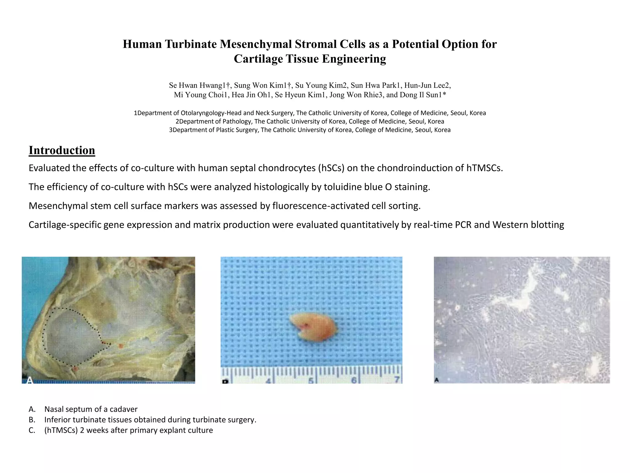
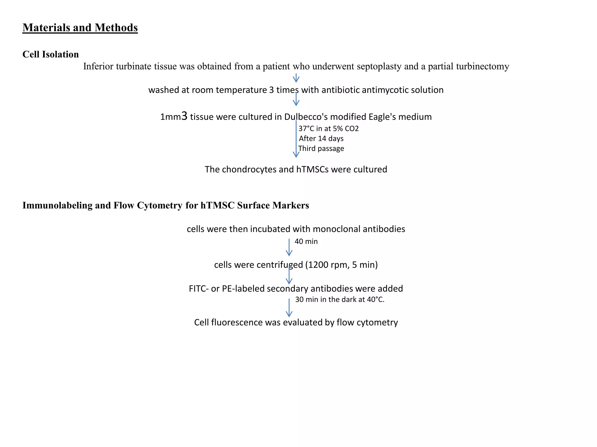
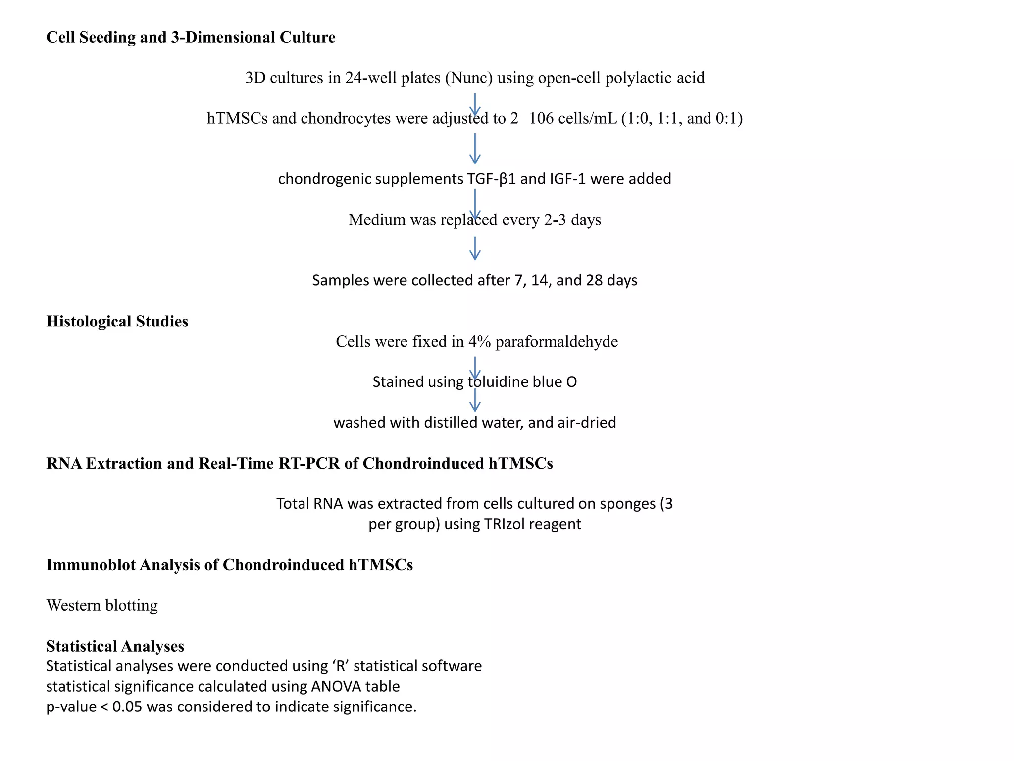
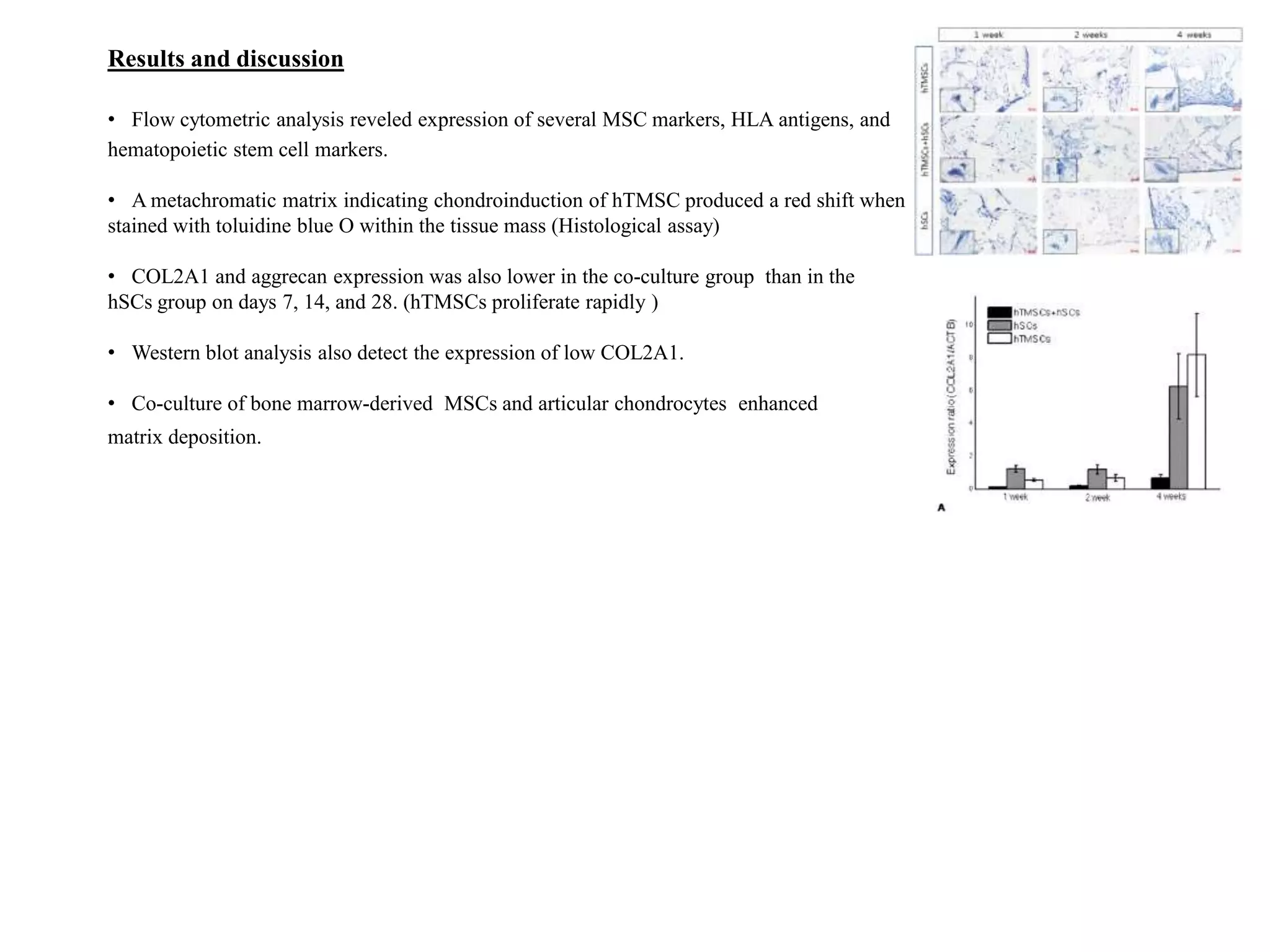

This study evaluated the effects of co-culturing human septal chondrocytes (hSCs) with human turbinate mesenchymal stromal cells (hTMSCs) to induce chondrogenesis of the hTMSCs. hTMSCs were isolated from inferior turbinate tissues and cultured with hSCs in 3D polylactic acid scaffolds. Histological staining showed metachromatic matrix formation, indicating chondroinduction of hTMSCs. However, expression of cartilage matrix genes COL2A1 and aggrecan was lower in the co-culture group compared to the hSCs group alone, as detected by real-time PCR and Western blotting


![Coded Agents – with UiPath SDK + LangGraph [Virtual Hands-on Workshop]](https://cdn.slidesharecdn.com/ss_thumbnails/codedagentsdeck-251215155422-5497c599-thumbnail.jpg?width=640&height=640&fit=bounds)


![Vibe Coding vs. Spec-Driven Development [Free Meetup]](https://cdn.slidesharecdn.com/ss_thumbnails/vibecodingvsspecdrivendevelopment-251209105622-43f455e7-thumbnail.jpg?width=640&height=640&fit=bounds)


