Determination and comparison rate of expression markers of osteoblast derived...
736-2319-1-PB
1. Journal of Biological Researches : 19 (26-31) 2013
EFFECT OF CALCUSOLTM
ON CuZnSOD EXPRESSION IN MICE
RENAL OF NEPHROLITHIASIS MODEL
Arief Azhari* and Sri Widyarti**
*
Graduate Program of Biology, Faculty of Mathematics and Natural Sciences, University of Brawijaya, Malang
**
Department of Biology, Faculty of Mathematics and Natural Sciences, University of Brawijaya, Malang
Corresponding Author email: swid@ub.ac.id
ABSTRACT
This study aimed to determine the effect of traditional medicine, CalcusolTM
, on number of cells expressing CuZnSOD in mice renal of nephrolithiasis
model. Eight weeks old Swiss strain male mice (Mus musculus) were divided into five groups: (A) control, (B) nephrolithiasis, (C) CalcusolTM
, (D)
nephrolithiasis & CalcusolTM
, and (E) nephrolithiasis & CalcusolTM
simultaneously. Nephrolithiasis was induced by applying porang tuber (Amor-
phophallus muelleri) flour 0.06 mg/g of body weight during 3 months. CalcusolTM
is traditional medicine, made of tempuyung leaves extract with
Saccharum lactis as additional substance. The dosage for CalcusolTM
treatment was 3.3 mg/g of body weight. After 3 months treatment, the mice
were killed by neck-dislocation, the kidneys were isolated and prepared for paraffin histology. CuZnSOD was analyzed by immunohistochemistry
(IHC), using rabbit policlonal antibody anti-SOD1 (Bioss, bs-1079R) as primary antibody. Tissues were observed under Olympus BX51, 400x mag-
nification. Images were documented with Olympus Digital Camera DP20. The histology images were analized in Immunoratio software online (URL:
http://153.1.200.58:8080/immunoratio/) to receive the percentage of number of cells expressing CuZnSOD. The result showed that CalcusolTM
administration could decrease number of cells expressing CuZnSOD in kidney significantly (P<0.05). It was supposed that antioxidant content in
CalcusolTM
could scavenge ROS directly with no induction of CuZnSOD production in cell.
Key words: Calcusol, CuZnSOD, nephrolithiasis.
INTRODUCTION
Nephrolithiasis is a disease with the formation of
stones or crystals in the kidney or in the urinary tract. The
formation of kidney stones can be caused by a super-
saturation of mineral salts in urine. Crystal sized less than
5 mm can be removed with urine. Accumulation of larger
crystals is able to close the urethra, which is able to cause
pain in the lower abdomen (Marieb, 2004).
According to data of Cipto Mangunkusumo Hospital,
an increase in the number of kidney stone patients occu-
rred in the range of 1997-2002, from 182 to 847 patients.
Although it did not threaten the life of patients, the dise-
ase could cause pain that was very disturbing. The costs
incurred for the care of kidney stone patients was not
cheap. In the United States annually spent two billion
U.S. dollars to serve patients with kidney stones (Syah-
putra, 2011).
Some studies suggested that the increase in crystal nu-
cleation in renal tubular cells was mediated by free
radicals. A previous research indicates that the adminis-
tration of porang (Amorphophallus muelleri) tuber flour
that containing oxalate continuously in mice was able to
increase levels of malondialdehyde (MDA) in the kidney
(Rosyidah, 2013). The accumulation of free radicals was
able to cause oxidative stress in renal tubular cells which
could ultimately lead to necrosis. Necrosis made oxalate
crystal nucleation going more quickly (Tsujihata, 2007).
To reduce oxidative stress, it requires active com-
pounds that can reduce free radicals, to break the chains
of peroxidation in the cell, thus oxalate crystal nucleation
can be decreased. The active compounds that reduce free
radicals are called antioxidants. Antioxidants that are pro-
duced by the body called endogenous antioxidants, while
derived from nutrients called exogenous antioxidants.
Endogenous antioxidants are usually in the form of en-
zyme or non-enzyme. One of the antioxidant enzymes
that catalyzes the reduction of levels of oxidants is super-
oxide dismutase (SOD) that reduce superoxide radicals
(O·-
). While exogenous antioxidants, such as flavonoids
which contained in many plants, diminish free radicals di-
rectly by providing some electrons then make it non-
radical molecules (Halliwell and Gutteridge, 1999).
CalcusolTM
is a product of traditional medicine made
of Sonchus arvensis L. or tempuyung leaves extract with
saccharum lactis as additional substance. This herbal
medicine can be a source of exogenous antioxidant that
can overcome oxidative stress in renal cells. Sonchus ar-
vensis or tempuyung (local name) contains a variety of
bioactive phenolic compounds and flavonoids which act
as free radical scavenger (Khan, 2012). A previous study
proved that the administration of CalcusolTM
to mice
orally were able to reduce levels of MDA in the kidney
(Rosyidah, 2013). Flavonoid compounds may also stimu-
late the production of antioxidant enzymes in the body,
such as the SOD (Akhlaghi and Bandy, 2009).
In animal cells, based on the active side, there are two
isoforms of SOD: CuZnSOD and MnSOD. In the human
kidney, CuZnSOD activity was higher than the MnSOD
(Halliwell and Gutteridge, 1999). Therefore, it is suspect-
ted that the antioxidants contained in the CalcusolTM
pro-
duct can affect the number of cells expressing CuZnSOD
which is able to affect oxalate crystal nucleation mediated
by free radicals. This study was to determine the effect of
CalcusolTM
on number of cells expressing CuZnSOD
enzyme in the mice kidneys of nephrolithiasis model and
to determine the significancy of CuZnSOD expression in
the cortex and medulla.
MATERIALS AND METHOD
The ethical clearence of this research was approved by
Research Ethic Committee of Brawijaya University No.
127-KEP-UB by March 19th
2013. This research was
done in two stages, treatment to experimental animals and
immunohistochemical analysis. Experimental animals
were used Swiss strain male mice (Mus musculus) eight
weeks old weighing about 25-30 grams in a healthy state.
2. Effect of CalcusolTM
on CuZnSOD Expression in Mice Renal of Nephrolithiasis Model
27
Mice were obtained from LPPT-UGM Yogyakarta.
Before being treated, mice were acclimatized for 7 days.
Mice were treated for three months.
Animal Treatment
Treatment groups were (A) control; (B) nephro-
lithiasis; (C) CalcusolTM
; (D) nephrolithiasis and Calcu-
solTM
; and (E) nephrolithiasis and CalcusolTM
simulta-
neously. Nephrolithiasis was induced by administering
porang tuber flour (Amorphophallus muelleri) orally with
the dosage 0.06 mg/g of body weight (Rosyidah, 2013).
CalcusolTM
were obtained from PT. Perusahaan Jamu
Tradisional (Traditional Herbal Company) DR. SAR-
DJITO Yogyakarta-Indonesia. Dosage of CalcusolTM
was
3.3 mg/g of body weight, administered for the last seven
days for D group and three months simultaneously with
porang administration for E group. After 3 months treat-
ment, the mice were killed by neck-dislocation, the kid-
neys were isolated and prepared for paraffin histology.
Immunohistochemical Analysis
The slides that had been deparrafinned,washed with
PBS 3 x 5 minutes, blocked with peroxidase block (Novo-
link RE7165) for 5 minutes, washed with PBS 3 x 5
minutes each, incubated with a protein block (Novolink
RE7166) for 5 minutes, washed with PBS 3 x 5 minutes,
incubated with primary antibody rabbit polyclonal anti-
SOD1 (Bioss, bs-1079R) with dilution 1:200 in 1% BSA
overnight at 4°C. Washed with PBS 3 x 5 minutes, incu-
bated with Novolink Polymer (RE7168) at room temp-
erature for 30 minutes, washed with PBS 3 x 5 minutes.
Incubated with DAB chromogen DAB substrate buffer-
Novolink 1:20 (Novolink-RE7171 RE7169) for 5 minutes
at room temperature, washed with distilled water until
clean. Furthermore, slides were counterstained with he-
matoxylin for 5 minutes, washed with distilled water until
clean, dried, and mounted with entellan. The slides were
observed with Olympus BX51 microscope in 400x mag-
nification to determine which cells express the enzyme
CuZnSOD. Images of each field were documented with
Olympus DP20 digital camera attached to the microscope.
The histology images were analized by using Immuno-
ratio software online (URL: http://153.1.200.58:8080/
immunoratio/) to receive the percentage of number of
cells expressing CuZnSOD.
Analysis Data
This study used a completely randomized design
with three replications, using one-way ANOVA test 95%
confidence interval for statistical analysis. Tukey HSD
test were used to performed for significancy. While the
analysis to determine significancy of CuZnSOD expre-
ssion in the cortex and medulla used independent-samples
t test. All data were analyzed with MS. Excel and SPSS
16.0 for Windows.
RESULTS
CuZnSOD expression in the kidneys could be
identified by the presence of brown color in the cytosol as
a result of immunohistochemistry with anti-SOD1 anti-
bodies. The intensity of brown color indicated the level of
CuZnSOD expressed in cell ( in fig. 3). In nephro-
lithiasis model (B), CuZnSOD enzyme was expressed in
almost every cell in the tissue with a thick brown color.
Mice kidney with treatment of control (A), CalcusolTM
(C), nephrolithiasis and CalcusolTM
(D), and nephro-
lithiasis and CalcusolTM
simultaneously (E) showed lower
expression of CuZnSOD than in nephrolithiasis model
(B), with a thinner brown color and also less number of
cells expressing CuZnSOD.
Figure 1. The percentage of number of cells expressing CuZnSOD in the
kidneys in each treatment (A) control; (B) nephrolithiasis; (c) Calcu-
solTM
; (D) nephrolithiasis and CalcusolTM
; and (E) nephrolithiasis and
CalcusolTM
simultaneously
Figure 2. The percentage of number of cells expressing CuZnSOD in the
cortex and medulla (ns = not significant).
The results of histologic observation of kidney of mice
(figure 1) showed that the percentage of number of cells
expressing CuZnSOD in the control (A); ne-
phrolithiasis (B); CalcusolTM
(C); nephrolithiasis and Cal-
cusolTM
(D); and nephrolithiasis and CalcusolTM
admi-
nistration simultaneously (E) respectively were 28.4 ±
7.9%; 60.6 ± 7.7%; 10.1 ± 2.3%; 13.8 ± 1.2% and 20.1 ±
7.4%. According to data, it is known that nephrolithiasis
induction by porang administration (B) could increase
significantly the number of cells expressing CuZnSOD
compared to the (A),(C),(D), and (E) treatment (P <0.05).
Calcusol treatment post nephrolithiasis (D) could reduce
the number of cells expressing CuZnSOD significantly (P
<0.05). Calcusol treatment post nephrolithiasis (D) and
nephrolithiasis and calcusol administration simulta-
neously treatment (E) could decrease the number of cells
3. Effect of CalcusolTM
on CuZnSOD Expression in Mice Renal of Nephrolithiasis Model
27
expressing CuZnSOD equal to control (A) (P> 0.05).
Calcusol (C) were able to decrease the number of cells
expressing CuZnSOD significantly compared to the
control (A) (P <0.05).
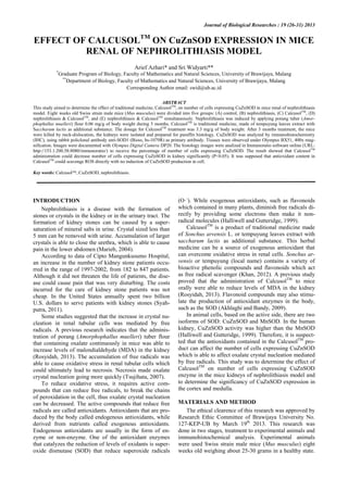
Figure 3. Distribution of CuZnSOD in kidney tissue of each treatment (magnification 400x). The right column shows the zooming 2x of
left image: (A) control; (B) nephrolithiasis; (C) CalcusolTM
; (D) nephrolithiasis and CalcusolTM
; and (E) nephrolithiasis and CalcusolTM
administration simultaneously. () indicates the CuZnSOD expression in kidney cells, ( ) tubules, and ( ) glomerulus. Bar scale: 30
µm.
A
B
E
D
C
28
4. Effect of CalcusolTM
on CuZnSOD Expression in Mice Renal of Nephrolithiasis Model
27
Cortex Medulla
Figure 4. Histology cortex and medulla in each treatment: (A) control; (B) nephrolithiasis; (C) CalcusolTM
; (D) nephrolithiasis and
CalcusolTM
; and (E) nephrolithiasis and CalcusolTM
administration simultaneously. Renal cortex looks more tightly with larger cells and
some glomerulus. Renal medulla composed of tubules with smaller cells. ( ) Tubules and ( ) glomerulus. The bar scale: 30 µm.
A
B
C
D
E
29
5. Effect of CalcusolTM
on CuZnSOD Expression in Mice Renal of Nephrolithiasis Model
27
CuZnSOD expression in the kidneys distributed
evenly (figure 2). Analysis of independent samples t test
found no significancy between the cortex and medulla
(P> 0.05).
DISCUSSION
The increase of CuZnSOD and MDA (Rosyidah, 20-
13) simultaneously after induction of nephrolithiasis indi-
cated that oxidative stress did not suppress the expression
of CuZnSOD in the kidney. Oxidative stress in this model
was triggered by excessive ROS production. Oxidative
stress can occur by two mechanisms: (1) reduction of in-
tracellular antioxidant activity, in this case is the anti-
oxidant enzymes such as superoxide dismutase, catalase,
and glutathione peroxidase produced in the cell; and (2)
the increase of ROS production caused by exposure to
high oxygen or the presence of the toxin compounds that
were metabolized to produce ROS, and excessive activity
[5]. In this study, induction of oxalate porang could in-
crease the production of free radicals that were marked by
the MDA (Rosyidah, 2013). However, oxidative stress
did not reduce the production of CuZnSOD enzyme in the
kidney.
Administration of CalcusolTM
after induction of neph-
rolithiasis could reduce the number of cells those expre-
ssed CuZnSOD and free radicals in the kidney. It sugges-
ted that the antioxidant compounds (such as flavonoids) in
Tempuyung extracts contained in CalcusolTM
could di-
rectly scevenge ROS by giving up electrons and/or pro-
tons to free radical molecules (Akhlaghi and Bandy,
2009) without inducing the cells to produce the CuZn-
SOD enzyme. Flavone, contained in Tempuyung leaves,
is one of the most powerful flavonoids in reducing ROS
produced by the body. The members of flavone are kaem-
pferol, quercetin, and luteolin. Flavonoids reduce free ra-
dical molecules to become more stable and less reactive.
Flavonoids stabilize ROS via the following reaction
(Nijveldt et al, 2001).
flavonoid(OH) + R· > flavonoid(O·) + RH
The ability of the active compounds in reducing free
radicals can be determined with IC50 values in the DPPH
test, which indicates the concentration needed to scavenge
50% DPPH radical. IC50 values of methanol fraction of
Tempuyung leaves extract in DPPH test was 3.4 ± 0.3
mg/ml (Khan, 2012). The lower the IC50 value, the stro-
nger the antioxidant capabilities. The ability of anti-
oxidants rated high if the IC50 between 10-20 μg/ml, mo-
derate in the range 21-100 μg/ml, low in the range 101-
200 μg/ml, and there is no active compound if IC50 is mo-
re than 200 μg/ml (Lubis et al, 2011). The strength of the
active flavonoid compounds are also determined by the
location and number of hydroxyl groups that owned the
compound (Amic et al, 2003).
The pathogenesis of kidney stones occurs through the
process of nucleation, crystal growth, crystal aggregation,
and attachment of crystals in the renal tubules (Zhai et al,
2013). The Increase of ROS production that causes oxi-
dative stress in renal epithelial cells can make cells un-
dergo necrosis. By necrosis, oxalate crystal nucleation can
take place more quickly (Tsujihata, 2007). It occurs when
cell debrises are more likely to attract calcium or other
mineral salts to precipitate through a process called dys-
trophy calcification (Kumar et al, 1992). By reducing free
radicals, lipid peroxidation can be inhibited and cell
necrosis can be terminated, so that the crystal nucleation
can be decreased.
The increase of number of cells expressing CuZnSOD
and MDA in synergy does not necessarily indicate that
CuZnSOD in high level could reduce superoxide radicals
much effectively. This is because hydrogen peroxide (H2-
O2), the result of superoxide convertion by CuZnSOD,
can become pro-oxidant through the fenton reaction into
the more reactive hydroxyl radical (Goode and Webster,
1993). Hydrogen peroxide should be directly catalyzed by
the catalase and/or glutathione peroxidase enzyme into
H2O and O2, whereas in this study there was no data on
the levels of catalase and glutathione peroxidase enzymes
which also act as intracellular antioxidants. If ROS dec-
reased these two antioxidant enzymes and other non-
enzymatic endogenous antioxidants, then oxidative stress
could occur. Superoxide radical can decrease the activity
of antioxidant enzymes such as catalase and glutathione
peroxidase (Halliwell and Gutteridge, 1999).
Proximal tubule is a functional part of the kidney
which is susceptible to oxidative stress, because it con-
tains many mitochondria (the site of oxidative phos-
phorylation). It shows that tubular cells have a high meta-
bolism activity. Metabolism yields ATP as a source of
energy to perform reabsorption of water, ions and glucose
for homeostasis. However, when mitochondria membrane
potential disrupted, the electron transport chain will pro-
duce more ROS (Ozbek, 2012).
Proximal tubules are mostly found in the renal cortex,
but its extension in the form of the loop of Henle located
in medulla and also acts in the reabsorption process
(Bloom and Flawcet, 2002). Medulla which consists of
the loop of Henle and collecting tubules is formed by cells
that actively metabolize. ATP yielded in the cell are
needed for reabsorption and secretion (Patton and Thibo-
deau, 2000).
Based on the result concluded that CalcusolTM
admi-
nistration could reduce the number of cells expressing
CuZnSOD in the kidneys of mice of nephrolithiasis mo-
del. There was no significancy found in the number of
cells expressing CuZnSOD in the renal cortex and me-
dulla. It was suspected that antioxidant compounds in
Calcusol™ could reduce ROS without inducing the cells
to produce the CuZnSOD enzyme.
AKNOWLEDGEMENT
Thanks to PT. Jamu Tradisional Dr. SARDJITO Yog-
yakarta that had funded for this work. We also thank to all
partners of Anatomy and Physiology Laboratory of Bio-
logy Department of UB.
Azhari and Widyarti 30
6. Effect of CalcusolTM
on CuZnSOD Expression in Mice Renal of Nephrolithiasis Model
27
REFERENCES
Akhlaghi, M. and B. Bandy. 2009. Mechanisms of Flavonoid Protection
against Myocardial Ischemia-Reperfusion Injury. Journal of
Molecular and Cellular Cardiology. 46: 309-317.
Amic, D., D. D. Amic, D. Beslo, and N. Trinajstic. 2003. Structure-
Radical Scavenging Activity Relationships of Flavonoids. Original
Scientific Paper. 76 (1): 55-61.
Bloom and D. W. Fawcett. 2002. Buku Ajar Histologi. Penerbit Buku
Kedokteran EGC. Jakarta.
Goode, H. F. and N. R. Webster. 1993. Free Radicals and Antioxidants
in Sepsis. Crit. Care Med. 21: 1770-1776.
Halliwell, B. and J. M. C. Gutteridge. 1999. Free Radicals in Biology
and Medicine. Oxford University Press. London.
Khan, R. A. 2012. Evaluation of Flavonoids and Diverse Antioxidant
Activities of Sonchus arvensis. Chemistry Central Journal. 6. 1-7.
Kumar, S.A., Citran, R. S., and Robbins, S. L. 1992. Basic Pathology 5th
Edition. W.B. Saunders Company. Harcourt Brace Javanovish, Inc.
Philadelphia.
Lubis, A. H., M. Nainggolan, K. R. Sinaga, Suryanto, and E. Sitompul.
2011. Pengujian Aktivitas Antioksidan dan Analisis Senyawa Kimia
Ekstrak Etanol serta Fraksi dari Kayu Secang (Caesalpinia sappan
L.), in Prosiding Seminar Nasional Biologi, Departemen Biologi
FMIPA Universitas Sumatera Utara, 22 Januari 2011, hlm. 236-
244.
Marieb, E. N. 2004. Human Anatomy & Physiology. Pearson Benjamin
Cummings. San Fransisco.
Nijveldt, R. J., E. V. Nood, D. EC. V. Hoorn, P. G. Boelens, K. V.
Norren, and P. AM. V. Leeuwen. 2001. Flavonoids: A Review of
Probable Mechanisms of Action and Potential Application. Am J
Clin Nutr. 74: 418-425.
Ozbek, E. 2012. Induction of Oxidative Stress in Kidney. International
Journal of Nephrology. 1-9.
Patton, K. T. and G. A. Thibodeau. 2000. Mosby’s Handbook of
Anatomy & Physiology. Mosby. Missouri.
Rosyidah, A. 2013. Studi Mekanisme Daun Tempuyung (Sonchus
arvensis) dalam Menurunkan Akumulasi Kalsium Oksalat pada
Mencit. Thesis. Biology Department. Faculty of Mathematics
Natural Sciences. University of Brawijaya. Malang.
Syahputra, F. A. 2011. Terapi Batu Ginjal: Dari Era Hippocrates ke Era
Minimal Invasif. Majalah Kedokteran Indonesia. 61 (3). 99-100.
Tsujihata, M. 2007. Mechanism of Calsium Oxalate Renal Stone
Formation and Renal Tubular Cell Injury. International Journal of
Urology. 15. 115-120.
Tsujihata, M. 2007. Mechanism of Calsium Oxalate Renal Stone
Formation and Renal Tubular Cell Injury. International Journal of
Urology. Vol 15: 115-120.
Zhai, W., J. Zheng, X. Yao, B. Peng, M. Liu, J. Huang, G. Wang, and Y.
Xu. 2013. Catechin Prevents the Calcium Oxalate Monohydrate
Induced Renal Calcium Crystallyzation in NRK-52E Cells and the
Ethylene Glycol Induced Renal Stone Formation in Rat. BMC
Complementary Alternative Medicine. 13: 228.
31