Cartilage Tissue Engineering Methods And Protocols Pauline M Doran
Cartilage Tissue Engineering Methods And Protocols Pauline M Doran
Cartilage Tissue Engineering Methods And Protocols Pauline M Doran
Cartilage Tissue Engineering Methods And Protocols Pauline M Doran
Cartilage Tissue Engineering Methods And Protocols Pauline M Doran
1.
Cartilage Tissue EngineeringMethods And
Protocols Pauline M Doran download
https://ebookbell.com/product/cartilage-tissue-engineering-
methods-and-protocols-pauline-m-doran-5221690
Explore and download more ebooks at ebookbell.com
2.
Here are somerecommended products that we believe you will be
interested in. You can click the link to download.
Cartilage Tissue Engineering Martin J Stoddart Elena Della Bella
https://ebookbell.com/product/cartilage-tissue-engineering-martin-j-
stoddart-elena-della-bella-47214780
Articular Cartilage Tissue Engineering K A Athanasiou Eric M Darling
Jerry C Hu
https://ebookbell.com/product/articular-cartilage-tissue-engineering-
k-a-athanasiou-eric-m-darling-jerry-c-hu-4116032
Gene Therapy For Cartilage And Bone Tissue Engineering 1st Edition
Yuchen Hu Auth
https://ebookbell.com/product/gene-therapy-for-cartilage-and-bone-
tissue-engineering-1st-edition-yuchen-hu-auth-4636150
Mimicked Tissue Engineering Scaffolds For Maxillofacial And Articular
Cartilage Surgery Jirut Meesane
https://ebookbell.com/product/mimicked-tissue-engineering-scaffolds-
for-maxillofacial-and-articular-cartilage-surgery-jirut-
meesane-48726212
3.
Tissue Engineering OfCartilage And Bone Novartis Foundation Symposium
249 Novartis Foundationeds
https://ebookbell.com/product/tissue-engineering-of-cartilage-and-
bone-novartis-foundation-symposium-249-novartis-foundationeds-4309124
Joint Homeostasis In Tissue Engineering For Cartilage Repair Danil Bf
Saris
https://ebookbell.com/product/joint-homeostasis-in-tissue-engineering-
for-cartilage-repair-danil-bf-saris-1423454
Stem Cell And Tissue Engineering Bone Cartilage And Associated Joint
Tissue Defects Krishna Pramanik
https://ebookbell.com/product/stem-cell-and-tissue-engineering-bone-
cartilage-and-associated-joint-tissue-defects-krishna-
pramanik-55926466
A Tissue Regeneration Approach To Bone And Cartilage Repair 1st
Edition Hala Zreiqat
https://ebookbell.com/product/a-tissue-regeneration-approach-to-bone-
and-cartilage-repair-1st-edition-hala-zreiqat-4973600
Regenerative Medicine And Plastic Surgery Skin And Soft Tissue Bone
Cartilage Muscle Tendon And Nerves 1st Ed 2019 Dominik Duscher
https://ebookbell.com/product/regenerative-medicine-and-plastic-
surgery-skin-and-soft-tissue-bone-cartilage-muscle-tendon-and-
nerves-1st-ed-2019-dominik-duscher-10797124
ME T HO D S I N MO L E C U L A R BI O L O G Y
Series Editor
John M. Walker
School of Life and Medical Sciences
University of Hertfordshire
Hatfield, Hertfordshire, AL10 9AB, UK
For further volumes:
http://www.springer.com/series/7651
8.
Cartilage Tissue Engineering
Methodsand Protocols
Edited by
Pauline M. Doran
FacultyofScience,EngineeringandTechnology,SwinburneUniversity
ofTechnology,Hawthorn,Melbourne,VIC,Australia
v
Cartilage in articularjoints is a relatively vulnerable tissue, being subject to common injuries
and degenerative conditions such as arthritis. Motivated by the need to develop new treat-
ment strategies, some of the earliest attempts at tissue engineering targeted cartilage as a
feasible goal for in vitro synthesis. For more than 20 years, interdisciplinary teams of biolo-
gists, engineers, materials scientists, and clinicians have studied the culture and differentia-
tion of cartilage cells and tissues. Many cornerstone technologies that distinguish tissue
engineering from routine cell culture, such as three-dimensional culture systems and the
use of scaffolds and bioreactors, were developed, tested, and widely adopted within the
context of cartilage tissue engineering.
So far, the goal of producing laboratory-grown functional cartilage has eluded us but
remains an active ambition. Irrespective of whether chondrocytes or stem cells are used as
starting material, exerting adequate control over cellular differentiation is a major chal-
lenge. We do not yet know how to integrate engineered constructs with host cartilage
in vivo and this continues to restrict clinical translation of cartilage engineering technology.
Other important areas requiring further research include the response of chondrogenic
cells to physical and mechanical stimuli, the heterogeneity of cell populations, and the com-
plex molecular networks and regulatory cascades that direct cell lineage commitment and
tissue development.
To answer all the outstanding questions in cartilage tissue engineering, further sig-
nificant creative and intellectual input is required. This should come not only from
established contributors but also, perhaps more importantly, from new and/or cross-
disciplinary researchers in the area. How does a newcomer to cartilage tissue engineer-
ing become familiar with the techniques that underpin this field? I hope this question
may be answered herein, as this book aims to describe clearly and in detail the key practi-
cal skills involved. Methods are outlined for isolation and expansion of chondrocytes and
stem cells, differentiation, synthesis and application of three-dimensional scaffolds,
design and operation of bioreactors, in vivo testing of engineered constructs, and molec-
ular and functional analysis of cartilage cells and tissues. Frequently used techniques are
covered, as well as more recent advances in inspirational areas such as “smart” biomaterial
development, novel bioreactor design, –omics analysis, and genetic manipulation of
matrix synthesis. The book does not attempt to be comprehensive; instead, it provides a
snapshot of selected practical technologies that are either responsible for the progress
already achieved in cartilage tissue engineering or indicative of the direction of future
related research.
The chapters have been written by 45 authors and coauthors who have personal practi-
cal experience in cartilage tissue engineering. In the interests of informing the scientific
community and expanding the engagement of researchers in this field, the contributors
have provided careful and detailed protocols for experimental work covering a broad range
of objectives for cartilage synthesis and regeneration. I thank all of the experts who have
Preface
11.
vi
generously contributed theirknowledge, insights, and valuable tips to prepare this volume.
We hope that readers will find it a useful resource. I would also like to acknowledge the kind
guidance and encouragement of Professor John Walker, Series Editor of Methods in
Molecular Biology, throughout the duration of this project.
Melbourne, VIC, Australia Pauline M. Doran
Preface
12.
vii
Preface. . .. . . . . . . . . . . . . . . . . . . . . . . . . . . . . . . . . . . . . . . . . . . . . . . . . . . . . . . v
Contributors. . . . . . . . . . . . . . . . . . . . . . . . . . . . . . . . . . . . . . . . . . . . . . . . . . . . . . ix
PART I OVERVIEW
1 Cartilage Tissue Engineering: What Have We Learned in Practice?. . . . . . . . . . 3
Pauline M. Doran
PART II CELL ISOLATION, EXPANSION, AND DIFFERENTIATION
2 Human Fetal and Adult Chondrocytes. . . . . . . . . . . . . . . . . . . . . . . . . . . . . . . 25
Kifah Shahin, Nastaran Mahmoudifar, and Pauline M. Doran
3 Mesenchymal Stem Cells Derived from Human Bone Marrow . . . . . . . . . . . . 41
Oliver F.W. Gardner, Mauro Alini, and Martin J. Stoddart
4 Mesenchymal Stem Cells Derived from Human Adipose Tissue. . . . . . . . . . . . 53
Nastaran Mahmoudifar and Pauline M. Doran
5 Derivation and Chondrogenic Commitment of Human Embryonic
Stem Cell-Derived Mesenchymal Progenitors . . . . . . . . . . . . . . . . . . . . . . . . . 65
Hicham Drissi, Jason D. Gibson, Rosa M. Guzzo, and Ren-He Xu
6 Differentiation of Human Induced Pluripotent Stem Cells
to Chondrocytes . . . . . . . . . . . . . . . . . . . . . . . . . . . . . . . . . . . . . . . . . . . . . . . 79
Rosa M. Guzzo and Hicham Drissi
7 Gene Transfer and Gene Silencing in Stem Cells
to Promote Chondrogenesis . . . . . . . . . . . . . . . . . . . . . . . . . . . . . . . . . . . . . . 97
Feng Zhang and Dong-An Wang
PART III BIOMATERIALS AND SCAFFOLDS
8 Hydrogels with Tunable Properties . . . . . . . . . . . . . . . . . . . . . . . . . . . . . . . . . 121
Peggy P.Y. Chan
9 Decellularized Extracellular Matrix Scaffolds for Cartilage Regeneration . . . . . 133
Shraddha Thakkar, Hugo Fernandes, and Lorenzo Moroni
10 Use of Interim Scaffolding and Neotissue Development to Produce
a Scaffold-Free Living Hyaline Cartilage Graft. . . . . . . . . . . . . . . . . . . . . . . . . 153
Ting Ting Lau, Wenyan Leong, Yvonne Peck, Kai Su,
and Dong-An Wang
11 Bioprinted Scaffolds for Cartilage Tissue Engineering . . . . . . . . . . . . . . . . . . . 161
Hyun-Wook Kang, James J. Yoo, and Anthony Atala
Contents
13.
viii
12 Scaffolds forControlled Release of Cartilage Growth Factors. . . . . . . . . . . . . . 171
Marie Morille, Marie-Claire Venier-Julienne,
and Claudia N. Montero-Menei
13 Nanostructured Capsules for Cartilage Tissue Engineering . . . . . . . . . . . . . . . 181
Clara R. Correia, Rui L. Reis, and João F. Mano
14 Stratified Scaffolds for Osteochondral Tissue Engineering . . . . . . . . . . . . . . . . 191
Patcharakamon Nooeaid, Gundula Schulze-Tanzil, and Aldo R. Boccaccini
PART IV BIOREACTORS
15 Mechanobioreactors for Cartilage Tissue Engineering . . . . . . . . . . . . . . . . . . . 203
Joanna F. Weber, Roman Perez, and Stephen D. Waldman
16 Shear and Compression Bioreactor for Cartilage Synthesis. . . . . . . . . . . . . . . . 221
Kifah Shahin and Pauline M. Doran
17 Microbioreactors for Cartilage Tissue Engineering. . . . . . . . . . . . . . . . . . . . . . 235
Yu-Han Chang and Min-Hsien Wu
PART V IN VIVO APPLICATION
18 Transplantation of Tissue-Engineered Cartilage in an Animal Model
(Xenograft and Autograft): Construct Validation. . . . . . . . . . . . . . . . . . . . . . . 247
Hitoshi Nemoto, Deborah Watson, and Koichi Masuda
PART VI EVALUATION
19 Proteomic Analysis of Engineered Cartilage. . . . . . . . . . . . . . . . . . . . . . . . . . . 263
Xinzhu Pu and Julia Thom Oxford
20 Mechanical Testing of Cartilage Constructs. . . . . . . . . . . . . . . . . . . . . . . . . . . 279
Dinorath Olvera, Andrew Daly, and Daniel John Kelly
Index. . . . . . . . . . . . . . . . . . . . . . . . . . . . . . . . . . . . . . . . . . . . . . . . . . . . . . . . . . . 289
Contents
14.
ix
MAURO ALINI •Musculoskeletal Regeneration, AO Research Institute Davos, Davos Platz,
Switzerland
ANTHONY ATALA • Wake Forest Institute for Regenerative Medicine, Wake Forest School
of Medicine, Winston-Salem, NC, USA
ALDO R. BOCCACCINI • Department of Materials Science and Engineering,
Institute of Biomaterials, University of Erlangen-Nuremberg, Erlangen, Germany
PEGGY P.Y. CHAN • Faculty of Science, Engineering and Technology, Swinburne University
of Technology, Melbourne, VIC, Australia
YU-HAN CHANG • Department of Orthopaedic Surgery, Chang Gung Memorial Hospital,
Linko, Taiwan
CLARA R. CORREIA • 3B’ s Research Group—Biomaterials, Biodegradables and Biomimetics,
University of Minho, Headquarters of the European Institute of Excellence on Tissue
Engineering and Regenerative Medicine, Guimarães, Portugal; ICVS/3B’s –PT
Government Associate Laboratory, Guimarães, Braga, Portugal
ANDREW DALY • Trinity Centre for Bioengineering, Trinity Biomedical Sciences Institute,
Trinity College Dublin, Dublin, Ireland; Department of Mechanical and Manufacturing
Engineering, School of Engineering, Trinity College Dublin, Dublin, Ireland;
Advanced Materials and Bioengineering Research Centre (AMBER),
Trinity College Dublin, Dublin, Ireland
PAULINE M. DORAN • Faculty of Science, Engineering and Technology,
Swinburne University of Technology, Melbourne, VIC, Australia
HICHAM DRISSI • Department of Orthopaedic Surgery, UConn Health, Farmington,
CT, USA; Department of Genetics and Genome Sciences, UConn Health,
Farmington, CT, USA; Stem Cell Institute, UConn Health, Farmington, CT, USA
HUGO FERNANDES • University of Maastricht – MERLN Institute for Technology Inspired
Regenerative Medicine, Complex Tissue Regeneration Department, The Netherlands
OLIVER F.W. GARDNER • Musculoskeletal Regeneration, AO Research Institute Davos,
Davos Platz, Switzerland; Cardiff University School of Biosciences, Cardiff, UK
JASON D. GIBSON • Department of Orthopaedic Surgery, UConn Health, Farmington, CT,
USA
ROSA M. GUZZO • Department of Orthopaedic Surgery, UConn Health, Farmington, CT,
USA; Stem Cell Institute, UConn Health, Farmington, CT, USA
HYUN-WOOK KANG • Wake Forest Institute for Regenerative Medicine, Wake Forest School
of Medicine, Winston-Salem, NC, USA
DANIEL JOHN KELLY • Trinity Centre for Bioengineering, Trinity Biomedical Sciences
Institute, Trinity College Dublin, Dublin, Ireland; Department of Mechanical and
Manufacturing Engineering, School of Engineering, Trinity College Dublin, Dublin,
Ireland; Advanced Materials and Bioengineering Research Center (AMBER), Trinity
College Dublin, Dublin, Ireland
TING TING LAU • Division of Bioengineering, School of Chemical and Biomedical
Engineering, Nanyang Technological University, Singapore
Contributors
15.
x
WENYAN LEONG •Division of Bioengineering, School of Chemical and Biomedical
Engineering, Nanyang Technological University, Singapore
NASTARAN MAHMOUDIFAR • School of Biotechnology and Biomolecular Sciences,
University of New South Wales, Sydney, NSW, Australia
JOÃO F. MANO • 3B’ s Research Group—Biomaterials, Biodegradables and Biomimetics,
University of Minho, Headquarters of the European Institute of Excellence on Tissue
Engineering and Regenerative Medicine, Guimarães, Portugal; ICVS/3B’s –PT
Government Associate Laboratory, Guimarães, Braga, Portugal
KOICHI MASUDA • Department of Orthopaedic Surgery, School of Medicine,
University of California San Diego, La Jolla, CA, USA
CLAUDIA N. MONTERO-MENEI • Micro et Nanomédecines Biomimétiques (MINT),
LUNAM Université, Angers, France; INSERM U1066, Angers, France
MARIE MORILLE • Institute Charles Gerhardt Montpellier, Equipe Matériaux Avancés
pour la Catalyse et la Santé, Montpellier, France
LORENZO MORONI • University of Maastricht – MERLN Institute for Technology Inspired
Regenerative Medicine, Complex Tissue Regeneration Department, The Netherlands
HITOSHI NEMOTO • Division of Otolaryngology-Head and Neck Surgery, Department of
Surgery, School of Medicine, University of California San Diego, La Jolla, CA, USA;
Department of Plastic and Reconstructive Surgery, Showa University Fujigaoka Hospital,
Yokohama, Kanagawa, Japan
PATCHARAKAMON NOOEAID • Department of Materials Science and Engineering, Institute of
Biomaterials, University of Erlangen-Nuremberg, Erlangen, Germany
DINORATH OLVERA • Trinity Centre for Bioengineering, Trinity Biomedical Sciences
Institute, Trinity College Dublin, Dublin, Ireland; Department of Mechanical
and Manufacturing Engineering, School of Engineering, Trinity College Dublin,
Dublin, Ireland; Advanced Materials and Bioengineering Research Centre (AMBER),
Trinity College Dublin, Dublin, Ireland
JULIA THOM OXFORD • Department of Biological Sciences, Biomolecular Research Center,
Boise State University, Boise, ID, USA
YVONNE PECK • Division of Bioengineering, School of Chemical and Biomedical
Engineering, Nanyang Technological University, Singapore
ROMAN PEREZ • Kennan Research Centre, Li Ka Shing Knowledge Institute,
St. Michael’s Hospital, Toronto, Canada; Institute of Tissue Regeneration
Engineering (ITREN), Dankook University, Cheonan, Republic of Korea
XINZHU PU • Department of Biological Sciences, Biomolecular Research Center, Boise State
University, Boise, ID, USA
RUI L. REIS • 3B’ s Research Group—Biomaterials, Biodegradables and Biomimetics,
University of Minho, Headquarters of the European Institute of Excellence on Tissue
Engineering and Regenerative Medicine, Guimarães, Portugal; ICVS/3B’s –PT
Government Associate Laboratory, Guimarães, Braga, Portugal
GUNDULA SCHULZE-TANZIL • Institute of Anatomy, Paracelsus Medical University,
Salzburg and Nuremberg, Nuremberg, Germany; Department for Orthopedic,
Trauma and Reconstructive Surgery, Charité-University of Medicine, Berlin,
Nuremberg, Germany
KIFAH SHAHIN • Westmead Millennium Institute for Medical Research,
University of Sydney, Westmead, NSW, Australia
Contents
16.
xi
MARTIN J. STODDART• Musculoskeletal Regeneration, AO Research Institute Davos, Davos
Platz, Switzerland
KAI SU • Division of Bioengineering, School of Chemical and Biomedical Engineering,
Nanyang Technological University, Singapore
SHRADDHA THAKKAR • Department of Biomedical Engineering, Eindhoven University of
Technology, Eindhoven, The Netherlands
MARIE-CLAIRE VENIER-JULIENNE • Micro et Nanomédecines Biomimétiques (MINT),
LUNAM Université, Angers, France; INSERM U1066, Angers, France
STEPHEN D. WALDMAN • Department of Mechanical and Materials Engineering,
Queen’s University, Kingston, Canada; Kennan Research Centre, Li Ka Shing
Knowledge Institute, St. Michael’s Hospital, Toronto, Canada; Department of Chemical
Engineering, Ryerson University, Toronto, ON, Canada
DONG-AN WANG • Division of Bioengineering, School of Chemical & Biomedical
Engineering, Nanyang Technological University, Singapore
DEBORAH WATSON • Division of Otolaryngology-Head and Neck Surgery,
Department of Surgery, School of Medicine, University of California San Diego,
La Jolla, CA, USA
JOANNA F. WEBER • Department of Mechanical and Materials Engineering,
Queen’s University, Kingston, Canada; Human Mobility Research Centre,
Kingston General Hospital and Queen’s University, Kingston, Canada;
Kennan Research Centre, Li Ka Shing Knowledge Institute, St. Michael’s Hospital,
Toronto, Canada
MIN-HSIEN WU • Graduate Institute of Biochemical and Biomedical Engineering,
Chang Gung University, Tao-Yuan, Taiwan
REN-HE XU • Department of Genetics and Genome Sciences, UConn Health, Farmington,
CT, USA; Stem Cell Institute, UConn Health, Farmington, CT, USA
JAMES J. YOO • Wake Forest Institute for Regenerative Medicine, Wake Forest School
of Medicine, Winston-Salem, NC, USA
FENG ZHANG • Division of Bioengineering, School of Chemical and Biomedical
Engineering, Nanyang Technological University, Singapore
Contents
4
supplied readily fromoutside the construct in liquid-based in vitro
culture systems.
To function successfully within joints, engineered cartilage
must satisfy the most exacting biochemical and mechanical require-
ments. The extraordinary load-bearing, resilience, and low-friction
properties of articular cartilage depend on its chemical composi-
tion and structure; tissues of inferior quality will not withstand the
very high shear and compressive forces generated during normal
joint movement. It is crucially important, therefore, that the prop-
erties of engineered cartilage match those of native articular carti-
lage. In adults, water accounts for approximately 70–80 % of the
weight of cartilage tissue. The principal constituents of the dry
matrix are collagen (50–75 % w/w) for tensile strength and pro-
teoglycan (15–30 % w/w) for compressive stiffness, load distribu-
tion, and resilience [5, 6]. Several different collagen types are
found; however, 90–95 % of the collagen in articular cartilage is
type II. The principal proteoglycan is aggrecan, which consists of a
core protein with many unbranched glycosaminoglycan (GAG)
side chains. Chondrocytes, the cells responsible for synthesizing
and maintaining cartilage, account for only about 1 % of the vol-
ume of mature tissue. In contrast, during fetal and early childhood
growth, the concentration of chondrocytes in developing cartilage
is 1–2 orders of magnitude greater than in adults, consistent with
the need for active matrix synthesis and deposition. The composi-
tion of developing cartilage also differs significantly from that of
adult tissue in terms of its collagen and GAG contents (Table 1).
Progress during the last 20–25 years of research into cartilage
tissue engineering has been substantial. Innovative and pioneering
work to develop new cell culture systems, scaffolds, bioreactors,
differentiation techniques, and analytical methods has brought us
closer to the goal of producing functional human cartilage in vitro.
Using cartilage as a model system, researchers have created a sub-
stantial body of knowledge and developed an impressive skill-set of
techniques for growing and regenerating tissues. Here, the most
important practical lessons learned from cartilage tissue engineer-
ing are summarized. Critical areas where further research is needed
to overcome the remaining barriers to clinical implementation are
also highlighted.
2 Tissue Development Depends on a Three-Dimensional Culture Environment
Chondrocytes isolated from cartilage matrix tend to dedifferenti-
ate when cultured in monolayer on flat, two-dimensional surfaces,
resulting in downregulation of aggrecan and collagen type II syn-
thesis and an increase in collagen type I production [7, 8]. Because
expression of collagen type I leads to the development of mechani-
cally inferior fibrocartilage, the consequences of cell attachment
Pauline M. Doran
6
and spreading ontwo-dimensional surfaces are highly undesirable
for articular cartilage engineering. The ability of dedifferentiated
chondrocytes to recover characteristics of the differentiated pheno-
type when returned to a favorable three-dimensional environment
[7, 9–11] has underpinned an extensive research effort to produce
scaffolds for cell attachment or entrapment that stimulate and sup-
port chondrogenesis and cartilage synthesis.
Many different scaffold materials have been studied for carti-
lage tissue engineering. These include porous foams and fibrous
meshes made of biodegradable polymers such as poly(glycolic
acid), poly(lactic acid), and poly(lactide-co-glycolide), and hydro-
gels based on polysaccharides such as alginate, agarose, hyaluro-
nan, and chitosan or proteins such as collagen, gelatin, and fibrin
(reviewed in [12, 13]). The focus of much scaffold development
has been to mimic the native extracellular matrix (ECM) of carti-
lage where chondrocytes normally reside. To this end, complex
scaffolds with physical and chemical gradients that imitate the
zonal organization of articular cartilage, and scaffolds based on
decellularized cartilage tissue itself, have been applied (reviewed in
[14, 15]). High levels of sophistication have been achieved with
the synthesis of advanced or “smart” scaffolds with features such as
tunable material, surface, and pore properties and degradation
rates. Multifunctional scaffolds can now be applied for delivery of
trophic factors, regulatory molecules, or genetic components to
control cellular differentiation, self-assembly of micro- and nano-
scale surface patterning to enhance cell–scaffold interactions, and
production of complex hierarchical structures for in situ optimiza-
tion of scaffold conditions (reviewed in [16]).
Biochemical interactions and the regulatory responses of cells
to surfaces have a major effect on cell attachment, orientation,
shape, movement, distribution, proliferation, and differentiation
(reviewed in [16]). The role of cell surface receptors to mediate
the effects of the external environment, and thus determine
whether cells grow, differentiate, switch between different lineages,
or undergo apoptosis, is now well recognized. The observed sen-
sitivity of chondrocytes and chondrogenic cells to cell–surface
interactions highlights the importance of rational scaffold design
and engineering to provide an appropriate material–biologic inter-
face for tissue regeneration. Cartilage tissue engineering has been
carried out using scaffolds with a wide variety of physicochemical
and biological characteristics, all of which have the potential to
influence cell behavior. A broad range of material and surface
properties such as strength, stiffness, hydrophobicity, electrostatic
charge, molecular functionality, and cell adhesiveness, and archi-
tectural properties such as porosity, pore size distribution, and
micro- and nano-topography, has been examined. Similarly, a wide
range of biodegradation mechanisms and kinetics, and ability to
elicit inflammatory or immunogenic reactions, has been tested.
Pauline M. Doran
24.
7
Despite these assiduousefforts, however, no single scaffold material
or manner of fabrication has been identified as offering a clearly
superior approach for cartilage synthesis. Development of new
scaffolds that enhance the outcomes of cartilage tissue engineering
continues to be an important research goal.
Extrinsic scaffolds are not essential for production of cartilagi-
nous tissue, as scaffold-free three-dimensional culture systems can
also be used. A potential disadvantage of using scaffolds is that they
may induce changes in cell morphology that are unfavorable for
chondrogenesis and cartilage synthesis [17]. To improve cellular
adhesion to fibrous or gel scaffolds, chondrocytes tend to elongate
and produce cytoplasmic extensions, thus destroying the spheroi-
dal shape associated with the fully differentiated phenotype. To
overcome this problem and more closely recapitulate the conden-
sation phase of embryonic chondrogenesis, self-assembled or
scaffold-free forms of three-dimensional cell culture have been
developed (reviewed in [18]). Many cell types, including chondro-
cytes, are self-adherent and spontaneously form small aggregates
under appropriate culture conditions; simple pellet or micromass
cultures have been used extensively in tissue engineering studies.
Close control is required over the size and cell density of low-
porosity pellets to avoid necrosis within the aggregates [19]: in the
absence of convective mass transfer networks such as vasculature,
diffusional restrictions limit the practical size of cell aggregates.
Although retention of the chondrocytic phenotype and ECM syn-
thesis have been reported using scaffold-free systems [20, 21],
other studies have shown reduced chondrogenic differentiation
and cartilage development in pellet cultures compared with
dynamic scaffold-based systems [22].
3 Dynamic Culture Is Better Than Static Culture
Static forms of cell culture provide a suboptimal environment for
cartilage development, leading to the production of tissues of rela-
tively poor quality in terms of their biochemical composition and
mechanical properties. Several studies have established that fluid
mixing enhances the development of cartilage tissues relative to
static culture methods [23–25]. These findings reflect the critical
role that liquid convection plays in cartilage maintenance and func-
tion in vivo, where joint movement during normal exercise drives
the exchange and mixing of components between the synovial
fluid bathing the joint and the interior of the cartilage tissue. The
mechanisms by which fluid flow improves cartilage synthesis
in vitro include physical effects, such as enhanced gas exchange and
convective mass transfer to and from the cells, and direct biological
effects as externally delivered hydrodynamic forces interact with
mechanoreceptors on the cells to influence gene expression,
What Have We Learned?
25.
8
cellular differentiation, andmatrix deposition [26–28]. Fluid
motion applied to developing tissue constructs needs to be regu-
lated and applied judiciously in culture systems, as flow that is too
vigorous or applied too early in the culture when there is little
ECM present can lead to loss of cells and/or matrix components
and poor construct quality [29–31].
Cell culture conditions incorporating some level of fluid flow
can be achieved using simple well plate, Petri dish, or tissue flask
systems if incubation is carried out on a shaking platform. However,
because bioreactors can be designed to give high levels of control
over fluid flow and mixing, mass transfer, gas exchange, and
mechanical stimuli, they offer many advantages for engineering of
cartilage tissue under reproducible conditions. A wide range of
bioreactor configurations has been applied for cartilage production
(reviewed in [5]), including spinner flask, rotating wall, perfusion,
and wavy walled vessels [32]. One of the simplest designs is the
perfusion system, in which recirculating medium is forced to flow
through porous cell-seeded scaffolds inserted in the flow path. As
long as the scaffold is fitted tightly against the walls of the bioreac-
tor so that medium flows through the construct and not around
the edges, direct contact is maintained between the moving fluid
and the cells. Under these conditions, internal as well as external
convective mass transport of nutrients and oxygen is achieved.
Flow of medium through the scaffold in perfusion systems also
generates hydrodynamic shear forces that provide mechanical stim-
ulus to the cells; for a given scaffold, the magnitude of this stimulus
is readily controlled by varying the medium recirculation flow rate.
In addition to the forces associated with fluids, such as hydro-
static pressure and hydrodynamic shear, cartilage cells and tissues
respond to a variety of other mechanical stimuli. Specialized mech-
anobioreactors are required to exert direct compressive, tensile,
mechanical shear and/or frictional forces on developing constructs
(reviewed in [5]). The most commonly applied mechanical treat-
ment in cartilage tissue engineering is uniaxial compression; how-
ever, because static compression has a detrimental effect on tissue
development [33], it is important that dynamic or cyclical com-
pression is applied. Dynamic compression enhances the synthesis
of cartilage matrix in three-dimensional chondrocyte cultures [34,
35]; stimulatory effects on chondrogenesis have also been observed
in scaffold-seeded stem cells [36–39]. The combination of dynamic
compression with transient shear forces mimics the mechanical
environment experienced by cartilage cells during the rolling and
squeezing action of articular joints [40]. Mechanobioreactors that
provide combined shear and compression stimuli have been shown
to improve chondrocytic gene expression, cartilage synthesis, and/
or construct mechanical properties compared with unstimulated
controls [40–43].
Pauline M. Doran
26.
9
Although it isclear that a dynamic culture environment,
including fluid flow and mixing, mechanical stimulus, and ade-
quate nutrient transport and gas exchange, produces higher quality
engineered cartilage than static cultures, there is no consensus in
the literature about the best culture system or bioreactor configu-
ration required. Many different bioreactor types have been demon-
strated to deliver culture conditions that support chondrogenesis
and cartilage development.
4 Starting Cell Types: Many Contenders
Many different cell types have been investigated as source materials
for engineering of human cartilage (Table 2). Comparative studies
aimed at determining the relative merits of these cell types in terms
of their proliferative capacity, chondrogenesis, and ability to syn-
thesize cartilage components have also been carried out (Table 3).
So far, however, no single cell type has been identified as a clearly
superior starting point for production of engineered cartilage. To
a large extent, this outcome reflects the wide diversity of isolation,
storage, culture, and analytical procedures used to assess cellular
performance, and the lack of standardized protocols for cell com-
parison. Given the major controlling influence of three-dimensional
and dynamic culture conditions on chondrogenesis and cartilage
deposition, it is questionable whether comparative studies carried
out under distinctly suboptimal or inhibitory conditions, such as in
static monolayer cultures, can provide useful results. A further
complicating factor is that the relative performance of cell cultures
in vitro may not be a reliable indicator of the performance achieved
after in vivo transplantation [44–46].
Embryonic stem cells, induced pluripotent stem cells, and mesen-
chymal stem cells are currently being investigated for cartilage tis-
sue engineering.
● Embryonic stem cells are pluripotent cells obtained from the
early mammalian embryo at the blastocyst stage, which occurs
a few days after fertilization. When maintained in an undiffer-
entiated state, embryonic stem cells can be propagated indefi-
nitely while retaining the ability to differentiate along all
primary differentiation lineages, viz. ectoderm, endoderm, and
mesoderm, into any cell type.
● Induced pluripotent stem cells are obtained by manipulating
adult somatic cells to produce selected transcription factors,
including Oct3/4, Sox2, c-Myc, and Klf4, that play key roles
in cell proliferation, pluripotency, and differentiation.
Fibroblasts are often used as the starting cell type for induction
of induced pluripotent stem cells; however, other types of
4.1 Stem Cells
What Have We Learned?
27.
10
somatic cell havealso been applied (Table 2). Like embryonic
stem cells, induced pluripotent stem cells have an unlimited
capacity for self-renewal as well as the ability to differentiate
into all three germ layers.
Table 2
Cell types used for human cartilage tissue engineering
Cell type Example reference
Embryonic stem cells Reviewed in [57]
Induced pluripotent stem cells Reviewed in [57]
From dermal fibroblasts [102]
From synovial cells [103]
From chondrocytes [104]
From fetal neural stem cells [105]
Mesenchymal stem cells Reviewed in [77, 106, 107]
From bone marrow Reviewed in [108]
From adipose tissue [22]
From umbilical cord blood [109]
From peripheral blood [110]
From amniotic fluid [111]
From placenta [112]
From umbilical cord matrix
(Wharton’s jelly)
[113]
From periosteum [114]
From dental pulp [115]
From synovium [116]
From muscle [117]
Fetal chondrocytes, from articular cartilage [61]
Neonatal chondrocytes [62]
Juvenile chondrocytes, from articular
cartilage
[63]
Adult chondrocytes
From articular cartilage [64]
From nasal septal cartilage [118]
From rib cartilage [118]
From external ear cartilage [119]
Pauline M. Doran
28.
11
● Mesenchymal stemcells are usually derived from adult tissues
and have a high capacity for self-renewal. They are considered
to be multipotent rather than pluripotent, with the ability
to differentiate along standard mesenchymal lineages into
Table 3
Examples of studies comparing the performance of different cell types for human cartilage tissue
engineering
Cells compared
In vitro
study
In vivo
study Reference
Embryonic stem cells and induced pluripotent stem cells from dermal
fibroblasts
Yes No [120]
Embryonic stem cells and induced pluripotent stem cells from dermal
fibroblasts
Yes No [102]
Embryonic stem cells and induced pluripotent stem cells from dermal
fibroblasts
Yes Yes [121]
Induced pluripotent stem cells from chondrocytes and adult
chondrocytes from articular cartilage
Yes Yes [104]
Mesenchymal stem cells from bone marrow and mesenchymal stem cells
from adipose tissue
Yes No [122]
Mesenchymal stem cells from bone marrow, mesenchymal stem cells
from adipose tissue, mesenchymal stem cells from periosteum,
mesenchymal stem cells from synovium, and mesenchymal stem
cells from skeletal muscle
Yes No [123]
Mesenchymal stem cells from bone marrow and mesenchymal stem
cells from umbilical cord matrix (Wharton’s jelly)
Yes No [113]
Mesenchymal stem cells from bone marrow, mesenchymal stem cells
from adipose tissue, and mesenchymal stem cells from synovium
Yes Yes [88]
Mesenchymal stem cells from bone marrow, neonatal chondrocytes,
and adult chondrocytes
Yes No [62]
Mesenchymal stem cells from bone marrow, mesenchymal stem cells
from adipose tissue, adult chondrocytes from articular cartilage, adult
chondrocytes from nasal cartilage, and adult chondrocytes from
external ear cartilage
Yes Yes [46]
Mesenchymal stem cells from adipose tissue and fetal chondrocytes
from articular cartilage
Yes No [69]
Adult chondrocytes from articular cartilage and adult chondrocytes
from nasal cartilage
Yes Yes [124]
Juvenile chondrocytes from articular cartilage and adult chondrocytes
from articular cartilage
Yes Yes [63]
Adult chondrocytes from external ear cartilage, adult chondrocytes
from nasal septal cartilage, and adult chondrocytes from rib cartilage
Yes No [118]
What Have We Learned?
29.
12
chondrocytes, osteoblasts, adipocytes,and myocytes. However,
differentiation into other cell types, including neural cells, car-
diomyocytes, hepatocytes, pancreatic cells, and endothelial
cells, has also been reported [47], suggesting that mesenchy-
mal stem cells, or cells co-purifying with mesenchymal stem
cells, may exhibit some tendency towards pluripotent charac-
teristics [48].
Disadvantages associated with human embryonic stem cells
include ethical concerns over the destruction of embryos to obtain
the cells, the tendency of the cells to form tumors after implanta-
tion into patients [49, 50], and the potential for immune rejection
of allogeneic grafts in vivo [51, 52]. Tumorigenicity is a major
obstacle limiting the clinical application of embryonic stem cells, as
formation of any type of tumor is unacceptable in medical practice.
Because terminally differentiated cells are not tumorigenic, lineage
commitment and completion of differentiation protocols prior to
transplantation may, in theory, overcome this problem. However,
because it is difficult to ensure that all residual pluripotent stem
cells are excluded or eliminated, for example, by cell sorting or
selective induction of apoptosis or necrosis, the risk of teratoma
formation due to carryover of tumorigenic cells remains [49, 50].
Immune rejection occurs when the donor and recipient of cells or
tissues are unrelated: the likelihood of rejection depends on allelic
differences in transplant antigens expressed by the two individuals.
Previously, because of their early stage of development, embryonic
stem cells have been considered “immune-privileged” or poten-
tially unrecognizable by the immune defences of recipient patients.
There is increasing evidence, however, that even in their undiffer-
entiated state, embryonic stem cells express enough antigens that
are recognized by the immune system to render them susceptible
to rejection mechanisms [51–53]. This is a serious impediment to
clinical applications and strategies are being developed to address
embryonic stem cell immunogenicity (reviewed in [51, 54]). For
example, in the future, cell banks of immunotyped stem cells may
be developed to provide a range of cell lines that are closely
matched or sufficiently immunocompatible with the majority of a
given population, so that only manageable, low-grade rejection
responses occur [54].
The use of induced pluripotent stem cells overcomes the ethi-
cal issues relating to harvesting of human embryos; however, prob-
lems with tumor formation and immune rejection remain. The
genetic and epigenetic characteristics of induced pluripotent stem
cells in addition to their pluripotency make these cells more tumor-
igenic than embryonic stem cells [49]. Techniques to reduce this
elevated risk of tumorigenesis are under investigation: these include
new approaches to cellular reprogramming to eliminate the use of
viral vectors, and new methods to either prevent permanent
Pauline M. Doran
30.
13
integration of transgenesor excise them from the host genome
after reprogramming (reviewed in [55–57]). In principle, because
induced pluripotent stem cells are generated using adult somatic
cells, any problems with immune rejection could be overcome by
using the patient’s own cells as starting material. Currently, how-
ever, this is not a practical option because of the low success rate,
inefficiency, and high cost of personalized cell line reprogramming
[54]. Accordingly, methods for cell banking and inducing immune
tolerance in transplant recipients that are currently being investi-
gated for embryonic stem cells are also relevant for clinical applica-
tion of induced pluripotent stem cells.
Mesenchymal stem cells offer several important advantages for
cartilage tissue engineering. They do not require embryo harvest-
ing, are not normally associated with tumor formation, and can be
obtained readily from the individuals requiring treatment so that
problems with immune rejection are avoided. Mesenchymal stem
cells are available from a range of human tissues (Table 2); how-
ever, those derived from either bone marrow or adipose tissue are
most commonly applied. As well as their capacity for chondrogenic
differentiation, mesenchymal stem cells are recognized for their
direct therapeutic value in vivo. After injection or infusion into
animals, the cells secrete bioactive molecules that induce multiple
panacrine effects with antiapoptotic, immunomodulatory, antiscar-
ring, and chemoattractant functions (reviewed in [58–60]). The
degree of immunomodulation achieved depends on the
environment, particularly the matrix and surface surroundings and
local inflammatory conditions [59, 60]. In tissue engineering
applications, the immunosuppressive and enhanced reparative
properties of mesenchymal stem cells may allow allogeneic con-
structs to be transplanted into patients without activating the full
immune reaction responsible for tissue rejection. Before this can be
implemented, however, further research is needed to understand
the limits and opportunities associated with the therapeutic func-
tions of mesenchymal stem cells in vivo.
Human chondrocytes from fetal [31, 61], neonatal [62], juvenile
(<13 years old: [63]), and adult [64, 65] tissues have been used for
cartilage tissue engineering. Of these, adult chondrocytes have most
clinical relevance: younger allogeneic cells are unlikely to be used
for treatment of patients without the need for immunosuppression
therapy to prevent rejection. Nevertheless, human chondrocytes
from cartilage at various stages of development represent a valuable
tool in tissue engineering research. Chondrocytes have been shown
in several studies to be better producers of cartilage matrix than
chondro-induced mesenchymal stem cells [46, 66–71].
An important disadvantage associated with chondrocytes is
that the number of cells available for isolation from native cartilage
is generally very limited, so that ex vivo expansion is required.
4.2 Chondrocytes
What Have We Learned?
31.
14
Inevitably, dedifferentiation occursduring monolayer expansion
prior to scaffold seeding [7, 8]. Subsequent three-dimensional cul-
ture has been reported to promote redifferentiation of expanded
chondrocytes [7, 9–11]; however, all aspects of the mature chon-
drocytic phenotype may not be recovered after monolayer culture
[72, 73].
5 Our Ability to Control Differentiation Is Currently Inadequate
Irrespective of the cell type employed, no tissue-engineered carti-
lage has yet been produced that replicates the biochemical and
functional properties of native articular cartilage. The best con-
structs have been generated using chondrocytes; however, although
these tissues contain the same or higher concentrations of GAG
compared with adult cartilage, their collagen type II contents are
substantially lower [74, 75]. Accordingly, collagen synthesis and
accumulation currently represent the greatest biochemical limita-
tions in cartilage tissue engineering.
The types of collagen found in cultured constructs are typically
uncharacteristic of articular cartilage. In chondrocytes and mesen-
chymal stem cells, relatively high levels of collagen type I and
relatively low levels of collagen type II are expressed [10, 76, 77].
This tendency towards production of fibrocartilage contributes to
the poor biomechanical properties and low durability of engi-
neered tissues. Overexpression of collagen type I is also often
observed during chondrogenic differentiation of embryonic and
induced pluripotent stem cells [78, 79]. Methods to suppress col-
lagen type I synthesis using gene silencing and/or application of
selected growth factors are being investigated [80, 81].
Chondrogenesis and the control of differentiation become a
major challenge when stem cells are used for tissue engineering.
Specific differentiation triggers are required in the culture environ-
ment to induce differentiation and maintain a chondrocytic pheno-
type. Members of the transforming growth factor-β (TGF-β) family
of cytokines, especially TGF-β1 and TGF-β3, play a primary role in
regulating cartilage development; however, many other growth
factors such as those in the bone morphogenetic protein (BMP)
and basic fibroblast growth factor (bFGF) groups also influence
chondro-induction, either alone or in combination with TGF-β
[82]. Indeed, a large number of biomolecules are known to modu-
late chondrogenesis [75], and specific genes must be transiently
upregulated or downregulated in the correct order to achieve
robust differentiation outcomes [83, 84]. With this in mind,
attempts to control stem cell differentiation by applying just one or
two growth factors have little prospect of long-term success.
When mesenchymal stem cells are used for chondrogenic dif-
ferentiation, although key cartilage markers such as aggrecan and
Pauline M. Doran
32.
15
collagen type IImay be expressed, the resulting cellular phenotype
is more typical of hypertrophic cartilage than functional articular
cartilage [77, 85]. Tissue mineralization and expression of hyper-
trophy markers such as collagen type X, matrix metalloproteinase
13, and alkaline phosphatase indicate that the differentiation path-
ways induced in vitro do not lead to a normal or stable chondro-
cytic state. Instead, differentiation proceeds towards endochondral
ossification rather than terminating at the chondrocytic stage.
Strategies investigated to control hypertrophy in chondro-induced
stem cells include choice of scaffold material and cell source tissue,
and application of regulatory factors, small molecule inhibitors,
and hypoxic culture conditions [86–91]. Several studies have dem-
onstrated that coculture of mesenchymal stem cells with differenti-
ated chondrocytes suppresses the hypertrophic phenotype
(reviewed in [92]). Exposure of the stem cells to conditioned
medium from chondrocyte culture reduced hypertrophy, but
stronger inhibition was achieved in cocultures providing direct
contact between the two cell types [93]. Our understanding of the
regulatory effects triggered by chondrocyte–stem cell interactions
[93] is currently very sketchy.
6 Integration: A Remaining Challenge
Most researchers working on cartilage tissue engineering have not
considered the problem of integration of the engineered tissue into
the recipient’s joint after implantation. Yet, strong and stable con-
nections between the graft and native tissues are essential for func-
tional integrity of the implant: poor integration compromises the
mechanical strength and durability of the joint and could result in
tissue degradation. Although vertical integration with subchondral
bone can be achieved for full-thickness cartilage implants, lateral
integration with the adjacent host cartilage and integration of
smaller grafts away from the underlying bone typically fail [94].
This is a major obstacle to the success of tissue engineering strate-
gies for cartilage repair.
Cell migration leading to cell and matrix accumulation at the
interfacial zone is a key factor influencing tissue integration.
Although chondrocytes migrating to the graft interface may origi-
nate from either the engineered or host tissue [95], chondrocyte
movement is restricted in native cartilage due to location of the
cells in lacunae. The dense networks of collagen fibrils and proteo-
glycans that make up cartilage matrix further impede cell migration
and, because adult cartilage is avascular, transfer of progenitor cells
from the blood stream or bone marrow to the graft site is also
severely impaired. Enhancement of chondrocyte migration using
gene- or protein-level induction of signaling pathways has been
suggested as a strategy to improve integration outcomes [96].
What Have We Learned?
33.
16
1. Birk GT,DeLee JC (2001) Osteochondral
injuries: clinical findings. Clin Sports Med
20:279–286
2. Friel NA, Chu CR (2013) The role of ACL
injury in the development of posttraumatic
knee osteoarthritis. Clin Sports Med 32:
1–12
3. Johnson VL, Hunter DJ (2014) The epide-
miology of osteoarthritis. Best Pract Res Clin
Rheumatol 28:5–15
Several other factors are known to contribute to poor cartilage
integration, including low chondrocyte viability at the graft and
host edges, differences in architecture and collagen cross-linking
between the graft and host tissues, and the activity of potentially
inhibitory compounds in native cartilage and synovial fluid [94].
Development of cartilage bioadhesive [97], application of lysyl-
oxidase enzyme to promote cross-linking between engineered and
native tissues [98], inhibition of chondrocyte death at the graft
edge [99], and formation of cartilage constructs with collagen-
based fibrous capsules [100] have been reported to improve inte-
gration outcomes in vitro. Application of a functionalized
chondroitin sulfate bioadhesive to covalently link engineered and
native cartilage has also been tested in vivo in animal joints [101].
Further research into improving the adhesion and integration of
cartilage implants is needed for the translation of tissue engineer-
ing technologies into clinical practice.
7 Conclusions
Because chondrocytes dedifferentiate when cultured on two-
dimensional surfaces, using three-dimensional culture systems to
retain the biosynthetic capacity of the cells is well established in
cartilage tissue engineering. This promoted a rapid expansion in
biomaterials science for development of appropriate scaffolds to
support cartilage production. Early work also showed that static
culture of cells without mixing or motion of the culture medium
generates constructs of much poorer quality than those produced in
dynamic culture environments. As a result, bioreactors suitable for
culture of three-dimensional cartilaginous tissues were designed
and are now used widely in tissue engineering. Since cartilage engi-
neering began as an active area of research, rapid developments in
techniques for identifying, isolating, and culturing stem cells led to
a wide range of cell types being used as starting material for cartilage
production. Although our understanding of cellular differentiation
has improved substantially over the period, knowledge in this area is
as yet inadequate for practical purposes. Control over differentia-
tion, and the development of new technologies for integrating engi-
neered and native cartilage, are currently the most important
challenges in the field. Research is ongoing into many aspects of
cartilage tissue engineering that need further improvement.
References
Pauline M. Doran
34.
17
4. Shepherd DET,Seedhom BB (1999)
Thickness of human articular cartilage in
joints of the lower limb. Ann Rheum Dis
58:27–34
5. Schulz RM, Bader A (2007) Cartilage tissue
engineering and bioreactor systems for the
cultivation and stimulation of chondrocytes.
Eur Biophys J 36:539–568
6. Athanasiou K, Darling EM, Hu JC (2009)
Articular cartilage tissue engineering. Morgan
and Claypool, San Rafael, USA
7. Aulthouse AL, Beck M, Griffey E et al (1989)
Expression of the human chondrocyte pheno-
type in vitro. In Vitro Cell Dev Biol 25:
659–668
8. Hardingham T, Tew S, Murdoch A (2002)
Tissue engineering: chondrocytes and carti-
lage. Arthritis Res 4(Suppl 3):S63–S68
9. Benya PD, Shaffer JD (1982) Dedifferentiated
chondrocytes reexpress the differentiated col-
lagen phenotype when cultured in agarose
gels. Cell 30:215–224
10. Murphy CL, Sambanis A (2001) Effect of
oxygen tension and alginate encapsulation on
restoration of the differentiated phenotype
of passaged chondrocytes. Tissue Eng 7:
791–803
11. Schulze-Tanzil G, Mobasheri A, De Souza P
et al (2004) Loss of chondrogenic potential in
dedifferentiated chondrocytes correlates with
deficient Shc–Erk interaction and apoptosis.
Osteoarthritis Cartilage 12:448–458
12. Pan Z, Ding J (2012) Poly(lactide-co-
glycolide) porous scaffolds for tissue engi-
neering and regenerative medicine. Interface
Focus 2:366–377
13. Zhao W, Jin X, Cong Y et al (2013)
Degradable natural polymer hydrogels for
articular cartilage tissue engineering. J Chem
Technol Biotechnol 88:327–339
14. Klein TJ, Malda J, Sah RL et al (2009) Tissue
engineering of articular cartilage with biomi-
metic zones. Tissue Eng Part B Rev 15:
143–157
15. Benders KEM, Van Weeren PR, Badylak SF
et al (2013) Extracellular matrix scaffolds for
cartilage and bone regeneration. Trends
Biotechnol 31:169–176
16. Barrère F, Mahmood TA, De Groot K et al
(2008) Advanced biomaterials for skeletal tis-
sue regeneration: instructive and smart func-
tions. Mat Sci Eng R 59:38–71
17. Nuernberger S, Cyran N, Albrecht C et al
(2011) The influence of scaffold architecture
on chondrocyte distribution and behavior in
matrix-associated chondrocyte transplanta-
tion grafts. Biomaterials 32:1032–1040
18. Athanasiou KA, Eswaramoorthy R, Hadidi P
et al (2013) Self-organization and the self-
assembling process in tissue engineering.
Annu Rev Biomed Eng 15:115–136
19. Ko EC, Fujihara Y, Ogasawara T et al (2011)
Administration of the insulin into the scaffold
atelocollagen for tissue-engineered cartilage.
J Biomed Mater Res A 97:186–192
20. Ofek G, Revell CM, Hu JC et al (2008)
Matrix development in self-assembly of artic-
ular cartilage. PLoS One 3(7):e2795
21. Mohanraj B, Farran AJ, Mauck RL et al
(2014) Time-dependent functional matura-
tion of scaffold-free cartilage tissue analogs. J
Biomech 47:2137–2142
22. Mahmoudifar N, Doran PM (2010)
Chondrogenic differentiation of human
adipose-derived stem cells in polyglycolic acid
mesh scaffolds under dynamic culture condi-
tions. Biomaterials 31:3858–3867
23. Pazzano D, Mercier KA, Moran JM et al
(2000) Comparison of chondrogenesis in
static and perfused bioreactor culture.
Biotechnol Prog 16:893–896
24. Gooch KJ, Kwon JH, Blunk T et al (2001)
Effects of mixing intensity on tissue-
engineered cartilage. Biotechnol Bioeng
72:402–407
25. Gemmiti CV, Guldberg RE (2006) Fluid flow
increases type II collagen deposition and ten-
sile mechanical properties in bioreactor-
grown tissue-engineered cartilage. Tissue
Eng 12:469–479
26. Mammoto A, Ingber DE (2009) Cytoskeletal
control of growth and cell fate switching.
Curr Opin Cell Biol 21:864–870
27. Arnsdorf EJ, Tummala P, Kwon RY et al
(2009) Mechanically induced osteogenic dif-
ferentiation—the role of RhoA, ROCKII and
cytoskeletal dynamics. J Cell Sci 122:546–553
28. Yeatts AB, Choquette DT, Fisher JP (2013)
Bioreactors to influence stem cell fate: aug-
mentation of mesenchymal stem cell signaling
pathways via dynamic culture systems.
Biochim Biophys Acta 1830:2470–2480
29. Davisson T, Sah RL, Ratcliffe A (2002)
Perfusion increases cell content and matrix
synthesis in chondrocyte three-dimensional
cultures. Tissue Eng 8:807–816
30. Freyria A-M, Yang Y, Chajra H et al (2005)
Optimization of dynamic culture conditions:
effects on biosynthetic activities of chondro-
cytes grown in collagen sponges. Tissue Eng
11:674–684
31. Shahin K, Doran PM (2011) Strategies
for enhancing the accumulation and retention
of extracellular matrix in tissue-engineered
What Have We Learned?
35.
18
cartilage cultured inbioreactors. PLoS One
6(8):e23119
32. Bueno EM, Bilgen B, Barabino GA (2005)
Wavy-walled bioreactor supports increased
cell proliferation and matrix deposition in
engineered cartilage constructs. Tissue Eng
11:1699–1709
33. Grodzinsky AJ, Levenston ME, Jin M et al
(2000) Cartilage tissue remodeling in
response to mechanical forces. Annu Rev
Biomed Eng 2:691–713
34. Kisiday JD, Jin M, DiMicco MA et al (2004)
Effects of dynamic compressive loading on
chondrocyte biosynthesis in self-assembling
peptide scaffolds. J Biomech 37:595–604
35. Waldman SD, Spiteri CG, Grynpas MD et al
(2004) Long-term intermittent compressive
stimulation improves the composition and
mechanical properties of tissue-engineered
cartilage. Tissue Eng 10:1323–1331
36. Mouw JK, Connelly JT, Wilson CG et al
(2007) Dynamic compression regulates the
expression and synthesis of chondrocyte-
specific matrix molecules in bone marrow
stromal cells. Stem Cells 25:655–663
37. Terraciano V, Hwang N, Moroni L et al
(2007) Differential response of adult and
embryonic mesenchymal progenitor cells to
mechanical compression in hydrogels. Stem
Cells 25:2730–2738
38. Pelaez D, Huang C-YC, Cheung HS (2009)
Cyclic compression maintains viability and
induces chondrogenesis of human mesenchy-
mal stem cells in fibrin gel scaffolds. Stem
Cells Dev 18:93–102
39. Haugh MG, Meyer EG, Thorpe SD et al
(2011) Temporal and spatial changes in
cartilage-matrix-specific gene expression in
mesenchymal stem cells in response to
dynamic compression. Tissue Eng Part A 17:
3085–3093
40. Shahin K, Doran PM (2012) Tissue engineer-
ing of cartilage using a mechanobioreactor
exerting simultaneous mechanical shear and
compression to simulate the rolling action of
articular joints. Biotechnol Bioeng 109:
1060–1073
41. Li Z, Yao S-J, Alini M et al (2010)
Chondrogenesis of human bone marrow
mesenchymal stem cells in fibrin–polyure-
thane composites is modulated by frequency
and amplitude of dynamic compression and
shear stress. Tissue Eng Part A 16:575–584
42. Grad S, Loparic M, Peter R et al (2012)
Sliding motion modulates stiffness and fric-
tion coefficient at the surface of tissue engi-
neered cartilage. Osteoarthritis Cartilage 20:
288–295
43. Huang AH, Baker BM, Ateshian GA et al
(2012) Sliding contact loading enhances the
tensile properties of mesenchymal stem cell-
seeded hydrogels. Eur Cells Mater 24:29–45
44. Vinardell T, Sheehy EJ, Buckley CT et al
(2012) A comparison of the functionality and
in vivo phenotypic stability of cartilaginous
tissues engineered from different stem cell
sources. Tissue Eng Part A 18:1161–1170
45. Phillips MD, Kuznetsov SA, Cherman N et al
(2014) Directed differentiation of human
induced pluripotent stem cells toward bone
and cartilage: in vitro versus in vivo assays.
Stem Cells Transl Med 3:867–878
46. Pleumeekers MM, Nimeskern L, Koevoet
WLM et al (2014) The in vitro and in vivo
capacity of culture-expanded human cells from
several sources encapsulated in alginate to
form cartilage. Eur Cells Mater 27:264–280
47. Huang S-J, Fu R-H, Shyu W-C et al (2013)
Adipose-derived stem cells: isolation, charac-
terization, and differentiation potential. Cell
Transplant 22:701–709
48. Jiang Y, Jahagirdar BN, Reinhardt RL et al
(2002) Pluripotency of mesenchymal stem
cells derived from adult marrow. Nature
418:41–49
49. Ben-David U, Benvenisty N (2011) The
tumorigenicity of human embryonic and
induced pluripotent stem cells. Nat Rev
Cancer 11:268–277
50. Ghosh Z, Huang M, Hu S et al (2011)
Dissecting the oncogenic and tumorigenic
potential of differentiated human induced
pluripotent stem cells and human embryonic
stem cells. Cancer Res 71:5030–5039
51. Lui KO, Waldmann H, Fairchild PJ (2009)
Embryonic stem cells: overcoming the immu-
nological barriers to cell replacement therapy.
Curr Stem Cell Res Ther 4:70–80
52. English K, Wood KJ (2011) Immunogenicity
of embryonic stem cell-derived progenitors
after transplantation. Curr Opin Organ
Transplant 16:90–95
53. Swijnenburg R-J, Schrepfer S, Govaert JA
et al (2008) Immunosuppressive therapy mit-
igates immunological rejection of human
embryonic stem cell xenografts. Proc Natl
Acad Sci USA 105:12991–12996
54. Taylor CJ, Bolton EM, Bradley JA (2011)
Immunological considerations for embryonic
and induced pluripotent stem cell banking.
Philos Trans R Soc Lond B Biol Sci
366:2312–2322
55. Zhou Y, Zeng F (2013) Integration-free meth-
ods for generating induced pluripotent stem
cells. Genomics Proteomics Bioinformatics
11:284–287
Pauline M. Doran
36.
19
56. Diecke S,Jung SM, Lee J et al (2014) Recent
technological updates and clinical applica-
tions of induced pluripotent stem cells.
Korean J Intern Med 29:547–557
57. Park S, Im G-I (2014) Embryonic stem cells
and induced pluripotent stem cells for skeletal
regeneration. Tissue Eng Part B Rev 20:
381–391
58. Da Silva Meirelles L, Fontes AM, Covas DT
et al (2009) Mechanisms involved in the ther-
apeutic properties of mesenchymal stem cells.
Cytokine Growth Factor Rev 20:419–427
59. Aronin CEP, Tuan RS (2010) Therapeutic
potential of the immunomodulatory activities
of adult mesenchymal stem cells. Birth
Defects Res C Embryo Today 90:67–74
60. Griffin MD, Ritter T, Mahon BP (2010)
Immunological aspects of allogeneic mesen-
chymal stem cell therapies. Hum Gene Ther
21:1641–1655
61. Mahmoudifar N, Doran PM (2005) Tissue
engineering of human cartilage in bioreactors
using single and composite cell-seeded scaf-
folds. Biotechnol Bioeng 91:338–355
62. Saha S, Kirkham J, Wood D et al (2010)
Comparative study of the chondrogenic
potential of human bone marrow stromal
cells, neonatal chondrocytes and adult chon-
drocytes. Biochem Biophys Res Commun
401:333–338
63. Adkisson HD, Martin JA, Amendola RL et al
(2010) The potential of human allogeneic
juvenile chondrocytes for restoration of articu-
lar cartilage. Am J Sports Med 38:1324–1333
64. Jakob M, Démarteau O, Suetterlin R et al
(2004) Chondrogenesis of expanded adult
human articular chondrocytes is enhanced by
specific prostaglandins. Rheumatology 43:
852–857
65. Schrobback K, Klein TJ, Schuetz M et al
(2011) Adult human articular chondrocytes
in a microcarrier-based culture system: expan-
sion and redifferentiation. J Orthop Res
29:539–546
66. Mauck RL, Yuan X, Tuan RS (2006)
Chondrogenic differentiation and functional
maturation of bovine mesenchymal stem cells
in long-term agarose culture. Osteoarthritis
Cartilage 14:179–189
67. Connelly JT, Wilson CG, Levenston ME
(2008) Characterization of proteoglycan pro-
duction and processing by chondrocytes and
BMSCs in tissue engineered constructs.
Osteoarthritis Cartilage 16:1092–1100
68. Huang AH, Stein A, Mauck RL (2010)
Evaluation of the complex transcrip-
tional topography of mesenchymal stem cell
chondrogenesis for cartilage tissue engineer-
ing. Tissue Eng Part A 16:2699–2708
69. Mahmoudifar N, Doran PM (2010) Extent of
cell differentiation and capacity for cartilage
synthesis in human adult adipose-derived
stem cells: comparison with fetal chondro-
cytes. Biotechnol Bioeng 107:393–401
70. Meretoja VV, Dahlin RL, Wright S et al
(2013) The effect of hypoxia on the chondro-
genic differentiation of co-cultured articular
chondrocytes and mesenchymal stem cells in
scaffolds. Biomaterials 34:4266–4273
71. Saha S, Kirkham J, Wood D et al (2013)
Informing future cartilage repair strategies: a
comparative study of three different human
cell types for cartilage tissue engineering. Cell
Tissue Res 352:495–507
72. Darling EM, Athanasiou KA (2005) Rapid
phenotypic changes in passaged articular
chondrocyte subpopulations. J Orthop Res
23:425–432
73. Rackwitz L, Djouad F, Janjanin S et al (2014)
Functional cartilage repair capacity of de-
differentiated, chondrocyte- and mesenchy-
mal stem cell-laden hydrogels in vitro.
Osteoarthritis Cartilage 22:1148–1157
74. Kafienah W, Cheung FL, Sims T et al (2008)
Lumican inhibits collagen deposition in tissue
engineered cartilage. Matrix Biol 27:526–534
75. Mahmoudifar N, Doran PM (2012)
Chondrogenesis and cartilage tissue engineer-
ing: the longer road to technology develop-
ment. Trends Biotechnol 30:166–176
76. Steck E, Bertram H, Abel R et al (2005)
Induction of intervertebral disc-like cells from
adult mesenchymal stem cells. Stem Cells
23:403–411
77. Somoza RA, Welter JF, Correa D et al (2014)
Chondrogenic differentiation of mesenchy-
mal stem cells: challenges and unfulfilled
expectations. Tissue Eng Part B Rev 20:
596–608
78. Hwang NS, Varghese S, Elisseeff J (2008)
Derivation of chondrogenically-committed
cells from human embryonic cells for cartilage
tissue regeneration. PLoS One 3(6):e2498
79. Qu C, Puttonen KA, Lindeberg H et al
(2013) Chondrogenic differentiation of
human pluripotent stem cells in chondrocyte
co-culture. Int J Biochem Cell Biol 45:
1802–1812
80. Yao Y, Zhang F, Zhou R et al (2010) Effects
of combinational adenoviral vector-mediated
TGFβ3 transgene and shRNA silencing type I
collagen on articular chondrogenesis of
synovium-derived mesenchymal stem cells.
Biotechnol Bioeng 106:818–828
What Have We Learned?
37.
20
81. Perrier-Groult E,Pasdeloup M, Malbouyres
M et al (2013) Control of collagen produc-
tion in mouse chondrocytes by using a combi-
nation of bone morphogenetic protein-2 and
small interfering RNA targeting Col1a1 for
hydrogel-based tissue-engineered cartilage.
Tissue Eng Part C Methods 19:652–664
82. Freyria A-M, Mallein-Gerin F (2012)
Chondrocytes or adult stem cells for cartilage
repair: the indisputable role of growth factors.
Injury 43:259–265
83. Sekiya I, Vuoristo JT, Larson BL et al (2002)
In vitro cartilage formation by human adult
stem cells from bone marrow stroma defines
the sequence of cellular and molecular events
during chondrogenesis. Proc Natl Acad Sci
USA 99:4397–4402
84. Pelttari K, Winter A, Steck E et al (2006)
Premature induction of hypertrophy during
in vitro chondrogenesis of human mesenchy-
mal stem cells correlates with calcification
and vascular invasion after ectopic transplan-
tation in SCID mice. Arthritis Rheum 54:
3254–3266
85. Pelttari K, Steck E, Richter W (2008) The use
of mesenchymal stem cells for chondrogene-
sis. Injury 39(Suppl 1):S58–S65
86. Kim Y-J, Kim H-J, Im G-I (2008) PTHrP
promotes chondrogenesis and suppresses
hypertrophy from both bone marrow-derived
and adipose tissue-derived MSCs. Biochem
Biophys Res Commun 373:104–108
87. Varghese S, Hwang NS, Canver AC et al
(2008) Chondroitin sulfate based niches for
chondrogenic differentiation of mesenchymal
stem cells. Matrix Biol 27:12–21
88. Dickhut A, Pelttari K, Janicki P et al (2009)
Calcification or dedifferentiation: require-
ment to lock mesenchymal stem cells in a
desired differentiation stage. J Cell Physiol
219:219–226
89. Weiss S, Hennig T, Bock R et al (2010)
Impact of growth factors and PTHrP on early
and late chondrogenic differentiation of
human mesenchymal stem cells. J Cell Physiol
223:84–93
90. Lee H-H, Chang C-C, Shieh M-J et al (2013)
Hypoxia enhances chondrogenesis and pre-
vents terminal differentiation through PI3K/
Akt/FoxO dependent anti-apoptotic effect.
Sci Rep 3:2683. doi:10.1038/srep02683
91. Lee JM, Kim JD, Oh EJ et al (2014)
PD98059-impregnated functional PLGA
scaffold for direct tissue engineering pro-
motes chondrogenesis and prevents hypertro-
phy from mesenchymal stem cells. Tissue Eng
Part A 20:982–991
92. Hubka KM, Dahlin RL, Meretoja VV et al
(2014) Enhancing chondrogenic phenotype
for cartilage tissue engineering: monoculture
and coculture of articular chondrocytes and
mesenchymal stem cells. Tissue Eng Part B
Rev 20:641–654
93. Fischer J, Dickhut A, Rickert M et al (2010)
Human articular chondrocytes secrete para-
thyroid hormone-related protein and inhibit
hypertrophy of mesenchymal stem cells in
coculture during chondrogenesis. Arthritis
Rheum 62:2696–2706
94. Khan IM, Gilbert SJ, Singhrao SK et al
(2008) Cartilage integration: evaluation of
the reasons for failure of integration during
cartilage repair. Eur Cells Mater 16:26–39
95. Pabbruwe MB, Esfandiari E, Kafienah W et al
(2009) Induction of cartilage integration
by a chondrocyte/collagen-scaffold implant.
Biomaterials 30:4277–4286
96. Lu Y, Xu Y, Yin Z et al (2013) Chondrocyte
migration affects tissue-engineered cartilage
integration by activating the signal transduc-
tion pathways involving Src, PLCγ1, and
ERK1/2. Tissue Eng Part A 19:2506–2516
97. Allon AA, Ng KW, Hammoud S et al (2012)
Augmenting the articular cartilage-implant
interface: functionalizing with a collagen
adhesion protein. J Biomed Mater Res A
100:2168–2175
98. Athens AA, Makris EA, Hu JC (2013)
Induced collagen cross-links enhance carti-
lage integration. PLoS One 8(4):e60719
99. Gilbert SJ, Singhrao SK, Khan IM et al
(2009) Enhanced tissue integration during
cartilage repair in vitro can be achieved by
inhibiting chondrocyte death at the wound
edge. Tissue Eng Part A 15:1739–1749
100. Yang Y-H, Ard MB, Halper JT et al (2014)
Type I collagen-based fibrous capsule
enhances integration of tissue-engineered car-
tilage with native articular cartilage. Ann
Biomed Eng 42:716–726
101. Wang D-A, Varghese S, Sharma B et al (2007)
Multifunctional chondroitin sulphate for car-
tilage tissue–biomaterial integration. Nat
Mater 6:385–392
102. Koyama N, Miura M, Nakao K et al (2013)
Human induced pluripotent stem cells differ-
entiated into chondrogenic lineage via gener-
ation of mesenchymal progenitor cells. Stem
Cells Dev 22:102–113
103. Kim M-J, Son MJ, Son M-Y et al (2011)
Generation of human induced pluripotent
stem cells from osteoarthritis patient-
derived synovial cells. Arthritis Rheum 63:
3010–3021
Pauline M. Doran
38.
21
104. Wei Y,Zeng W, Wan R et al (2012)
Chondrogenic differentiation of induced plu-
ripotent stem cells from osteoarthritic chon-
drocytes in alginate matrix. Eur Cells Mater
23:1–12
105. Medvedev SP, Grigor’eva EV, Shevchenko AI
et al (2011) Human induced pluripotent stem
cells derived from fetal neural stem cells suc-
cessfully undergo directed differentiation into
cartilage. Stem Cells Dev 20:1099–1112
106. Gardner OFW, Archer CW, Alini M et al
(2013) Chondrogenesis of mesenchymal
stem cells for cartilage tissue engineering.
Histol Histopathol 28:23–42
107. Ahmed TAE, Hincke MT (2014)
Mesenchymal stem cell-based tissue engineer-
ing strategies for repair of articular cartilage.
Histol Histopathol 29:669–689
108. Caplan AI (2007) Adult mesenchymal stem
cells for tissue engineering versus regenerative
medicine. J Cell Physiol 213:341–347
109. Pievani A, Scagliotti V, Russo FM et al (2014)
Comparative analysis of multilineage proper-
ties of mesenchymal stromal cells derived
from fetal sources shows an advantage of mes-
enchymal stromal cells isolated from cord
blood in chondrogenic differentiation poten-
tial. Cytotherapy 16:893–905
110. Tondreau T, Meuleman N, Delforge A et al
(2005) Mesenchymal stem cells derived from
CD133-positive cells in mobilized peripheral
blood and cord blood: proliferation, Oct4
expression, and plasticity. Stem Cells 23:
1105–1112
111. Kolambkar YM, Peister A, Soker S et al
(2007) Chondrogenic differentiation of
amniotic fluid-derived stem cells. J Mol Histol
38:405–413
112. Li F, Chen Y-Z, Miao Z-N et al (2012)
Human placenta-derived mesenchymal stem
cells with silk fibroin biomaterial in the repair
of articular cartilage defects. Cell Reprogram
14:334–341
113. Baksh D, Yao R, Tuan RS (2007) Comparison
of proliferative and multilineage differentia-
tion potential of human mesenchymal stem
cells derived from umbilical cord and bone
marrow. Stem Cells 25:1384–1392
114. Jansen EJP, Emans PJ, Guldemond NA et al
(2008) Human periosteum-derived cells from
elderly patients as a source for cartilage tissue
engineering? J Tissue Eng Regen Med 2:
331–339
115. Barachini S, Danti S, Pacini S et al (2014)
Plasticity of human dental pulp stromal
cells with bioengineering platforms: a versa-
tile tool for regenerative medicine. Micron
67:155–168
116. Chang C-H, Chen C-C, Liao C-H et al
(2014) Human acellular cartilage matrix
powders as a biological scaffold for cartilage
tissue engineering with synovium-derived
mesenchymal stem cells. J Biomed Mater Res
A 102:2248–2257
117. Andriamanalijaona R, Duval E, Raoudi M
et al (2008) Differentiation potential of
human muscle-derived cells towards chon-
drogenic phenotype in alginate beads culture.
Osteoarthritis Cartilage 16:1509–1518
118. Tay AG, Farhadi J, Suetterlin R et al (2004)
Cell yield, proliferation, and postexpansion
differentiation capacity of human ear, nasal,
and rib chondrocytes. Tissue Eng 10:
762–770
119. Park SS, Jin H-R, Chi DH et al (2004)
Characteristics of tissue-engineered cartilage
from human auricular chondrocytes.
Biomaterials 25:2363–2369
120. Liu Y, Goldberg AJ, Dennis JE et al (2012)
One-step derivation of mesenchymal stem cell
(MSC)-like cells from human pluripotent
stem cells on a fibrillar collagen coating. PLoS
One 7(3):e33225
121. Cheng A, Kapacee Z, Peng J et al (2014)
Cartilage repair using human embryonic stem
cell-derived chondroprogenitors. Stem Cells
Transl Med 3:1287–1294
122. Kim H-J, Im G-I (2009) Chondrogenic dif-
ferentiation of adipose tissue-derived mesen-
chymal stem cells: greater doses of growth
factor are necessary. J Orthop Res 27:
612–619
123. Sakaguchi Y, Sekiya I, Yagishita K et al (2005)
Comparison of human stem cells derived
from various mesenchymal tissues: superiority
of synovium as a cell source. Arthritis Rheum
52:2521–2529
124. Kafienah W, Jakob M, Démarteau O et al
(2002) Three-dimensional tissue engineering
of hyaline cartilage: comparison of adult nasal
and articular chondrocytes. Tissue Eng 8:
817–826
What Have We Learned?
26
a low-weight-
bearing location:200–300-mg slivers of partial- or
full-
thickness articular cartilage are obtained routinely from
patients to provide cells for autologous chondrocyte implantation
[3]. Other options include harvesting articular cartilage from
adult donors after death [4, 5] or limb amputation [6, 7], and col-
lecting cartilage tissue removed during arthroplasty or joint sur-
gery [7, 8]. Because most joint surgery is carried out in response
to damage of articular cartilage by osteoarthritis or physical
trauma, it is important that the cartilage tissues and chondrocytes
recovered from arthroplasty or similar procedures are not detri-
mentally affected by disease or injury. When cultured in three-
dimensional scaffolds, articular chondrocytes from patients with
osteoarthritis were found to differentially express 184 genes com-
pared with chondrocytes from normal donors [9]. In other stud-
ies, collagen synthesis by chondrocytes isolated from
osteoarthritis-affected cartilage was significantly lower than for
chondrocytes from unaffected tissue [10].
Alternatives to adult chondrocytes for human cartilage tissue
engineering are fetal [11, 12], neonatal [13], and juvenile (<13
years old) [14] chondrocytes. These cells are suitable principally
for research purposes: allogeneic chondrocytes and the engineered
tissues derived from them may be expected to produce undesirable
immune responses if used clinically, although there is evidence that
fresh osteochondral allografts and juvenile allogeneic chondrocytes
do not elicit graft rejection reactions [14]. Chondrocytes isolated
from young (neonatal or juvenile) human cartilage expand in cul-
ture at a considerably faster rate than adult chondrocytes [13, 14].
Juvenile-derived chondrocytes also out-perform chondrocytes
from adult donors in terms of their ability to synthesize cartilage
matrix components, suggesting that chondrocytes from young tis-
sues have a more strongly enhanced chondrogenic potential com-
pared with adult cells [14]. Several studies have demonstrated that
fetal, neonatal, and adult chondrocytes are more highly productive
of cartilage matrix in three-dimensional cultures in vitro than
chondro-induced mesenchymal stem cells [15–17].
Irrespective of whether fetal, neonatal, juvenile, or adult cartilage
is used as a source of human chondrocytes, the number of cells that
can be isolated from the available tissue is likely to be too low for
direct tissue engineering applications, particularly when scaffold
seeding is required. Accordingly, expansion of the cells must be
carried out, typically using monolayer culture. It has been known
for several decades that monolayer expansion is detrimental to
chondrocyte differentiation [18–20]. Dedifferentiation to a
fibroblast-
like phenotype, including development of a flat
spindle-
like morphology, downregulation of cartilage-specific
genes, and reduction in cartilage matrix synthesis, constitutes the
usual response to monolayer culture. Representative gene expres-
siondataillustratingthiseffectareshowninFig.1.Dedifferentiation
1.2 Practical
Aspects
Kifah Shahin et al.
44.
27
Fig. 1 Geneexpression levels relative to β-actin as housekeeping gene for (a)
collagen type II, (b) aggrecan, and (c) collagen type I. Gene expression levels
were measured using PCR for freshly isolated fetal chondrocytes, fetal chondro-
cytes after one passage (P1: 12 days) of monolayer culture, and fetal chondro-
cytes after two passages (P2: 20 days) of monolayer culture. The error bars
represent standard errors from four fetuses and three to five cultures. The typi-
cal response of chondrocytes to monolayer culture is downregulation of chon-
drocytic markers such as collagen type II and aggrecan, and upregulation of
collagen type I
Fetal and Adult Chondrocytes
45.
28
is highly undesirablebecause dedifferentiated chondrocytes
produce fibrocartilage, a less resilient type of extracellular matrix
lacking the vital functional properties of articular cartilage that give
joints their strength and durability. Practically, it means that
expanded chondrocytes must be redifferentiated for tissue engi-
neering purposes.
Several investigators have observed that dedifferentiated chon-
drocytes are capable of redifferentiating when cultured in three-
dimensional hydrogels [18, 19, 21, 22]. However, results indicating
that full chondrocytic function is irreversibly compromised after
monolayer expansion have also been reported [23, 24]. Because
cartilage synthesis capacity decreases as the number of population
doublings increases [23, 24], in our work, we limit monolayer pas-
saging of chondrocytes to Passage 2. Alternatives to monolayer
expansion have been sought to allow better retention of the chon-
drocytic phenotype. Although cell differentiation is maintained to
a greater extent in three-dimensional systems compared with sur-
face culture, three-dimensional culture is not a feasible option for
increasing cell numbers. As shown in Fig. 2, cell proliferation is
greatly reduced when the cells are seeded in scaffolds compared
with the level of expansion achieved under monolayer conditions.
Chondrocytes isolated from individual donors can differ sub-
stantially in their ability to synthesize cartilage matrix. As an exam-
ple, levels of glycosaminoglycan (GAG) production by human
chondrocytes from 52 different individuals varied by a factor of
Fig. 2 Cell number after expansion of fetal chondrocytes in monolayer culture for
one passage (12 days), monolayer culture for two passages (20 days), polygly-
colic acid (PGA) scaffolds for 20 days, and PGA–alginate scaffolds [12] for 20
days. The initial number of freshly isolated cells in each culture was 1.5×10⁶
.
The error bars represent standard errors from triplicate cultures. Expansion in
monolayer culture for two passages generated 4.4–5.2 times the number of cells
obtained using three-dimensional scaffold systems over the same period
Kifah Shahin et al.
46.
29
five- to six-foldwhen the cells were cultured as pellets in medium
with growth factors [25]. Significant inter-individual variation was
found between donors within the same age group as well as across
different ages. The observed variations in chondrogenic potential
may reflect genetic differences between individuals as well as life-
style diversity and pharmaceutical history. When chondrocytes
from individual patients are used for cartilage tissue engineering,
there is a risk that the experimental findings will reflect the proper-
ties of the particular chondrocytes studied and will not be relevant
more generally. To overcome this problem, chondrocytes isolated
from as many individuals as possible are pooled for use in experi-
ments. In this way, the results obtained reflect an average cellular
phenotype, thus dampening the effects of individual variation.
Here we describe methods for isolation, expansion, cryopreserva-
tion, and characterization of chondrocytes from human fetal and
adult cartilage. The methods for cell isolation are based on enzy-
matic digestion of cartilage tissue using collagenase type II. General
protocols for cell expansion are outlined using monolayer culture
and serum-containing medium; cryopreservation in liquid nitro-
gen is carried out using dimethyl sulfoxide (DMSO) as cryoprotec-
tant. Techniques for quantitative polymerase chain reaction
(Q-PCR) analysis are provided for examining the expression of
cartilage-specific genes in freshly isolated or expanded chondro-
cytes. The cells isolated and characterized using these methods are
suitable for use in tissue engineering culture systems. As well as
direct applications for production of cartilage matrix using porous
scaffolds and bioreactors [11, 26], chondrocytes may also be used
in coculture systems to improve the chondrogenic differentiation
of mesenchymal stem cells [27] and for in vitro development of
composite osteochondral constructs suitable for joint repair [28].
2 Materials
1. Human fetal knee or hip joint tissues obtained with informed
parental consent (see Note 1).
2. Adult articular cartilage tissue pieces obtained with informed con-
sent from patients undergoing knee or hip surgery (see Note 1).
1. DMEM base medium: Dulbecco’s modified Eagle’s medium
(DMEM) containing 4.5 g/L of glucose and 584 mg/L of
l-glutamine with 3.7 g of sodium hydrogen carbonate, 2.39 g
of N-2-(hydroxyethyl)piperazine-N′-2-ethane sulfonic acid
(HEPES), 0.046 g of l-proline, and 10 mL of 100× nones-
sential amino acid solution (Sigma-Aldrich) made up to
895 mL in Milli-Q water. Sterilize by filtration using 0.2-μm
pressure-filtration units.
1.3 Chondrocyte
Isolation,
Characterization,
and Application
2.1 Clinical Samples
2.2 Chondrocyte
Isolation, Expansion,
and Cryopreservation
Fetal and Adult Chondrocytes
47.
30
2. Antibiotic solution:10,000 U/mL penicillin, 10 mg/mL
streptomycin, and 25 μg/mL amphotericin B.
3. PBS (Dulbecco’s phosphate buffered saline): 137 mM NaCl,
9.5 mM phosphate, and 2.7 mM KCl. Dissolve 8 g of NaCl,
0.2 g of KCl, 0.2 g of KH2PO4, and 1.14 g of Na2HPO4 in
Milli-Q water to make up 1 L of solution. Sterilize by autoclav-
ing and store at 4 °C.
4. Tissue dissection solution: 1 % (v/v) antibiotic solution in
PBS.
5. Type II collagenase stock solution: for cartilage tissue diges-
tion. Dissolve type II clostridial collagenase (Sigma-Aldrich) in
DMEM base medium at a concentration equivalent to
3000 U/mL for fetal cartilage, or 10 mg/mL (approx.
4300 U/mL) for adult cartilage (see Note 2). Filter the solu-
tion using a 0.45-μm syringe filter to remove any undissolved
materials, and then sterilize using a 0.2-μm single-use syringe
filter. Aliquot and store at −20 °C for up to 1 year (see Note 3).
6. Digestion solution: 0.5 mL of fetal bovine serum (FBS),
0.1 mL of antibiotic solution, and 1 mL of Type II collagenase
stock solution made up to 10 mL using DMEM base medium.
Prepare the digestion solution fresh before use, making up
10 mL of digestion solution for every 300 mg of cartilage to
be digested. Digestion solution contains FBS unless otherwise
stated. The final concentration of collagenase in the digestion
solution is equivalent to 300 U/mL for fetal cartilage, or 0.1 %
w/v (approx. 430 U/mL) for adult cartilage (see Note 2).
7. Cell expansion culture medium (CECM): 895 mL of DMEM
base medium with 100 mL of FBS and 5 mL of antibiotic solu-
tion added before use (see Note 4).
8. Viability stain: 0.4 % Trypan Blue in 0.81 % NaCl. Store at
room temperature.
9. Trypsin–EDTA solution: Dissolve 0.04 % ethylenediaminetet-
raacetic acid disodium dihydrate (EDTA-Na2·2H2O) in PBS,
and add to it an equal volume of DMEM base medium con-
taining 0.1 % trypsin. Sterilize using a 0.2-μm single-use
syringe filter and store at −20 °C.
10. Chondrocyte freezing solution: a sterile solution of 20 %
dimethyl sulfoxide (DMSO) in FBS (see Note 5).
1. RNeasy®
Mini kit, for total RNA extraction (Qiagen).
2. Superscript™ III First-Strand kit (Invitrogen), for reverse tran-
scription of messenger RNA (mRNA) into complementary
DNA (cDNA).
3. Primers for PCR amplification: Suitable primers for chondro-
cyte characterization are listed in Table 1 (see Note 6).
2.3 Q-PCR
Kifah Shahin et al.
48.
31
Prepare primers inautoclaved Milli-Q water at a concentration
of 7.5 μM for collagen type I, aggrecan, versican, and GAPDH
primers, 15 μM for the β-actin primers, and 22.5 μM for the
collagen type II primers. Aliquot and store at −20 °C.
4. Platinum®
SYBR®
Green kit (Invitrogen). This kit includes a
pre-mixed PCR reaction mixture (SuperMix-UDG) containing
SYBR®
Green I fluorescent dye, Platinum®
Taq DNA poly-
merase, Mg2+
, uracil-DNA glycosylase (UDG), and deoxyribo-
nucleotide triphosphates (dNTPs), with 2′-deoxyuridine
5′-triphosphate (dUTP) instead of 2′-deoxythymidine
5′-
triphosphate (dTTP). The kit also includes a passive refer-
ence dye containing a glycine conjugate of 5-carboxy-X-
rhodamine (ROX) (see Note 7).
3 Methods
1. Transport the fetal joint tissue to the laboratory on ice in
DMEM base medium containing 1 % (v/v) antibiotic solution
(see Note 8).
2. Working in a biosafety cabinet, place the tissue sections, one by
one, in disposable 90-mm Petri dishes. Wash with 2–4 mL of
cold tissue dissection solution and dissect using sterile forceps
and scalpels to expose the glossy white epiphyseal cartilage.
3. Cut out the soft cartilage tissue to separate it from the bone.
4. Carefully clean the cartilage and remove any attached fibrous
tissue. Place the cartilage in a Petri dish containing cold tis-
sue dissection solution while the digestion solution
(Subheading 2.2) is being prepared.
5. Transfer the cartilage pieces to a new Petri dish and chop into
fine pieces using scalpels in the presence of 1 mL of digestion
solution (without FBS, see Note 9).
3.1 Isolation,
Expansion,
and Cryopreservation
of Human Fetal
Chondrocytes
3.1.1 Cell Isolation
Table 1
Primers for Q-PCR analysis of chondrocytes
Gene of interest Forward primer Reverse primer
Collagen type I CAGCCGCTTCACCTACAGC TTTTGTATTCAATCACTGTCTTGCC
Collagen type II GGCAATAGCAGGTTCACGTACA CGATAACAGTCTTGCCCCACTT
Aggrecan TCGAGGACAGCGAGGCC TCGAGGGTGTAGCGTGTAGAGA
Versican TGGAATGATGTTCCCTGCAA AAGGTCTTGGCATTTTCTACAACAG
GAPDH ATGGGGAAGGTGAAGGTCG TAAAAGCAGCCCTGGTGACC
β-actin TGAGCGCGGCTACAGCTT TCCTTAATGTCACGCACGATTT
Fetal and Adult Chondrocytes
49.
32
6. Transfer theminced tissue to 50-mL centrifuge tubes contain-
ing digestion solution (300 mg of tissue per 10 mL of solution
per tube) using scalpels. Using a 5-mL pipette, rinse the Petri
dish with 1–2 mL of digestion solution and aspirate to transfer
the remaining tissue pieces (see Note 10).
7. Place the centrifuge tubes horizontally on their sides in a CO2
incubator, making sure that all tissue pieces are in the solution.
Incubate at 37 °C for 14–16 h.
8. Filter the digest through a sterile 150-μm nylon sieve to
remove tissue debris and collect the released cells.
9. Wash the cells twice in DMEM base medium by centrifuging
for 10 min at 450×g (see Note 11). Resuspend in 20–40 mL
of CECM for every 300 mg of cartilage.
10. To count the cells, mix 10 μL of cell suspension with an equal
volume of viability stain and wait for 1 min. Load about
5–10 μL of the stained cell suspension into a hemocytometer
and examine under a microscope to determine the cell number
and viability (see Note 12).
1. Adjust the concentration of viable cells to 1.25×10⁵
mL−1
of
CECM. Culture the cells in vented-cap treated T-flasks at an
area concentration of 2×10⁴
cells per cm2
.
2. Place the T-flasks in a CO2 incubator at 37 °C and leave undis-
turbed for 4 days to allow the chondrocytes to attach to the
growth surface.
3. On Day 4, replace the medium with fresh CECM. Replace the
medium every 3 days thereafter.
4. Examine the cultured cells for confluency using an inverted
microscope. When the cells reach 90–100 % confluency (see
Note 13), harvest the cells as described in steps 5–9 below.
5. Remove the culture medium from the flask completely using a
pipette, and then wash the cells three times with 5–10 mL of
PBS (pre-warmed in a water bath to 37 °C).
6. Add 1 mL of trypsin–EDTA solution (pre-warmed to 37 °C)
for every 20 cm2
of flask surface area. Incubate for 7 min at
37 °C.
7. Collect the trypsin–EDTA from the flask using a pipette and
add to a centrifuge tube containing at least an equal volume of
cold CECM (see Note 14).
8. Return the flask to the incubator and incubate for another
10 min.
9. Add 5–10 mL of CECM to the flask and tap gently at the bot-
tom of the flask to detach the remaining cells. Collect the
detached cells using a 10-mL pipette and combine with the
cells removed earlier.
3.1.2 Cell Expansion
and Cryopreservation
Kifah Shahin et al.
50.
33
10. Wash thecells twice in DMEM base medium by centrifuging
for 10 min at 450×g. Resuspend in DMEM base medium and
count the cells as described in Subheading 3.1.1.
11. To expand the cells further, repeat steps 1–10, or cryopreserve
the cells for later use.
12. To cryopreserve the chondrocytes, suspend them at a concen-
tration of 10–20×106
mL−1
in DMEM base medium. Add an
equal volume of chondrocyte freezing solution and mix quickly
using a pipette. Aliquot into 2-mL cryovials, place in a freezing
container, e.g., Nalgene™ Cryo 1 °C, and store at −70 °C
overnight. Transfer the cells to liquid nitrogen for long-
term
storage (see Note 15).
1. Place slices of adult articular cartilage obtained from knee or
hip surgery into 50-mL plastic centrifuge tubes containing
DMEM base medium supplemented with 1 % (v/v) antibiotic
solution. Transfer the tissue to the laboratory on ice. Store the
centrifuge tubes at 4 °C and process the cartilage samples on
the same day or within 24 h (see Note 8).
2. Using sterile forceps, place the cartilage slices in a sterile
90-mm disposable Petri dish in a biosafety cabinet and rinse
the slices twice with cold tissue dissection solution. Transfer
the slices to a 50-mL centrifuge tube containing tissue dissec-
tion solution and place the tube on ice.
3. Transfer approximately 500 mg of cartilage to a fresh sterile
90-mm disposable Petri dish. Add 1 mL of digestion solution
(without FBS, see Note 9) to the Petri dish and finely mince the
cartilage into 1 mm3
pieces using size 10 and 21 surgical blades.
4. Using a sterile spatula, transfer the minced cartilage to a 50-mL
centrifuge tube containing 5 mL of digestion solution. Rinse
the Petri dish with 4 mL of digestion solution and use a pre-
wetted sterile 5-mL disposable pipette (see Note 10) to trans-
fer all the remaining material to the centrifuge tube. The final
volume of digestion solution in the centrifuge tube is 10 mL.
5. Place the centrifuge tube containing minced cartilage and
digestion solution in a horizontal position on an orbital shaker
operated at 40 rpm (see Note 16). Digest at 37 °C for 18–24 h
until the cartilage matrix is digested and cells are free in sus-
pension (see Note 17).
6. Repeat steps 3–5 for all the cartilage slices.
7. Aspirate the cell suspension using a pre-wetted sterile 5-mL
disposable pipette to break up the cell clumps. Filter the cell
suspension through a sterile 150-μm nylon sieve to remove tis-
sue debris.
8. Centrifuge the cell suspension at 450×g for 10 min using a
swing-bucket centrifuge (see Note 11) at room temperature or
3.2 Isolation
and Expansion
of Human Adult
Chondrocytes
Fetal and Adult Chondrocytes
51.
34
4 °C. Acreamy-white pellet is obtained in this way. Wash the
pellet twice with DMEM base medium, resuspending the cells
each time to remove the collagenase.
9. Count the isolated chondrocytes using a hemocytometer and
viability stain as described in Subheading 3.1.1. Calculate the
viable cell yield obtained from the adult cartilage slices
(see Note 18).
10. Expand the cells by culturing in T-flasks as described in
Subheading 3.1.2. The cells may also be cryopreserved as
described in Subheading 3.1.2.
1. Extract total RNA from cell pellets using an RNeasy®
Mini kit
according to the manufacturer’s instructions. Use pellets of
3–5×10⁶
chondrocytes, either freshly isolated or expanded in
culture (see Note 19).
2. Measure the RNA concentration and RNA purity by reading
the sample optical density at 260 and 280 nm using a micro-
volume spectrophotometer, e.g., NanoDrop®
(NanoDrop
Technologies) (see Note 20).
3. Synthesize cDNA from up to 5 μg of total RNA by reverse
transcription using a Superscript™ III First-Strand kit accord-
ing to the manufacturer’s instructions (see Note 21). Include a
negative (non-template) control containing water only and
process it in the same way as for the test samples.
4. Dilute the resulting cDNA samples to a total volume of 200 μL
with autoclaved Milli-Q water. Aliquot into 10-μL volumes
and store at −20 °C.
5. Prepare a template of a 96-well PCR plate (see Note 22).
Designate duplicate PCR wells for each test you plan to per-
form. Each cDNA sample should be amplified using all primer
pairs to determine expression of the corresponding genes.
Include a duplicate non-template control using the negative
control from step 3.
6. For the first time only, determine the amplification efficiency of
each target sequence: create a representative sample by mixing
equal volumes of the cDNA test samples, and then serially dilute
(1:2) with autoclaved Milli-Q water. Allocate duplicate reaction
wells for each serially diluted sample for each primer pair.
7. Prepare a reagent master mixture for each primer pair. Each
test reaction should contain 25 μL of Platinum®
SYBR®
Green
qPCR SuperMix-UDG, 1 μL of ROX reference dye, 2 μL of
forward primer, 2 μL of reverse primer and 10 μL of auto-
claved Milli-Q water.
8. Load 40 μL of master mixture per PCR reaction.
9. Dilute aliquots of cDNA samples 1:100 with autoclaved
Milli-
Q water. Add 10 μL of each diluted sample to their
3.3 Determining
the Chondrocytic
Phenotype Using
Q-PCR
Kifah Shahin et al.
52.
35
designated wells andseal using cap strips to prevent contami-
nation and evaporation.
10. Mix the PCR plate for 2 min on a plate mixer, and then centri-
fuge briefly at 600×g.
11. Program a Q-PCR thermal cycler to hold samples for 2 min at
50 °C followed by 10 min at 95 °C and then perform 45
amplification cycles, each consisting of 95 °C for 15 s and then
60 °C for 30 s.
12. Program the thermal cycler to generate melting curves at the
end of the amplification cycles in three stages: 95 °C for 15 s
followed by 60 °C for 20 s and then 95 °C for 15 s, with
20 min ramping time between stages 2 and 3.
13. Analyze the amplification and melting curves directly using the
instrument computer or transfer the data to a separate com-
puter and analyze using third party software.
14. Set a baseline fluorescence for your experiment (see Note 23).
15. Set a fluorescence threshold in such a way that all samples cross
the threshold during their exponential amplification phase.
16. Use the software to calculate a threshold cycle number (Ct) for
each sample reaction: Ct is the number of cycles required for
the fluorescence intensity to reach the threshold value.
17. Melting curves should show one peak per reaction. Each peak
corresponds to the melting temperature of the amplified prod-
uct (see Note 24).
18. Plot efficiency curves as the measured increase in Ct after each
1:2 dilution (ΔCt) versus the logarithm (base 2) of the dilution
factor. Calculate the amplification efficiency as the slope of the
best linear fit of the efficiency curve (see Note 25).
19. To compare the expression levels of one gene between differ-
ent samples, the expression level must first be normalized to
the expression level of β-actin or GAPDH within each sample
according to the following equation:
R
E
E
GOI
HKG
GOI
HKG
GOI
=
+
( )
+
( )
1
1
Ct
Ct
where RGOI is the expression of the gene of interest (GOI)
normalized to that of the housekeeping gene (HKG), EHKG
and EGOI are the amplification efficiencies of the HKG and
GOI target sequences, respectively, and CtHKG and CtGOI are
the Ct values for the HKG and GOI, respectively. If the
amplification efficiencies are 1 or close to 1, the formula may
be simplified to (see Note 26):
DCt Ct Ct
GOI HKG GOI
= -
Fetal and Adult Chondrocytes
53.
36
4 Notes
1. Approvalfrom an appropriate local institutional human
research ethics committee must be obtained before human tis-
sues can be collected for research purposes. Because of the
potential variability between individual patients, tissues from as
many donors as possible should be obtained to allow pooling
of the isolated chondrocytes before application in tissue engi-
neering experiments. The age of the donor or gestational age
of the fetus should be recorded. Fetal tissues after 16–20 weeks
of gestation provide sufficient joint material for chondrocyte
isolation.
2. The activity of type II clostridial collagenase purchased from
Sigma-Aldrich varies: we have observed batch variations from
350 to 450 U/mg solid. Accordingly, the mass concentration
of collagenase in the stock solution should be adjusted to
obtain 3000 U/mL for digestion of fetal cartilage or approxi-
mately 4300 U/mL for digestion of adult cartilage. The unit
of collagenase enzyme activity used by Sigma is defined as the
amount of enzyme that liberates peptides from collagen from
bovine achilles tendon equivalent in ninhydrin color to
1.0 μmol of leucine in 5 h at 37 °C and pH 7.4 in the presence
of calcium ions. The conversion factor from this Sigma unit to
the Mandl unit is 1000 to 1.
3. Aliquot the stock solution into the volumes required before
freezing, as repeated freeze–thaw cycles reduce enzyme activ-
ity. One milliliter of collagenase stock solution is required to
digest 300 mg of fetal cartilage tissue or 500 mg of adult car-
tilage tissue.
4. Ascorbic acid is often added to chondrocyte culture media in
various contexts. However, we have found that including
ascorbic acid in the medium used to culture freshly isolated
fetal chondrocytes results in complete cell death.
5. DMSO can be sterilized by filtration using a 0.2-μm single-use
syringe filter.
6. GAPDH and β-actin are housekeeping genes. They are used as
internal reference genes with the assumption that their expres-
sion levels are not affected by the experimental treatment. The
use of up to five housekeeping genes is common and produces
better validated results than when only a single housekeeping
gene is applied [29].
7. ROX reference dye normalizes the fluorescent reporter signal.
It normalizes non-PCR-related fluctuations in fluorescence
from well-to-well that may occur due to artifacts such as pipet-
ting errors or instrument limitations. Because ROX dye is for-
mulated to work on Applied Biosystems (ABI) and Stratagene
Kifah Shahin et al.
54.
37
real-time PCR instruments,it may not work in other Q-PCR
systems.
8. The tissue samples should be processed as soon as possible
after surgical excision. The cartilage may be processed within
up to 48 h post operation; however, the cell viability drops if
processing is carried out after 24 h post operation.
9. Addition of 1 mL of digestion solution without FBS helps in
the mincing process by reducing the tendency of the fine car-
tilage pieces to stick to the Petri dish surface.
10. Pre-wet the pipette by aspirating DMEM base medium several
times to prevent minced tissue sticking to the inside of the
pipette.
11. Centrifugation using a swing-bucket centrifuge will result in a
firm, compact cell pellet, which is easy to handle during subse-
quent washing steps. Use of a fixed-angle centrifuge causes
smearing of the cell pellet at the lower end of the tube wall,
which may lead to cell loss during repeated washing steps.
12. Dead cells are stained blue. The percentage viable cells can be
calculated as:
% . /
viable cells number of blue cells total number of cells
= - ( )
é
ë1 00 ù
ù
û ´100
A typical cell yield from human fetal cartilage is 5×10⁴
cells per
mg of cartilage with >90 % viability.
13. Cells expand in number about fivefold during each passage.
Cells in Passage 1 take about 12 days to become confluent.
Passage 2 cells grow faster and reach confluency in about 8
days.
14. The FBS present in CECM is a trypsin inhibitor and protects
cells from the damaging effects of prolonged exposure to
trypsin.
15. The recovery of expanded cells following cryopreservation and
thawing exceeds 90 %, while the recovery of freshly isolated
chondrocytes is <40 %. Therefore, it is recommended to
expand the cells before cryostorage unless the cells are specifi-
cally required in a non-expanded state.
16. Mixing during digestion of adult cartilage helps with mass
transfer and disruption of the cartilage extracellular matrix
(ECM). If a temperature-controlled orbital shaker is not avail-
able, digestion can be performed using a small orbital shaker
placed inside a 37 °C incubator. Mixing is not required during
fetal tissue digestion.
17. Collagenase disrupts collagen fibers and releases chondrocytes
from the cartilage matrix. The solution in the centrifuge tubes
becomes turbid due to released cells and the cartilage pieces
become soft at the end of the digestion.
Fetal and Adult Chondrocytes
BEHIND CLOSED DOORS.No. 23.
BONANZA.
CHEYENNE.
GAMBLE WITH DEATH. See
DEATH VALLEY DAYS. 620.
THE GAMBLER. See
THE FLINTSTONES.
THE WEB. No. 3.
THE GAMBLERS. See
THE REAL MCCOYS.
GAMBLING IS A SINN. See
THE REAL MCCOYS. 16.
THE GAME. See
BOB HOPE PRESENTS THE CRYSLER THEATRE.
THE MILLIONAIRE. Jim Hayes.
PETER GUNN.
GAME AT THE BEACON CLUB. See
BRONCO.
DICK TRACY.
GANS VS.... See
TURN OF THE CENTURY FIGHTS.
THE GANTLET. See
COMBAT!
THE GAR STORY. See
SILENT SERVICE. Series no. 1, 5628.
THE GARAGE PAINTERS. See
LEAVE IT TO BEAVER.
THE GARAGE STORY. See
PETE AND GLADYS.
THE GARDEN CLUB. See
THE REAL MCCOYS.
A GARDEN FOR EVERYONE. See
THE SKYLINE SERIES.
THE GARDEN HOUSE. See
THE FUGITIVE.
GARDEN NOTEBOOK, BEST IN THE WORLD,
YOUR CAMERA AND KODAK COLOR FILM.
Welcome to ourwebsite – the perfect destination for book lovers and
knowledge seekers. We believe that every book holds a new world,
offering opportunities for learning, discovery, and personal growth.
That’s why we are dedicated to bringing you a diverse collection of
books, ranging from classic literature and specialized publications to
self-development guides and children's books.
More than just a book-buying platform, we strive to be a bridge
connecting you with timeless cultural and intellectual values. With an
elegant, user-friendly interface and a smart search system, you can
quickly find the books that best suit your interests. Additionally,
our special promotions and home delivery services help you save time
and fully enjoy the joy of reading.
Join us on a journey of knowledge exploration, passion nurturing, and
personal growth every day!
ebookbell.com


![3
Pauline M. Doran (ed.), Cartilage Tissue Engineering: Methods and Protocols, Methods in Molecular Biology,
vol. 1340, DOI 10.1007/978-1-4939-2938-2_1, © Springer Science+Business Media New York 2015
Chapter 1
Cartilage Tissue Engineering: What Have
We Learned in Practice?
Pauline M. Doran
Abstract
Many technologies that underpin tissue engineering as a research field were developed with the aim of
producing functional human cartilage in vitro. Much of our practical experience with three-dimensional
cultures, tissue bioreactors, scaffold materials, stem cells, and differentiation protocols was gained using
cartilage as a model system. Despite these advances, however, generation of engineered cartilage matrix
with the composition, structure, and mechanical properties of mature articular cartilage has not yet been
achieved. Currently, the major obstacles to synthesis of clinically useful cartilage constructs are our inability
to control differentiation to the extent needed, and the failure of engineered and host tissues to integrate
after construct implantation. The aim of this chapter is to distil from the large available body of literature
the seminal approaches and experimental techniques developed for cartilage tissue engineering and to
identify those specific areas requiring further research effort.
Key words Bioreactor, Dedifferentiation, Hypertrophy, Scaffold, Stem cell, Three-dimensional
culture, Tissue integration
1 Introduction
As one of the first tissues to be studied for tissue engineering, car-
tilage has played a crucial role in the development of methods and
techniques now considered integral to the discipline. Because car-
tilage is generated by only one type of cell and contains neither
blood vessels nor nerves, it was considered a suitable target for the
earliest attempts at producing living, functional tissue constructs
outside of the body. A compelling argument for engineering artic-
ular cartilage from both human health and commercial perspec-
tives is that a large percentage of the population suffers chronic
pain or disability at some stage in their life due to joint injury or
degeneration [1–3]. This creates a significant potential market for
tissue products suitable for implantation. Articular cartilage has the
added advantage of being relatively thin (e.g., 0.9–3.0 mm in
human lower joints: [4]), so that nutrients and oxygen can be](https://image.slidesharecdn.com/2610845-250620031004-75acac37/85/Cartilage-Tissue-Engineering-Methods-And-Protocols-Pauline-M-Doran-20-320.jpg)
![4
supplied readily from outside the construct in liquid-based in vitro
culture systems.
To function successfully within joints, engineered cartilage
must satisfy the most exacting biochemical and mechanical require-
ments. The extraordinary load-bearing, resilience, and low-friction
properties of articular cartilage depend on its chemical composi-
tion and structure; tissues of inferior quality will not withstand the
very high shear and compressive forces generated during normal
joint movement. It is crucially important, therefore, that the prop-
erties of engineered cartilage match those of native articular carti-
lage. In adults, water accounts for approximately 70–80 % of the
weight of cartilage tissue. The principal constituents of the dry
matrix are collagen (50–75 % w/w) for tensile strength and pro-
teoglycan (15–30 % w/w) for compressive stiffness, load distribu-
tion, and resilience [5, 6]. Several different collagen types are
found; however, 90–95 % of the collagen in articular cartilage is
type II. The principal proteoglycan is aggrecan, which consists of a
core protein with many unbranched glycosaminoglycan (GAG)
side chains. Chondrocytes, the cells responsible for synthesizing
and maintaining cartilage, account for only about 1 % of the vol-
ume of mature tissue. In contrast, during fetal and early childhood
growth, the concentration of chondrocytes in developing cartilage
is 1–2 orders of magnitude greater than in adults, consistent with
the need for active matrix synthesis and deposition. The composi-
tion of developing cartilage also differs significantly from that of
adult tissue in terms of its collagen and GAG contents (Table 1).
Progress during the last 20–25 years of research into cartilage
tissue engineering has been substantial. Innovative and pioneering
work to develop new cell culture systems, scaffolds, bioreactors,
differentiation techniques, and analytical methods has brought us
closer to the goal of producing functional human cartilage in vitro.
Using cartilage as a model system, researchers have created a sub-
stantial body of knowledge and developed an impressive skill-set of
techniques for growing and regenerating tissues. Here, the most
important practical lessons learned from cartilage tissue engineer-
ing are summarized. Critical areas where further research is needed
to overcome the remaining barriers to clinical implementation are
also highlighted.
2 Tissue Development Depends on a Three-Dimensional Culture Environment
Chondrocytes isolated from cartilage matrix tend to dedifferenti-
ate when cultured in monolayer on flat, two-dimensional surfaces,
resulting in downregulation of aggrecan and collagen type II syn-
thesis and an increase in collagen type I production [7, 8]. Because
expression of collagen type I leads to the development of mechani-
cally inferior fibrocartilage, the consequences of cell attachment
Pauline M. Doran](https://image.slidesharecdn.com/2610845-250620031004-75acac37/85/Cartilage-Tissue-Engineering-Methods-And-Protocols-Pauline-M-Doran-21-320.jpg)
![5
Table
1
Measured
composition
of
human
adult
and
fetal
cartilage.
(Data
from
[
61
])
Cartilage
tissue
Water
content
(weight
%)
Cell
concentration
×
10
−5
(per
mg
dry
weight)
Glycosaminoglycan
(GAG)
concentration
(%
dry
weight)
Total
collagen
concentration
(%
dry
weight)
Collagen
type
II
concentration
(%
dry
weight)
Collagen
type
II
as
a
%
of
total
collagen
Adult
79.0
±
0.3
0.4
±
0.1
17.3
±
0.51
54.0
±
0.39
46.6
±
6.3
86.3
±
11
Fetal
91.2
±
0.6
17.1
±
0.8
46.9
±
1.9
40.5
±
1.8
24.7
±
2.4
60.8
±
3.5
What Have We Learned?](https://image.slidesharecdn.com/2610845-250620031004-75acac37/85/Cartilage-Tissue-Engineering-Methods-And-Protocols-Pauline-M-Doran-22-320.jpg)
![6
and spreading on two-dimensional surfaces are highly undesirable
for articular cartilage engineering. The ability of dedifferentiated
chondrocytes to recover characteristics of the differentiated pheno-
type when returned to a favorable three-dimensional environment
[7, 9–11] has underpinned an extensive research effort to produce
scaffolds for cell attachment or entrapment that stimulate and sup-
port chondrogenesis and cartilage synthesis.
Many different scaffold materials have been studied for carti-
lage tissue engineering. These include porous foams and fibrous
meshes made of biodegradable polymers such as poly(glycolic
acid), poly(lactic acid), and poly(lactide-co-glycolide), and hydro-
gels based on polysaccharides such as alginate, agarose, hyaluro-
nan, and chitosan or proteins such as collagen, gelatin, and fibrin
(reviewed in [12, 13]). The focus of much scaffold development
has been to mimic the native extracellular matrix (ECM) of carti-
lage where chondrocytes normally reside. To this end, complex
scaffolds with physical and chemical gradients that imitate the
zonal organization of articular cartilage, and scaffolds based on
decellularized cartilage tissue itself, have been applied (reviewed in
[14, 15]). High levels of sophistication have been achieved with
the synthesis of advanced or “smart” scaffolds with features such as
tunable material, surface, and pore properties and degradation
rates. Multifunctional scaffolds can now be applied for delivery of
trophic factors, regulatory molecules, or genetic components to
control cellular differentiation, self-assembly of micro- and nano-
scale surface patterning to enhance cell–scaffold interactions, and
production of complex hierarchical structures for in situ optimiza-
tion of scaffold conditions (reviewed in [16]).
Biochemical interactions and the regulatory responses of cells
to surfaces have a major effect on cell attachment, orientation,
shape, movement, distribution, proliferation, and differentiation
(reviewed in [16]). The role of cell surface receptors to mediate
the effects of the external environment, and thus determine
whether cells grow, differentiate, switch between different lineages,
or undergo apoptosis, is now well recognized. The observed sen-
sitivity of chondrocytes and chondrogenic cells to cell–surface
interactions highlights the importance of rational scaffold design
and engineering to provide an appropriate material–biologic inter-
face for tissue regeneration. Cartilage tissue engineering has been
carried out using scaffolds with a wide variety of physicochemical
and biological characteristics, all of which have the potential to
influence cell behavior. A broad range of material and surface
properties such as strength, stiffness, hydrophobicity, electrostatic
charge, molecular functionality, and cell adhesiveness, and archi-
tectural properties such as porosity, pore size distribution, and
micro- and nano-topography, has been examined. Similarly, a wide
range of biodegradation mechanisms and kinetics, and ability to
elicit inflammatory or immunogenic reactions, has been tested.
Pauline M. Doran](https://image.slidesharecdn.com/2610845-250620031004-75acac37/85/Cartilage-Tissue-Engineering-Methods-And-Protocols-Pauline-M-Doran-23-320.jpg)
![7
Despite these assiduous efforts, however, no single scaffold material
or manner of fabrication has been identified as offering a clearly
superior approach for cartilage synthesis. Development of new
scaffolds that enhance the outcomes of cartilage tissue engineering
continues to be an important research goal.
Extrinsic scaffolds are not essential for production of cartilagi-
nous tissue, as scaffold-free three-dimensional culture systems can
also be used. A potential disadvantage of using scaffolds is that they
may induce changes in cell morphology that are unfavorable for
chondrogenesis and cartilage synthesis [17]. To improve cellular
adhesion to fibrous or gel scaffolds, chondrocytes tend to elongate
and produce cytoplasmic extensions, thus destroying the spheroi-
dal shape associated with the fully differentiated phenotype. To
overcome this problem and more closely recapitulate the conden-
sation phase of embryonic chondrogenesis, self-assembled or
scaffold-free forms of three-dimensional cell culture have been
developed (reviewed in [18]). Many cell types, including chondro-
cytes, are self-adherent and spontaneously form small aggregates
under appropriate culture conditions; simple pellet or micromass
cultures have been used extensively in tissue engineering studies.
Close control is required over the size and cell density of low-
porosity pellets to avoid necrosis within the aggregates [19]: in the
absence of convective mass transfer networks such as vasculature,
diffusional restrictions limit the practical size of cell aggregates.
Although retention of the chondrocytic phenotype and ECM syn-
thesis have been reported using scaffold-free systems [20, 21],
other studies have shown reduced chondrogenic differentiation
and cartilage development in pellet cultures compared with
dynamic scaffold-based systems [22].
3 Dynamic Culture Is Better Than Static Culture
Static forms of cell culture provide a suboptimal environment for
cartilage development, leading to the production of tissues of rela-
tively poor quality in terms of their biochemical composition and
mechanical properties. Several studies have established that fluid
mixing enhances the development of cartilage tissues relative to
static culture methods [23–25]. These findings reflect the critical
role that liquid convection plays in cartilage maintenance and func-
tion in vivo, where joint movement during normal exercise drives
the exchange and mixing of components between the synovial
fluid bathing the joint and the interior of the cartilage tissue. The
mechanisms by which fluid flow improves cartilage synthesis
in vitro include physical effects, such as enhanced gas exchange and
convective mass transfer to and from the cells, and direct biological
effects as externally delivered hydrodynamic forces interact with
mechanoreceptors on the cells to influence gene expression,
What Have We Learned?](https://image.slidesharecdn.com/2610845-250620031004-75acac37/85/Cartilage-Tissue-Engineering-Methods-And-Protocols-Pauline-M-Doran-24-320.jpg)
![8
cellular differentiation, and matrix deposition [26–28]. Fluid
motion applied to developing tissue constructs needs to be regu-
lated and applied judiciously in culture systems, as flow that is too
vigorous or applied too early in the culture when there is little
ECM present can lead to loss of cells and/or matrix components
and poor construct quality [29–31].
Cell culture conditions incorporating some level of fluid flow
can be achieved using simple well plate, Petri dish, or tissue flask
systems if incubation is carried out on a shaking platform. However,
because bioreactors can be designed to give high levels of control
over fluid flow and mixing, mass transfer, gas exchange, and
mechanical stimuli, they offer many advantages for engineering of
cartilage tissue under reproducible conditions. A wide range of
bioreactor configurations has been applied for cartilage production
(reviewed in [5]), including spinner flask, rotating wall, perfusion,
and wavy walled vessels [32]. One of the simplest designs is the
perfusion system, in which recirculating medium is forced to flow
through porous cell-seeded scaffolds inserted in the flow path. As
long as the scaffold is fitted tightly against the walls of the bioreac-
tor so that medium flows through the construct and not around
the edges, direct contact is maintained between the moving fluid
and the cells. Under these conditions, internal as well as external
convective mass transport of nutrients and oxygen is achieved.
Flow of medium through the scaffold in perfusion systems also
generates hydrodynamic shear forces that provide mechanical stim-
ulus to the cells; for a given scaffold, the magnitude of this stimulus
is readily controlled by varying the medium recirculation flow rate.
In addition to the forces associated with fluids, such as hydro-
static pressure and hydrodynamic shear, cartilage cells and tissues
respond to a variety of other mechanical stimuli. Specialized mech-
anobioreactors are required to exert direct compressive, tensile,
mechanical shear and/or frictional forces on developing constructs
(reviewed in [5]). The most commonly applied mechanical treat-
ment in cartilage tissue engineering is uniaxial compression; how-
ever, because static compression has a detrimental effect on tissue
development [33], it is important that dynamic or cyclical com-
pression is applied. Dynamic compression enhances the synthesis
of cartilage matrix in three-dimensional chondrocyte cultures [34,
35]; stimulatory effects on chondrogenesis have also been observed
in scaffold-seeded stem cells [36–39]. The combination of dynamic
compression with transient shear forces mimics the mechanical
environment experienced by cartilage cells during the rolling and
squeezing action of articular joints [40]. Mechanobioreactors that
provide combined shear and compression stimuli have been shown
to improve chondrocytic gene expression, cartilage synthesis, and/
or construct mechanical properties compared with unstimulated
controls [40–43].
Pauline M. Doran](https://image.slidesharecdn.com/2610845-250620031004-75acac37/85/Cartilage-Tissue-Engineering-Methods-And-Protocols-Pauline-M-Doran-25-320.jpg)
![9
Although it is clear that a dynamic culture environment,
including fluid flow and mixing, mechanical stimulus, and ade-
quate nutrient transport and gas exchange, produces higher quality
engineered cartilage than static cultures, there is no consensus in
the literature about the best culture system or bioreactor configu-
ration required. Many different bioreactor types have been demon-
strated to deliver culture conditions that support chondrogenesis
and cartilage development.
4 Starting Cell Types: Many Contenders
Many different cell types have been investigated as source materials
for engineering of human cartilage (Table 2). Comparative studies
aimed at determining the relative merits of these cell types in terms
of their proliferative capacity, chondrogenesis, and ability to syn-
thesize cartilage components have also been carried out (Table 3).
So far, however, no single cell type has been identified as a clearly
superior starting point for production of engineered cartilage. To
a large extent, this outcome reflects the wide diversity of isolation,
storage, culture, and analytical procedures used to assess cellular
performance, and the lack of standardized protocols for cell com-
parison. Given the major controlling influence of three-dimensional
and dynamic culture conditions on chondrogenesis and cartilage
deposition, it is questionable whether comparative studies carried
out under distinctly suboptimal or inhibitory conditions, such as in
static monolayer cultures, can provide useful results. A further
complicating factor is that the relative performance of cell cultures
in vitro may not be a reliable indicator of the performance achieved
after in vivo transplantation [44–46].
Embryonic stem cells, induced pluripotent stem cells, and mesen-
chymal stem cells are currently being investigated for cartilage tis-
sue engineering.
● Embryonic stem cells are pluripotent cells obtained from the
early mammalian embryo at the blastocyst stage, which occurs
a few days after fertilization. When maintained in an undiffer-
entiated state, embryonic stem cells can be propagated indefi-
nitely while retaining the ability to differentiate along all
primary differentiation lineages, viz. ectoderm, endoderm, and
mesoderm, into any cell type.
● Induced pluripotent stem cells are obtained by manipulating
adult somatic cells to produce selected transcription factors,
including Oct3/4, Sox2, c-Myc, and Klf4, that play key roles
in cell proliferation, pluripotency, and differentiation.
Fibroblasts are often used as the starting cell type for induction
of induced pluripotent stem cells; however, other types of
4.1 Stem Cells
What Have We Learned?](https://image.slidesharecdn.com/2610845-250620031004-75acac37/85/Cartilage-Tissue-Engineering-Methods-And-Protocols-Pauline-M-Doran-26-320.jpg)
![10
somatic cell have also been applied (Table 2). Like embryonic
stem cells, induced pluripotent stem cells have an unlimited
capacity for self-renewal as well as the ability to differentiate
into all three germ layers.
Table 2
Cell types used for human cartilage tissue engineering
Cell type Example reference
Embryonic stem cells Reviewed in [57]
Induced pluripotent stem cells Reviewed in [57]
From dermal fibroblasts [102]
From synovial cells [103]
From chondrocytes [104]
From fetal neural stem cells [105]
Mesenchymal stem cells Reviewed in [77, 106, 107]
From bone marrow Reviewed in [108]
From adipose tissue [22]
From umbilical cord blood [109]
From peripheral blood [110]
From amniotic fluid [111]
From placenta [112]
From umbilical cord matrix
(Wharton’s jelly)
[113]
From periosteum [114]
From dental pulp [115]
From synovium [116]
From muscle [117]
Fetal chondrocytes, from articular cartilage [61]
Neonatal chondrocytes [62]
Juvenile chondrocytes, from articular
cartilage
[63]
Adult chondrocytes
From articular cartilage [64]
From nasal septal cartilage [118]
From rib cartilage [118]
From external ear cartilage [119]
Pauline M. Doran](https://image.slidesharecdn.com/2610845-250620031004-75acac37/85/Cartilage-Tissue-Engineering-Methods-And-Protocols-Pauline-M-Doran-27-320.jpg)
![11
● Mesenchymal stem cells are usually derived from adult tissues
and have a high capacity for self-renewal. They are considered
to be multipotent rather than pluripotent, with the ability
to differentiate along standard mesenchymal lineages into
Table 3
Examples of studies comparing the performance of different cell types for human cartilage tissue
engineering
Cells compared
In vitro
study
In vivo
study Reference
Embryonic stem cells and induced pluripotent stem cells from dermal
fibroblasts
Yes No [120]
Embryonic stem cells and induced pluripotent stem cells from dermal
fibroblasts
Yes No [102]
Embryonic stem cells and induced pluripotent stem cells from dermal
fibroblasts
Yes Yes [121]
Induced pluripotent stem cells from chondrocytes and adult
chondrocytes from articular cartilage
Yes Yes [104]
Mesenchymal stem cells from bone marrow and mesenchymal stem cells
from adipose tissue
Yes No [122]
Mesenchymal stem cells from bone marrow, mesenchymal stem cells
from adipose tissue, mesenchymal stem cells from periosteum,
mesenchymal stem cells from synovium, and mesenchymal stem
cells from skeletal muscle
Yes No [123]
Mesenchymal stem cells from bone marrow and mesenchymal stem
cells from umbilical cord matrix (Wharton’s jelly)
Yes No [113]
Mesenchymal stem cells from bone marrow, mesenchymal stem cells
from adipose tissue, and mesenchymal stem cells from synovium
Yes Yes [88]
Mesenchymal stem cells from bone marrow, neonatal chondrocytes,
and adult chondrocytes
Yes No [62]
Mesenchymal stem cells from bone marrow, mesenchymal stem cells
from adipose tissue, adult chondrocytes from articular cartilage, adult
chondrocytes from nasal cartilage, and adult chondrocytes from
external ear cartilage
Yes Yes [46]
Mesenchymal stem cells from adipose tissue and fetal chondrocytes
from articular cartilage
Yes No [69]
Adult chondrocytes from articular cartilage and adult chondrocytes
from nasal cartilage
Yes Yes [124]
Juvenile chondrocytes from articular cartilage and adult chondrocytes
from articular cartilage
Yes Yes [63]
Adult chondrocytes from external ear cartilage, adult chondrocytes
from nasal septal cartilage, and adult chondrocytes from rib cartilage
Yes No [118]
What Have We Learned?](https://image.slidesharecdn.com/2610845-250620031004-75acac37/85/Cartilage-Tissue-Engineering-Methods-And-Protocols-Pauline-M-Doran-28-320.jpg)
![12
chondrocytes, osteoblasts, adipocytes, and myocytes. However,
differentiation into other cell types, including neural cells, car-
diomyocytes, hepatocytes, pancreatic cells, and endothelial
cells, has also been reported [47], suggesting that mesenchy-
mal stem cells, or cells co-purifying with mesenchymal stem
cells, may exhibit some tendency towards pluripotent charac-
teristics [48].
Disadvantages associated with human embryonic stem cells
include ethical concerns over the destruction of embryos to obtain
the cells, the tendency of the cells to form tumors after implanta-
tion into patients [49, 50], and the potential for immune rejection
of allogeneic grafts in vivo [51, 52]. Tumorigenicity is a major
obstacle limiting the clinical application of embryonic stem cells, as
formation of any type of tumor is unacceptable in medical practice.
Because terminally differentiated cells are not tumorigenic, lineage
commitment and completion of differentiation protocols prior to
transplantation may, in theory, overcome this problem. However,
because it is difficult to ensure that all residual pluripotent stem
cells are excluded or eliminated, for example, by cell sorting or
selective induction of apoptosis or necrosis, the risk of teratoma
formation due to carryover of tumorigenic cells remains [49, 50].
Immune rejection occurs when the donor and recipient of cells or
tissues are unrelated: the likelihood of rejection depends on allelic
differences in transplant antigens expressed by the two individuals.
Previously, because of their early stage of development, embryonic
stem cells have been considered “immune-privileged” or poten-
tially unrecognizable by the immune defences of recipient patients.
There is increasing evidence, however, that even in their undiffer-
entiated state, embryonic stem cells express enough antigens that
are recognized by the immune system to render them susceptible
to rejection mechanisms [51–53]. This is a serious impediment to
clinical applications and strategies are being developed to address
embryonic stem cell immunogenicity (reviewed in [51, 54]). For
example, in the future, cell banks of immunotyped stem cells may
be developed to provide a range of cell lines that are closely
matched or sufficiently immunocompatible with the majority of a
given population, so that only manageable, low-grade rejection
responses occur [54].
The use of induced pluripotent stem cells overcomes the ethi-
cal issues relating to harvesting of human embryos; however, prob-
lems with tumor formation and immune rejection remain. The
genetic and epigenetic characteristics of induced pluripotent stem
cells in addition to their pluripotency make these cells more tumor-
igenic than embryonic stem cells [49]. Techniques to reduce this
elevated risk of tumorigenesis are under investigation: these include
new approaches to cellular reprogramming to eliminate the use of
viral vectors, and new methods to either prevent permanent
Pauline M. Doran](https://image.slidesharecdn.com/2610845-250620031004-75acac37/85/Cartilage-Tissue-Engineering-Methods-And-Protocols-Pauline-M-Doran-29-320.jpg)
![13
integration of transgenes or excise them from the host genome
after reprogramming (reviewed in [55–57]). In principle, because
induced pluripotent stem cells are generated using adult somatic
cells, any problems with immune rejection could be overcome by
using the patient’s own cells as starting material. Currently, how-
ever, this is not a practical option because of the low success rate,
inefficiency, and high cost of personalized cell line reprogramming
[54]. Accordingly, methods for cell banking and inducing immune
tolerance in transplant recipients that are currently being investi-
gated for embryonic stem cells are also relevant for clinical applica-
tion of induced pluripotent stem cells.
Mesenchymal stem cells offer several important advantages for
cartilage tissue engineering. They do not require embryo harvest-
ing, are not normally associated with tumor formation, and can be
obtained readily from the individuals requiring treatment so that
problems with immune rejection are avoided. Mesenchymal stem
cells are available from a range of human tissues (Table 2); how-
ever, those derived from either bone marrow or adipose tissue are
most commonly applied. As well as their capacity for chondrogenic
differentiation, mesenchymal stem cells are recognized for their
direct therapeutic value in vivo. After injection or infusion into
animals, the cells secrete bioactive molecules that induce multiple
panacrine effects with antiapoptotic, immunomodulatory, antiscar-
ring, and chemoattractant functions (reviewed in [58–60]). The
degree of immunomodulation achieved depends on the
environment, particularly the matrix and surface surroundings and
local inflammatory conditions [59, 60]. In tissue engineering
applications, the immunosuppressive and enhanced reparative
properties of mesenchymal stem cells may allow allogeneic con-
structs to be transplanted into patients without activating the full
immune reaction responsible for tissue rejection. Before this can be
implemented, however, further research is needed to understand
the limits and opportunities associated with the therapeutic func-
tions of mesenchymal stem cells in vivo.
Human chondrocytes from fetal [31, 61], neonatal [62], juvenile
(<13 years old: [63]), and adult [64, 65] tissues have been used for
cartilage tissue engineering. Of these, adult chondrocytes have most
clinical relevance: younger allogeneic cells are unlikely to be used
for treatment of patients without the need for immunosuppression
therapy to prevent rejection. Nevertheless, human chondrocytes
from cartilage at various stages of development represent a valuable
tool in tissue engineering research. Chondrocytes have been shown
in several studies to be better producers of cartilage matrix than
chondro-induced mesenchymal stem cells [46, 66–71].
An important disadvantage associated with chondrocytes is
that the number of cells available for isolation from native cartilage
is generally very limited, so that ex vivo expansion is required.
4.2 Chondrocytes
What Have We Learned?](https://image.slidesharecdn.com/2610845-250620031004-75acac37/85/Cartilage-Tissue-Engineering-Methods-And-Protocols-Pauline-M-Doran-30-320.jpg)
![14
Inevitably, dedifferentiation occurs during monolayer expansion
prior to scaffold seeding [7, 8]. Subsequent three-dimensional cul-
ture has been reported to promote redifferentiation of expanded
chondrocytes [7, 9–11]; however, all aspects of the mature chon-
drocytic phenotype may not be recovered after monolayer culture
[72, 73].
5 Our Ability to Control Differentiation Is Currently Inadequate
Irrespective of the cell type employed, no tissue-engineered carti-
lage has yet been produced that replicates the biochemical and
functional properties of native articular cartilage. The best con-
structs have been generated using chondrocytes; however, although
these tissues contain the same or higher concentrations of GAG
compared with adult cartilage, their collagen type II contents are
substantially lower [74, 75]. Accordingly, collagen synthesis and
accumulation currently represent the greatest biochemical limita-
tions in cartilage tissue engineering.
The types of collagen found in cultured constructs are typically
uncharacteristic of articular cartilage. In chondrocytes and mesen-
chymal stem cells, relatively high levels of collagen type I and
relatively low levels of collagen type II are expressed [10, 76, 77].
This tendency towards production of fibrocartilage contributes to
the poor biomechanical properties and low durability of engi-
neered tissues. Overexpression of collagen type I is also often
observed during chondrogenic differentiation of embryonic and
induced pluripotent stem cells [78, 79]. Methods to suppress col-
lagen type I synthesis using gene silencing and/or application of
selected growth factors are being investigated [80, 81].
Chondrogenesis and the control of differentiation become a
major challenge when stem cells are used for tissue engineering.
Specific differentiation triggers are required in the culture environ-
ment to induce differentiation and maintain a chondrocytic pheno-
type. Members of the transforming growth factor-β (TGF-β) family
of cytokines, especially TGF-β1 and TGF-β3, play a primary role in
regulating cartilage development; however, many other growth
factors such as those in the bone morphogenetic protein (BMP)
and basic fibroblast growth factor (bFGF) groups also influence
chondro-induction, either alone or in combination with TGF-β
[82]. Indeed, a large number of biomolecules are known to modu-
late chondrogenesis [75], and specific genes must be transiently
upregulated or downregulated in the correct order to achieve
robust differentiation outcomes [83, 84]. With this in mind,
attempts to control stem cell differentiation by applying just one or
two growth factors have little prospect of long-term success.
When mesenchymal stem cells are used for chondrogenic dif-
ferentiation, although key cartilage markers such as aggrecan and
Pauline M. Doran](https://image.slidesharecdn.com/2610845-250620031004-75acac37/85/Cartilage-Tissue-Engineering-Methods-And-Protocols-Pauline-M-Doran-31-320.jpg)
![15
collagen type II may be expressed, the resulting cellular phenotype
is more typical of hypertrophic cartilage than functional articular
cartilage [77, 85]. Tissue mineralization and expression of hyper-
trophy markers such as collagen type X, matrix metalloproteinase
13, and alkaline phosphatase indicate that the differentiation path-
ways induced in vitro do not lead to a normal or stable chondro-
cytic state. Instead, differentiation proceeds towards endochondral
ossification rather than terminating at the chondrocytic stage.
Strategies investigated to control hypertrophy in chondro-induced
stem cells include choice of scaffold material and cell source tissue,
and application of regulatory factors, small molecule inhibitors,
and hypoxic culture conditions [86–91]. Several studies have dem-
onstrated that coculture of mesenchymal stem cells with differenti-
ated chondrocytes suppresses the hypertrophic phenotype
(reviewed in [92]). Exposure of the stem cells to conditioned
medium from chondrocyte culture reduced hypertrophy, but
stronger inhibition was achieved in cocultures providing direct
contact between the two cell types [93]. Our understanding of the
regulatory effects triggered by chondrocyte–stem cell interactions
[93] is currently very sketchy.
6 Integration: A Remaining Challenge
Most researchers working on cartilage tissue engineering have not
considered the problem of integration of the engineered tissue into
the recipient’s joint after implantation. Yet, strong and stable con-
nections between the graft and native tissues are essential for func-
tional integrity of the implant: poor integration compromises the
mechanical strength and durability of the joint and could result in
tissue degradation. Although vertical integration with subchondral
bone can be achieved for full-thickness cartilage implants, lateral
integration with the adjacent host cartilage and integration of
smaller grafts away from the underlying bone typically fail [94].
This is a major obstacle to the success of tissue engineering strate-
gies for cartilage repair.
Cell migration leading to cell and matrix accumulation at the
interfacial zone is a key factor influencing tissue integration.
Although chondrocytes migrating to the graft interface may origi-
nate from either the engineered or host tissue [95], chondrocyte
movement is restricted in native cartilage due to location of the
cells in lacunae. The dense networks of collagen fibrils and proteo-
glycans that make up cartilage matrix further impede cell migration
and, because adult cartilage is avascular, transfer of progenitor cells
from the blood stream or bone marrow to the graft site is also
severely impaired. Enhancement of chondrocyte migration using
gene- or protein-level induction of signaling pathways has been
suggested as a strategy to improve integration outcomes [96].
What Have We Learned?](https://image.slidesharecdn.com/2610845-250620031004-75acac37/85/Cartilage-Tissue-Engineering-Methods-And-Protocols-Pauline-M-Doran-32-320.jpg)
![16
1. Birk GT, DeLee JC (2001) Osteochondral
injuries: clinical findings. Clin Sports Med
20:279–286
2. Friel NA, Chu CR (2013) The role of ACL
injury in the development of posttraumatic
knee osteoarthritis. Clin Sports Med 32:
1–12
3. Johnson VL, Hunter DJ (2014) The epide-
miology of osteoarthritis. Best Pract Res Clin
Rheumatol 28:5–15
Several other factors are known to contribute to poor cartilage
integration, including low chondrocyte viability at the graft and
host edges, differences in architecture and collagen cross-linking
between the graft and host tissues, and the activity of potentially
inhibitory compounds in native cartilage and synovial fluid [94].
Development of cartilage bioadhesive [97], application of lysyl-
oxidase enzyme to promote cross-linking between engineered and
native tissues [98], inhibition of chondrocyte death at the graft
edge [99], and formation of cartilage constructs with collagen-
based fibrous capsules [100] have been reported to improve inte-
gration outcomes in vitro. Application of a functionalized
chondroitin sulfate bioadhesive to covalently link engineered and
native cartilage has also been tested in vivo in animal joints [101].
Further research into improving the adhesion and integration of
cartilage implants is needed for the translation of tissue engineer-
ing technologies into clinical practice.
7 Conclusions
Because chondrocytes dedifferentiate when cultured on two-
dimensional surfaces, using three-dimensional culture systems to
retain the biosynthetic capacity of the cells is well established in
cartilage tissue engineering. This promoted a rapid expansion in
biomaterials science for development of appropriate scaffolds to
support cartilage production. Early work also showed that static
culture of cells without mixing or motion of the culture medium
generates constructs of much poorer quality than those produced in
dynamic culture environments. As a result, bioreactors suitable for
culture of three-dimensional cartilaginous tissues were designed
and are now used widely in tissue engineering. Since cartilage engi-
neering began as an active area of research, rapid developments in
techniques for identifying, isolating, and culturing stem cells led to
a wide range of cell types being used as starting material for cartilage
production. Although our understanding of cellular differentiation
has improved substantially over the period, knowledge in this area is
as yet inadequate for practical purposes. Control over differentia-
tion, and the development of new technologies for integrating engi-
neered and native cartilage, are currently the most important
challenges in the field. Research is ongoing into many aspects of
cartilage tissue engineering that need further improvement.
References
Pauline M. Doran](https://image.slidesharecdn.com/2610845-250620031004-75acac37/85/Cartilage-Tissue-Engineering-Methods-And-Protocols-Pauline-M-Doran-33-320.jpg)
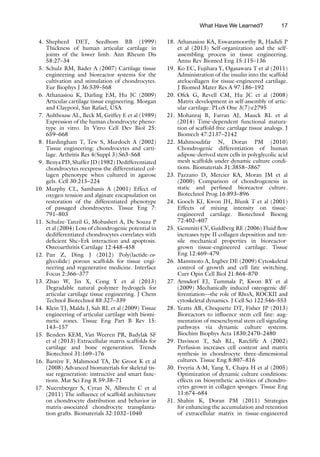


![25
Pauline M. Doran (ed.), Cartilage Tissue Engineering: Methods and Protocols, Methods in Molecular Biology,
vol. 1340, DOI 10.1007/978-1-4939-2938-2_2, © Springer Science+Business Media New York 2015
Chapter 2
Human Fetal and Adult Chondrocytes
Kifah Shahin, Nastaran Mahmoudifar, and Pauline M. Doran
Abstract
As the only cell type found in healthy adult cartilage, chondrocytes are the obvious and most direct starting
point for cartilage tissue engineering. Human adult, juvenile, neonatal, and fetal chondrocytes have all
been demonstrated to produce cartilage matrix components in vitro for production of engineered tissues.
In this chapter, procedures are outlined for isolation of chondrocytes from human fetal and adult cartilage.
Methods for expansion and cryopreservation of the cells and characterization of gene expression using
quantitative polymerase chain reaction (Q-PCR) analysis are also described.
Key words Chondrocyte, Collagenase, Cryopreservation, Dedifferentiation, Human adult and fetal
tissue, Q-PCR
1 Introduction
Human adult cartilage is an avascular and aneural connective tissue
containing chondrocytes in lacunae embedded within a dense solid
matrix. Chondrocytes are the cells that synthesize the biochemical
and structural elements of cartilage. They have a characteristic
rounded morphology and secrete an extracellular matrix rich in
collagen type II and the proteoglycan, aggrecan. To generate car-
tilage in vitro, chondrocytic cells are needed for application in
three-dimensional culture systems. Although human embryonic,
induced pluripotent and mesenchymal stem cells have been inves-
tigated for chondrogenesis and cartilage production [1, 2], differ-
entiated chondrocytes isolated from native cartilage tissue remain
the most direct effectors of cartilage synthesis.
Because cartilage has a very limited capacity for self-repair, obtain-
ing healthy human adult chondrocytes for tissue engineering
applications presents some difficulties, as there is a risk of inflicting
an unacceptable level of irreparable injury when cartilage is har-
vested from the joints of living donors. Nevertheless, surgical pro-
cedures may be used to remove a biopsy of articular cartilage from
1.1 Availability
and Properties
of Human
Chondrocytes
for Tissue Engineering](https://image.slidesharecdn.com/2610845-250620031004-75acac37/85/Cartilage-Tissue-Engineering-Methods-And-Protocols-Pauline-M-Doran-42-320.jpg)
![26
a low-weight-
bearing location: 200–300-mg slivers of partial- or
full-
thickness articular cartilage are obtained routinely from
patients to provide cells for autologous chondrocyte implantation
[3]. Other options include harvesting articular cartilage from
adult donors after death [4, 5] or limb amputation [6, 7], and col-
lecting cartilage tissue removed during arthroplasty or joint sur-
gery [7, 8]. Because most joint surgery is carried out in response
to damage of articular cartilage by osteoarthritis or physical
trauma, it is important that the cartilage tissues and chondrocytes
recovered from arthroplasty or similar procedures are not detri-
mentally affected by disease or injury. When cultured in three-
dimensional scaffolds, articular chondrocytes from patients with
osteoarthritis were found to differentially express 184 genes com-
pared with chondrocytes from normal donors [9]. In other stud-
ies, collagen synthesis by chondrocytes isolated from
osteoarthritis-affected cartilage was significantly lower than for
chondrocytes from unaffected tissue [10].
Alternatives to adult chondrocytes for human cartilage tissue
engineering are fetal [11, 12], neonatal [13], and juvenile (<13
years old) [14] chondrocytes. These cells are suitable principally
for research purposes: allogeneic chondrocytes and the engineered
tissues derived from them may be expected to produce undesirable
immune responses if used clinically, although there is evidence that
fresh osteochondral allografts and juvenile allogeneic chondrocytes
do not elicit graft rejection reactions [14]. Chondrocytes isolated
from young (neonatal or juvenile) human cartilage expand in cul-
ture at a considerably faster rate than adult chondrocytes [13, 14].
Juvenile-derived chondrocytes also out-perform chondrocytes
from adult donors in terms of their ability to synthesize cartilage
matrix components, suggesting that chondrocytes from young tis-
sues have a more strongly enhanced chondrogenic potential com-
pared with adult cells [14]. Several studies have demonstrated that
fetal, neonatal, and adult chondrocytes are more highly productive
of cartilage matrix in three-dimensional cultures in vitro than
chondro-induced mesenchymal stem cells [15–17].
Irrespective of whether fetal, neonatal, juvenile, or adult cartilage
is used as a source of human chondrocytes, the number of cells that
can be isolated from the available tissue is likely to be too low for
direct tissue engineering applications, particularly when scaffold
seeding is required. Accordingly, expansion of the cells must be
carried out, typically using monolayer culture. It has been known
for several decades that monolayer expansion is detrimental to
chondrocyte differentiation [18–20]. Dedifferentiation to a
fibroblast-
like phenotype, including development of a flat
spindle-
like morphology, downregulation of cartilage-specific
genes, and reduction in cartilage matrix synthesis, constitutes the
usual response to monolayer culture. Representative gene expres-
siondataillustratingthiseffectareshowninFig.1.Dedifferentiation
1.2 Practical
Aspects
Kifah Shahin et al.](https://image.slidesharecdn.com/2610845-250620031004-75acac37/85/Cartilage-Tissue-Engineering-Methods-And-Protocols-Pauline-M-Doran-43-320.jpg)
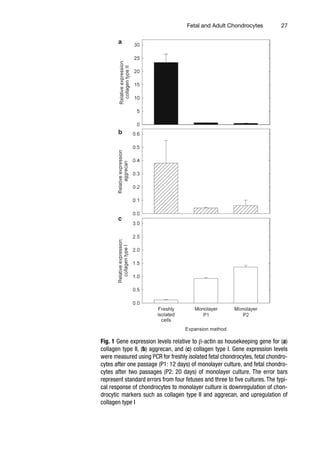
![28
is highly undesirable because dedifferentiated chondrocytes
produce fibrocartilage, a less resilient type of extracellular matrix
lacking the vital functional properties of articular cartilage that give
joints their strength and durability. Practically, it means that
expanded chondrocytes must be redifferentiated for tissue engi-
neering purposes.
Several investigators have observed that dedifferentiated chon-
drocytes are capable of redifferentiating when cultured in three-
dimensional hydrogels [18, 19, 21, 22]. However, results indicating
that full chondrocytic function is irreversibly compromised after
monolayer expansion have also been reported [23, 24]. Because
cartilage synthesis capacity decreases as the number of population
doublings increases [23, 24], in our work, we limit monolayer pas-
saging of chondrocytes to Passage 2. Alternatives to monolayer
expansion have been sought to allow better retention of the chon-
drocytic phenotype. Although cell differentiation is maintained to
a greater extent in three-dimensional systems compared with sur-
face culture, three-dimensional culture is not a feasible option for
increasing cell numbers. As shown in Fig. 2, cell proliferation is
greatly reduced when the cells are seeded in scaffolds compared
with the level of expansion achieved under monolayer conditions.
Chondrocytes isolated from individual donors can differ sub-
stantially in their ability to synthesize cartilage matrix. As an exam-
ple, levels of glycosaminoglycan (GAG) production by human
chondrocytes from 52 different individuals varied by a factor of
Fig. 2 Cell number after expansion of fetal chondrocytes in monolayer culture for
one passage (12 days), monolayer culture for two passages (20 days), polygly-
colic acid (PGA) scaffolds for 20 days, and PGA–alginate scaffolds [12] for 20
days. The initial number of freshly isolated cells in each culture was 1.5×10⁶
.
The error bars represent standard errors from triplicate cultures. Expansion in
monolayer culture for two passages generated 4.4–5.2 times the number of cells
obtained using three-dimensional scaffold systems over the same period
Kifah Shahin et al.](https://image.slidesharecdn.com/2610845-250620031004-75acac37/85/Cartilage-Tissue-Engineering-Methods-And-Protocols-Pauline-M-Doran-45-320.jpg)
![29
five- to six-fold when the cells were cultured as pellets in medium
with growth factors [25]. Significant inter-individual variation was
found between donors within the same age group as well as across
different ages. The observed variations in chondrogenic potential
may reflect genetic differences between individuals as well as life-
style diversity and pharmaceutical history. When chondrocytes
from individual patients are used for cartilage tissue engineering,
there is a risk that the experimental findings will reflect the proper-
ties of the particular chondrocytes studied and will not be relevant
more generally. To overcome this problem, chondrocytes isolated
from as many individuals as possible are pooled for use in experi-
ments. In this way, the results obtained reflect an average cellular
phenotype, thus dampening the effects of individual variation.
Here we describe methods for isolation, expansion, cryopreserva-
tion, and characterization of chondrocytes from human fetal and
adult cartilage. The methods for cell isolation are based on enzy-
matic digestion of cartilage tissue using collagenase type II. General
protocols for cell expansion are outlined using monolayer culture
and serum-containing medium; cryopreservation in liquid nitro-
gen is carried out using dimethyl sulfoxide (DMSO) as cryoprotec-
tant. Techniques for quantitative polymerase chain reaction
(Q-PCR) analysis are provided for examining the expression of
cartilage-specific genes in freshly isolated or expanded chondro-
cytes. The cells isolated and characterized using these methods are
suitable for use in tissue engineering culture systems. As well as
direct applications for production of cartilage matrix using porous
scaffolds and bioreactors [11, 26], chondrocytes may also be used
in coculture systems to improve the chondrogenic differentiation
of mesenchymal stem cells [27] and for in vitro development of
composite osteochondral constructs suitable for joint repair [28].
2 Materials
1. Human fetal knee or hip joint tissues obtained with informed
parental consent (see Note 1).
2. Adult articular cartilage tissue pieces obtained with informed con-
sent from patients undergoing knee or hip surgery (see Note 1).
1. DMEM base medium: Dulbecco’s modified Eagle’s medium
(DMEM) containing 4.5 g/L of glucose and 584 mg/L of
l-glutamine with 3.7 g of sodium hydrogen carbonate, 2.39 g
of N-2-(hydroxyethyl)piperazine-N′-2-ethane sulfonic acid
(HEPES), 0.046 g of l-proline, and 10 mL of 100× nones-
sential amino acid solution (Sigma-Aldrich) made up to
895 mL in Milli-Q water. Sterilize by filtration using 0.2-μm
pressure-filtration units.
1.3 Chondrocyte
Isolation,
Characterization,
and Application
2.1 Clinical Samples
2.2 Chondrocyte
Isolation, Expansion,
and Cryopreservation
Fetal and Adult Chondrocytes](https://image.slidesharecdn.com/2610845-250620031004-75acac37/85/Cartilage-Tissue-Engineering-Methods-And-Protocols-Pauline-M-Doran-46-320.jpg)
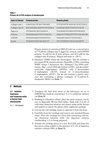
![36
4 Notes
1. Approval from an appropriate local institutional human
research ethics committee must be obtained before human tis-
sues can be collected for research purposes. Because of the
potential variability between individual patients, tissues from as
many donors as possible should be obtained to allow pooling
of the isolated chondrocytes before application in tissue engi-
neering experiments. The age of the donor or gestational age
of the fetus should be recorded. Fetal tissues after 16–20 weeks
of gestation provide sufficient joint material for chondrocyte
isolation.
2. The activity of type II clostridial collagenase purchased from
Sigma-Aldrich varies: we have observed batch variations from
350 to 450 U/mg solid. Accordingly, the mass concentration
of collagenase in the stock solution should be adjusted to
obtain 3000 U/mL for digestion of fetal cartilage or approxi-
mately 4300 U/mL for digestion of adult cartilage. The unit
of collagenase enzyme activity used by Sigma is defined as the
amount of enzyme that liberates peptides from collagen from
bovine achilles tendon equivalent in ninhydrin color to
1.0 μmol of leucine in 5 h at 37 °C and pH 7.4 in the presence
of calcium ions. The conversion factor from this Sigma unit to
the Mandl unit is 1000 to 1.
3. Aliquot the stock solution into the volumes required before
freezing, as repeated freeze–thaw cycles reduce enzyme activ-
ity. One milliliter of collagenase stock solution is required to
digest 300 mg of fetal cartilage tissue or 500 mg of adult car-
tilage tissue.
4. Ascorbic acid is often added to chondrocyte culture media in
various contexts. However, we have found that including
ascorbic acid in the medium used to culture freshly isolated
fetal chondrocytes results in complete cell death.
5. DMSO can be sterilized by filtration using a 0.2-μm single-use
syringe filter.
6. GAPDH and β-actin are housekeeping genes. They are used as
internal reference genes with the assumption that their expres-
sion levels are not affected by the experimental treatment. The
use of up to five housekeeping genes is common and produces
better validated results than when only a single housekeeping
gene is applied [29].
7. ROX reference dye normalizes the fluorescent reporter signal.
It normalizes non-PCR-related fluctuations in fluorescence
from well-to-well that may occur due to artifacts such as pipet-
ting errors or instrument limitations. Because ROX dye is for-
mulated to work on Applied Biosystems (ABI) and Stratagene
Kifah Shahin et al.](https://image.slidesharecdn.com/2610845-250620031004-75acac37/85/Cartilage-Tissue-Engineering-Methods-And-Protocols-Pauline-M-Doran-53-320.jpg)


![Argentine gaucho today) Eastman
color. © Edward Larocque Tinker &
Tinker Foundation; 29Jun64; MP14215.
THE GAUCHO OF SALTA. Tinker Foundation
& Edward Larocque Tinker. 45 min.,
sd., color, 16 mm. (The Argentine
gaucho today) Eastman color.
© Edward Larocque Tinker & Tinker
Foundation; 29Jun64; MP14217.
THE GAUCHO OF THE PAMPAS. Tinker
Foundation & Edward Larocque Tinker.
45 min., sd., color, 16 mm. (The
Argentine gaucho today) Eastman
color. © Edward Larocque Tinker &
Tinker Foundation; 29Jun64; MP14216.
GAUGUIN IN TAHITI: THE SEARCH FOR
PARADISE. See
CBS NEWS SPECIAL.
LES GAULOISES BLEUES. Les Films Ariane
[Paris] Released by Artistes Associes;
in the U.S. by Lopert Pictures Corp.
93 min., sd., color, 35 mm. © Les
Films Ariane, S.A.; 28Aug68; LF49.
THE GAUNTLET. See
THE VIRGINIAN.
GAVEA, RIO DE JANEIRO. See
SHELL'S WONDERFUL WORLD OF GOLF.](https://image.slidesharecdn.com/2610845-250620031004-75acac37/85/Cartilage-Tissue-Engineering-Methods-And-Protocols-Pauline-M-Doran-72-320.jpg)