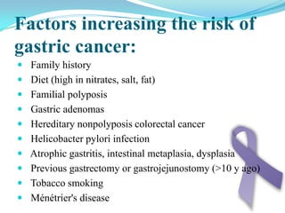
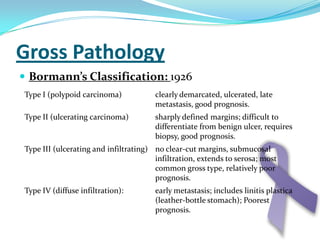
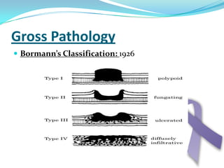
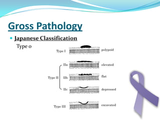
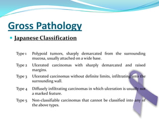
Gastric Cancer EpidemiologyAmr Mohamed Helmy 	 734
Features of Gastric Cancer1) Second most common cancer 2) Dramatic decline worldwide 3) Wide variation in incidence4) Altered risk among migrants
Age and Sex DistributionMen > womenMost are elderly at diagnosis. Median age 65 yearsYounger patients may represent a more aggressive variant.Cigarettes
Geographic Distribution
Geographic DistributionHighest rates (over 40 per 100,000 in males) are reported from Japan, China, the former USSR, and certain countries in Latin America.The lowest rates (< 15 per 100,000) are seen in North America (specifically, its white population), India, the Philippines, most African countries, some countries in Western Europe, and Australia.
Change In Histology PatternIntestinal gastric cancer common in males older age more prevalent in high-risk areas linked to environmental factors The diffuse or infiltrative typefrequent in both sexescommon in younger age groupsworse prognosis
Change In Histology PatternWorldwide decline in the incidence of the intestinal type.By contrast, the decline in the diffuse type has been more gradual (now accounts for approximately 30 percent of gastric carcinoma)Despite the decline in gastric cancer overall, there has been an explosive increase in incidence of cancer of the gastric cardia.
LymphomaGIT is the predominant site of extranodal non-Hodgkin lymphomas.Primary NHLs of the GI tract are rare, accounting for only 1 to 4 percent of malignancies arising in the stomach, small intestine, or colon. Secondary GI involvement is relatively common, occurring in approximately 10 percent of patients with limited stage NHL at the time of diagnosis, and up to 60 percent of those dying from advanced NHL.In the United States (US), gastric lymphoma is the most common extranodal site of lymphoma.
CarcinoidTumourThe overall age-adjusted incidence rates were 2.0 for men and 2.4/100,000 for women in 1983-1998.
GISTThe most common nonepithelial benign neoplasm involving the GI tract.Constitute only 1 percent of primary GI cancers. The annual incidence of GIST in the United States is at least 4000 to 6000 new cases (roughly 7 to 20 cases per million population per year).
EtiologyAmr Mohamed Samir 	735Amr Mohamed Dawood	737
Gastric cancer is more common in patients with pernicious anemia, blood group A, or a family history of gastric cancer.Environmental factors appear to be more related etiologically to the intestinal form than the diffuse form.
Factors increasing the risk of gastric cancer:  Family history  Diet (high in nitrates, salt, fat)  Familial polyposis  Gastric adenomas  Hereditary nonpolyposis colorectal cancer  Helicobacter pylori infection  Atrophic gastritis, intestinal metaplasia, dysplasia  Previous gastrectomy or gastrojejunostomy (>10 y ago)  Tobacco smokingMénétrier's disease
Factors decreasing the risk of gastric cancer:  Aspirin  Diet (high fresh fruit and vegetable intake)  Vitamin C
Helicobacter Pylori:Chronic helicobacter pylori infection increases the risk of gastric cancer about threefold when compared to uninfected patients.Patients with history of gastric ulcer are more likely to  develop gastric cancer, and patients with a history of duodenal ulcer are at decreased risk for gastric cancer.Premalignant conditions of the stomach1. PolypsHyperplastic polyps can be associated with carcinoma.Patients with familial adenomatouspolyposis have a high prevalence of gastric adenocarcinoma.
2. Atrophic gastritisChronic atrophic gastritis is the most common precursor for gastric cancer particularly the intestinal subtype and is usually associated with H. Pylori infection.
3. Intestinal metaplasiaGastric carcinoma often occurs in an area of intestinal metaplasia.Type III intestinal metaplasia is most commonly associated with gastric cancer, usually of the intestinal subtype.Eradication of helicobacter pylori infection leads to significant regression of intestinal metaplasia and improvement in atrophic gastritis.
4. Gastric remnant cancer stomach cancer can develop, usually years following distal gastrectomy or gastroenterostomy.5. Other premalignant states:Patients with hereditary, non polyposis colorectal cancer have a 10% risk of developing gastric cancer.Ménétrier's disease: gastric mucosal hypertrophy.
Gross PathologyAmr Maher Basiony	733Amr Mohamed Helmy	734
Premalignant Conditions of the StomachPolypsHyperplastic Polyps75 percent of gastric polyps
	Associated with H. pylori is common
	Small, dome-shaped, or stalked polyps
	Average size 1.0 cm, range 0.1 to 12 cm
	Single or multiple
	They primarily occur in the antrum
	They often regress following H. pylori eradicationPremalignant Conditions of the StomachPolypsFundic Gland Polyps	Small (0.1 to 0.8 cm), hyperemic, sessile, flat, nodular 	lesions
	Occur exclusively in the gastric corpus
	Sometimes be large
 	Removal of polyps greater than 1 cm in size is 	recommended
	Associated with Long-term therapy with proton pump 	inhibitors (PPIs)
	Regression has been reported following an H. pylori 	infection.Premalignant Conditions of the StomachPolypsGastric AdenomasAdenomatous polyps
6 to 10 percent of gastric polyps
Found in the antrum
May be flat or polypoid
Range in size from a few millimeters to several centimetersPremalignant Conditions of the StomachGastric CarcinoidTumoursMay grow as a polypoid lesion
Most commonly they are present in the corpus
Sessile, broad-based nodules, with a smooth surface contourPremalignant Conditions of the StomachAtrophic GastritisThe most common precursor for gastric cancer
Prevalence is higher in older age groups
H. pylori is involved in the pathogenesis of atrophic gastritis.
Three distinct patterns have been descriped
Autoimmune (involves the acid-secreting proximal stomach),
Hypersecretory (involving the distal stomach)
Environmental (involving multiple random areas at the junction of the oxyntic and antral mucosa)Premalignant Conditions of the StomachIntestinal MetaplasiaA precursor lesion to gastric cancer.
Eradication of H. pylori infection leads to significant regression of intestinal metaplasia and improvement in atrophic gastritis.
Treatment of H. pylori infection is a reasonable recommendationPremalignant Conditions of the StomachBenign Gastric UlcerInadequately biopsied ulcers  was mistakenly labeled as "benign"
Now all gastric ulcers are cancer until proven otherwise with adequate biopsy and follow-up.Premalignant Conditions of the StomachGastric Remnant CancerStomach cancer can develop in the gastric remnant
Most tumors develop > 10 years following the initial operation
Arise often near the stoma
Quite large at presentation
Equally divided between intestinal and diffuse subtypes
Most cases have been reported following Billroth II gastroenterostomy
Prognosis is similar to proximal gastric cancerPremalignant Conditions of the StomachDysplasiagastric dysplasia is the universal precursor to gastric adenocarcinoma
Patients with severe dysplasia should be considered for gastric resection
EMR is recommended if the severe dysplasia is localized
Patients should be followed with endoscopic biopsy surveillance, and Helicobacter eradicationPremalignant Conditions of the Stomach Early Gastric CancerIt is adenocarcinoma limited to the mucosa and submucosa of the stomach, regardless of lymph node status
Approximately 10% of patients with early gastric cancer will have lymph node metastases
There are several types and subtypes:
Approximately 70% of early gastric cancers are well differentiated, 30% are poorly differentiated
Small intramucosal lesions can be treated with EMRGross PathologyBormann’s Classification: 1926
Gross PathologyBormann’s Classification: 1926
Gross PathologyJapanese ClassificationType 0
Gross PathologyJapanese Classification
Microscopic picture of adenocarcinomaAmr Mohamed Mosa	735Amr Mohamed Halawa	738
There are a number of classifications proposed forgastric  adenocarcinoma.
Lauren classification Intestinal typeGlandular cells .(large , abundant cytoplasm , hyperchromic nuclei)Intracellular mucin polarized to surface .Pushed margins , presence of lymphocytes .Metastasizes to nodes, liverDiffuse typeSmall clamps or single .(small . less cytoplasm , hypochromic nuclei)No polarized mucin .Diffuse and infiltrative .Mixtures of above two types
WHO classification TubularTubules and aciniBecomes solid if poorly differentiatedPapillaryFibrovascular stalksMucinous>50% of tumor is mucinSignet ring>50% of carcinoma is composed of signet ring cells
  Special types Clear cell variant described as havingclear to pale eosinophilic cytoplasm Tubulo-papillary architectureDysplasia ranges from minimal to severe Round basal or mid-level nuclei in lower gradesLocation in cardia and pylorus
HepatoidClosely resembles hepatocellular carcinoma Frequently associated with intestinal type / tubulo-papillary gastric adenocarcinomaEosinophilic to clear cytoplasmRound to oval nuclei with prominent nucleoliFrequent vascular invasion and liver metastasis
AdenosquamousMixture of two patterns, more than just focal
GradingWHO (applies only to tubular pattern in WHO classification)Well differentiated – well formed glandsModerately differentiated - may be combined as well (low grade)Poorly differentiated (high grade)Highly irregular glandsorSingle cells and clusters
Signs and symptomsAmr Mohamed Halawa738
DyspepsiaHighly suspicion for any elderly man above 40 complaining of dyspepsia which does not clear completely under simple medical treatment to stop medication and be investigated for  gastric carcinoma .
Pain:it differs from that of peptic ulcer that it is not usually induced after meals but more to be induced after meat and protein meals .it is not responsive to medical treatment , vomiting or alkali .it is usually due to high tone of the stomach wall or involving of nerve or peritoneum.It is visceral type of pain , vague , located in upper abdomen specially in epigastric region . N.B In gross picture type 2 and 3 (non infiltrating – infiltrating ulcer) there is peptic ulcer like pain and even relieved by antacid and this is dangerous cause it delay the diagnosis but usually there is achlorhydra .
Vomiting:Feature of organic stomach diseases like peptic ulcer or gastric carcinoma, it occurs in the moment of highest pain ,induced by tension and hyper tonicity of the stomach .it is a good sign if there is relief after vomiting which exclude out the malignancy potential
Nausea:Sickness sensation without actual vomit + sweating + feeling to faint .It happen due to hypotonia of gastric musculature , It is usually psychic in origin but as well in organic diseases like carcinoma or chronic gastritis .Nausea usually precede vomiting
Flatulence:Distention of stomach with gas which usually tend to be belshed but it is more in functional stomach disease more than organic ones .
Disturbance of appetite:Anorexia specially to protein meals also anorexia due to other diseases like gastritis ,TB , Anaemia , psychogenic .
Others:Heart burn , waterbrash ( water and mucus in mouth) and erucation (acidic gastric contents in mouth)
2- Anemia and Cachexia:Progressive anaemia , loss of weight , dehydration and general weakness and this can be the only presenting picture .
3- Mass:Mass in epigastrium or left hypochondrium , it may be the only presentation and it indicate a late inoperable tumor .It is irregular , tender , mobile or fixed.
Complication:Haematemesis , may be few in amount or copious associated with food particles , in peptic ulcer the amount is always higher than that of gastric carcinoma.Secondaries to the liver , pyloric obstruction …..
Spread:-Direct :It extends to seroperitonium then to the near by organs (pancreas , spleen , liver , transverse colon ) .Tumor in cardia : spread to esophagus Tumor in pylorus : to duodenum .Lymphatic : (very aggressive )to lymphnodes in greater and lesser curvature .celiac and para aortic mediastinal (for tumors of the cardia)virschaw lymph nodes
SpreadBlood :from portal vein to liver and maybe lung or bone .transperitoneal :1-krunkenburg syndrome (or maybe by blood) .2-sister mary Josef nodules .3-malignant ascites .4-Blumer’s shelf (mass during rectal examination) .
Paraneoplastic manifestations:-rarely seen at initial presentation. Dermatologic findings may include the sudden appearance of diffuse seborrheic keratoses or acanthosis nigricans  which is characterized by velvety and darkly pigmented patches on skin folds. Neither finding is specific for gastric cancer.Other paraneoplastic abnormalities that can occur in gastric cancer include a microangiopathic hemolytic anemia , membranous nephropathy , and hypercoagulable states (Trousseau's syndrome) . Polyarteritis nodosa has been reported as the single manifestation of an early and surgically curable gastric cancer
InvestigationsAmr Mohamed Dawood	737
I. Laboratory StudiesCBC : reveals anemia, due to bleeding, liver   dysfunction, or poor nutrition.Serum electrolytes.Coagulation studies.Liver and Kidney function tests.Tumor markers:Carcinoembryonic antigen (CEA)         Cancer antigen (CA) 19-9
II. EndoscopyRoom set-up and patient positioning for endoscopyUpper GI endoscopy is the diagnostic procedure of choice in the work-up of gastric carcinoma.Multiple biopsy specimens (7 or more) should be obtained from any visually suspicious areas.
Repeated sampling at the same tissue site, reaching deeper into the gastric wall.
Multiple samples should be obtained around the ulcer crater. Lesions may be:Fungating cauliflower lesion.Mucosal ulceration.Diffuse infiltrative (linitisplastica).
Adenocarcinoma of the cardia.  Large, lobulated, ulcerated mass at the gastroesophageal junction
Adenocarcinoma in the antrum manifested by ulcerated, circumferential mass and gastric outlet obstruction
Endoscopic view of an ulcerating adenocarcinoma
III. Endoscopic ultrasonography (EUS)EUS helps to determine the depth of tumor invasion.
Can be used to guide for biopsy.
Has the ability to detect sarcomas and other tumors arising from the submucosa and the musculosa (GIST).The gastric wall is visualized as 5 concentric bands:
Mucosa
Muscularis mucosa
Submucosa
Muscularispropria
SerosaEUS cannot permit assessment of tissue beyond a depth of about 5 cm.
Can not be used to assess distant nodal or liver metastases.
Can not differentiates between malignant and benign ulcers.IV. RadiologyBarium studyProvides preliminary information that may help to determine if a gastric lesion has benign or malignant features.
It may show thickened or enlarged gastric folds, filling defects, mass or ulcer.Barim study of upper gastrointestinal study shows a superficial ulcer in the gastric antrum (arrow) with thickened folds radiating towards the ulcer
Fungating mass of the body of the stomach
Barium meal showing infiltrating gastric carcinoma in the region of the incisura. There is irregular narrowing affecting both the lesser and greater curvatures (arrow) ‘Apple core sign”
Linitisplastica "leather-flask" appearing stomachThe tumor tend to infiltrate the submucosa and muscularispropria, superficial mucosal biopsies may be falsely negative.Computed Tomograohy (CT)It is widely used for tumor staging.
Demonstrates accurately  the size and location of the tumor.
Helps to assess the presence of nodal or visceral spread and involvement of other peritoneal structures (e.g., ovaries, liver).
Can not detect metastases smaller than 5 mm.T StagingCT scan showing infiltrating carcinoma                       Carcinoma of the cardia with liver 						                        metastasis
N StagingCarcinoma of the body of the stomach associated with regional lymphadenopathy and ascites
M StagingThe liver is the most common site for hematogenous metastases. Less common sites are the lungs, adrenal glands, kidneys, bone and brain.Pulmonary metastases and left pleural effusion
IV. Staging laparoscopyMore invasive modality.Laparoscopic evaluation of metastasis.
Has the advantage of directly visualizing the liver surface, the peritoneum,  local lymph nodes and other abdominal metastases.
Peritoneal cytology may be done, which increases the sensitivity of laparoscopy.TNM staging The staging schema of the AJCC (American Joint Committee on Cancer)   is based upon: Tumor (T) Nodal involvement (N) Metastasis (M)
T stage is dependent on depth of tumor invasion and not size.
Nodal stage is based upon the number of positive lymph nodes and not the proximity of the nodes to the primary tumor.Regional nodes include those located:
Along the greater curvature (greater curvature, gastroduodenal, gastroepiploic)
Along the lesser curvature (lesser curvature, left gastric, common hepatic, celiac)
Pancreatic and splenic area
Involvement of intraabdominal nodal groups (e.g. retropancreatic, portal, mesenteric, and paraaortic)    is classified as distant metastases.
The 2002 AJCCTNM staging of gastric carcinomaPrimary tumor (T)TX - Primary tumor (T) cannot be assessedT0 - No evidence of primary tumorTis - Carcinoma in situ, intraepithelial tumor without invasion of      	lamina propriaT1 - Tumor invades lamina propria or submucosaT2 - Tumor invades muscularispropria or subserosaT3 - Tumor penetrates serosa (ie, visceral peritoneum) without 	invasion of adjacent structuresT4 - Tumor invades adjacent structures
Regional lymph nodes (N)NX - Regional lymph nodes (N) cannot be assessedN0 - No regional lymph node metastasesN1 - Metastasis in 1-6 regional lymph nodesN2 - Metastasis in 7-15 regional lymph nodesN3 - Metastasis in more than 15 regional lymph nodesDistant metastasis (M)MX - Distant metastasis cannot be assessedM0 - No distant metastasisM1 - Distant metastasis
The AJCC 2010 modificationsTumors arising at the gastroesophageal junction (GEJ) or at the cardia that extend to the GEJ or esophegus are staged using the TNM system for esophageal cancer rather than that of the gastric cancer.N categories were modified as follow:
N1 = 1-2 positive nodes (compared to 1-6 in the  	        2002 criteria)
N2 = 3-6 positive nodes (compared to 7 - 15 in            	       2002)
N3 = >7 positive nodes (compared to >15 in 2002)
Positive peritoneal cytology is classified as M1 disease.
Stage grouping have been changed.REFERENCES Bailey and Love’s, short practice of surgery, 25th edition.
Schwartz’s principles of surgery, 9th edition.
AJCC (American Joint Committee on Cancer) Cancer Staging Manual, 7th ed, Edge, SB, Byrd, DR, Compton, CC, et al (Eds), Springer, New York 2010. p. 117.
Pollack, BJ, Chak, A, Sivak MV, Jr. Endoscopic ultrasonography. SeminOncol 1996; 23:336.
Byrne, MF, Jowell, PS. Gastrointestinal imaging: endoscopic ultrasound. Gastroenterology 2002; 122:1631.
Karita, M, Tada, M. Endoscopic and histologic diagnosis of submucosal tumors of the gastrointestinal tract using combined strip biopsy and bite biopsy.
Kienle, P, Buhl, K, Kuntz, C, et al. Prospective comparison of endoscopy, endosonography and computed tomography for staging of gastric tumors. Digestion 2002; 66:230.TreatmentAmr Mohamed Samir	735
Operability  Patients with incurable disease should not be subjected to radical surgery that cannot help them.Incurable patients are those with:Haematogenous metastasesInvolvement of the distant peritoneumN4 nodal disease and disease beyond the N4 nodesFixation to structures that cannot be removed. Involvement of another organ does not indicate incurability, provided that it can be removed.
Controversies with respect to operability include patients with:N3 nodal involvementInvolvement of the adjacent peritoneumRemaining patients are considered operable and should undergo curative resection. Most of the patients should have neoadjuvant chemotherapy as it improves survival.Total gastrectomyThe operation is best performed through a long upper midline incision. The stomach is removed en bloc, including the tissues of the entire greater omentum and lesser omentum. The transverse colon is completely separated from the greater omentum.
The subpyloric nodes are dissected and the first part of the duodenum is divided, usually with a surgical stapler.The hepatic nodes are dissected down to clear the hepatic artery. This dissection also includes the suprapyloric lymph nodes.The lymph node dissection is continued to the origin of the left gastric artery.
The dissection is then continued along the splenic artery taking all of the nodes at the superior aspect of the pancreas and in the splenichilum.The access to the nodal tissues around the upper stomach and oesophagogastric junction is achieved by separation of the stomach from the spleen, if the spleen is not going to be removed.
The oesophegus is divided at an appropriate point using a combination of stay sutures and a soft non crushing clamp, usually of the right angled variety.
It is important that the proximal and distal resection margins are well clear of the tumour as their involvement carries an appalling prognosis. If in doubt, a frozen section should be performed. Gastrointestinal continuity is reconstituted by means of a Roux loop.
Effective oesophagojejunostomyis achieved by:A purse-string is placed in the cut end of the oesophagus.The side of the oesophagus is stapled onto the side of the Roux loop by means of a circular stapler. The anastomosis can also be fashioned end to end.
The blind open end of the Roux loop may then be closed either with sutures or with a linear stapler. The Roux loop may be placed in either, an anticolic or retrocolic position.The jejunojejunostomy is undertaken in the usual fashion(end to side).
Extent of lymphadenectomyThere remains some controversy about the extent of the lymphadenectomy required for the optimal treatment of curable gastric cancers. In Japan, at least a D2 gastrectomy (removal of second tier of lymph nodes) is performed on all operable gastric cancers. They usually preserve the spleen and pancreas.
The Japanese research society for gastric cancer has numbered the lymph node stations that potentially drain the stomach. Generally, these are grouped into level D1 nodes which are perigastric, D2 nodes which are along the hepatic and splenic arteries, and D3 nodes which are the most distant.
Overall, it seems that the oncological outcome may be better following a D2 gastrectomy. The traditional radical gastrectomy removes the spleen and the distal pancreas en bloc with the stomach which is adequate for the clearance of the lymph nodes around the splenicartery. However, there now seems doubt that this substantially increases the complication rates. Therefore, it’s no longer routinely done as part of the D2 gatrectomy.
The difference between D1 and D2 operations depend upon the tiers of lymph node removed. In general, D1 resection involves the removal of perigastricnodes while D2 resection involves removal of the lymph nodes along the major arterial trunks along with the perigastric nodes.
Subtotal gastrectomyFor tumours distally placed in the stomach it seems unnecessary to remove the whole stomach. The operation is very similar to total gastrectomy except that the most proximal part of the stomach is preserved, the blood supply being derived from the short gastric arteries.
After the resection the gastrointestinal continuity is restored by:The simplest form of reconstruction is to close the stomach from the lesser curve, near the oesophagogastric junction with staples or sutures and then perform an anastomosis of the greater curve to the jejunum as in Billroth II/ polya type gastrectomy.Reconstruction using a Roux loop to avoid marked enterogastric reflux and bile reflux oesophagitis that occurs with Billroth II/ polya type gastrectomy.
Palliative surgery:In inoperable patients with resectabletumours having significant symptoms of, either obstruction or bleeding, palliative resection is appropriate. A palliative gastrectomy need not be radical and it is sufficient to remove the tumour and reconstruct the gastrointestinal tract.In inoperable patients with non resectabletumours obstructing the distal stomach, other palliative procedures need to be considered. Palliative procedures include:High gastroenterostomy which is a poor operation that very frequently doesn’t allow the stomach to empty adequately and may produce the additional problem of bile reflux.A Roux loop with wide anastomosis between the stomach and the jejunum may be a better option, even though this may not allow the stomach to empty well.
3.  Gastric exclusion and oesophagojejunostomy is practiced by some surgeons.In inoperable patients with non resectabletumours situated in the cardia, either palliative intubation, stenting or another form of recanalisation can be used.Endoscopic resection:In has been demonstrated in some centers that some patients with early gastric cancer can be adequately treated by an endoscopic mucosal resection. The addition of laparoscopic lymph node sampling may be considered in selected patients. This procedure should be limited to patients with:Tumors less than 2 cm in sizeNegative lymph node samplingConfined to the mucosa on endoscopic ultrasonogrophyAbsence of other gastric lesions
If the resected specimen shows no ulceration, no penetration of the muscularis mucosa, no lymphatic invasion, and size less than 3 cm, then the risk of lymph node metastases is less than 1%.

Gastric cancer presentation final

  • 1.
  • 2.
    Features of GastricCancer1) Second most common cancer 2) Dramatic decline worldwide 3) Wide variation in incidence4) Altered risk among migrants
  • 3.
    Age and SexDistributionMen > womenMost are elderly at diagnosis. Median age 65 yearsYounger patients may represent a more aggressive variant.Cigarettes
  • 4.
  • 5.
    Geographic DistributionHighest rates(over 40 per 100,000 in males) are reported from Japan, China, the former USSR, and certain countries in Latin America.The lowest rates (< 15 per 100,000) are seen in North America (specifically, its white population), India, the Philippines, most African countries, some countries in Western Europe, and Australia.
  • 6.
    Change In HistologyPatternIntestinal gastric cancer common in males older age more prevalent in high-risk areas linked to environmental factors The diffuse or infiltrative typefrequent in both sexescommon in younger age groupsworse prognosis
  • 7.
    Change In HistologyPatternWorldwide decline in the incidence of the intestinal type.By contrast, the decline in the diffuse type has been more gradual (now accounts for approximately 30 percent of gastric carcinoma)Despite the decline in gastric cancer overall, there has been an explosive increase in incidence of cancer of the gastric cardia.
  • 8.
    LymphomaGIT is thepredominant site of extranodal non-Hodgkin lymphomas.Primary NHLs of the GI tract are rare, accounting for only 1 to 4 percent of malignancies arising in the stomach, small intestine, or colon. Secondary GI involvement is relatively common, occurring in approximately 10 percent of patients with limited stage NHL at the time of diagnosis, and up to 60 percent of those dying from advanced NHL.In the United States (US), gastric lymphoma is the most common extranodal site of lymphoma.
  • 9.
    CarcinoidTumourThe overall age-adjustedincidence rates were 2.0 for men and 2.4/100,000 for women in 1983-1998.
  • 10.
    GISTThe most commonnonepithelial benign neoplasm involving the GI tract.Constitute only 1 percent of primary GI cancers. The annual incidence of GIST in the United States is at least 4000 to 6000 new cases (roughly 7 to 20 cases per million population per year).
  • 11.
    EtiologyAmr Mohamed Samir 735Amr Mohamed Dawood 737
  • 12.
    Gastric cancer ismore common in patients with pernicious anemia, blood group A, or a family history of gastric cancer.Environmental factors appear to be more related etiologically to the intestinal form than the diffuse form.
  • 13.
    Factors increasing therisk of gastric cancer: Family history Diet (high in nitrates, salt, fat) Familial polyposis Gastric adenomas Hereditary nonpolyposis colorectal cancer Helicobacter pylori infection Atrophic gastritis, intestinal metaplasia, dysplasia Previous gastrectomy or gastrojejunostomy (>10 y ago) Tobacco smokingMénétrier's disease
  • 14.
    Factors decreasing therisk of gastric cancer:  Aspirin  Diet (high fresh fruit and vegetable intake)  Vitamin C
  • 15.
    Helicobacter Pylori:Chronic helicobacterpylori infection increases the risk of gastric cancer about threefold when compared to uninfected patients.Patients with history of gastric ulcer are more likely to develop gastric cancer, and patients with a history of duodenal ulcer are at decreased risk for gastric cancer.Premalignant conditions of the stomach1. PolypsHyperplastic polyps can be associated with carcinoma.Patients with familial adenomatouspolyposis have a high prevalence of gastric adenocarcinoma.
  • 16.
    2. Atrophic gastritisChronicatrophic gastritis is the most common precursor for gastric cancer particularly the intestinal subtype and is usually associated with H. Pylori infection.
  • 17.
    3. Intestinal metaplasiaGastriccarcinoma often occurs in an area of intestinal metaplasia.Type III intestinal metaplasia is most commonly associated with gastric cancer, usually of the intestinal subtype.Eradication of helicobacter pylori infection leads to significant regression of intestinal metaplasia and improvement in atrophic gastritis.
  • 18.
    4. Gastric remnantcancer stomach cancer can develop, usually years following distal gastrectomy or gastroenterostomy.5. Other premalignant states:Patients with hereditary, non polyposis colorectal cancer have a 10% risk of developing gastric cancer.Ménétrier's disease: gastric mucosal hypertrophy.
  • 19.
    Gross PathologyAmr MaherBasiony 733Amr Mohamed Helmy 734
  • 20.
    Premalignant Conditions ofthe StomachPolypsHyperplastic Polyps75 percent of gastric polyps
  • 21.
    Associated with H.pylori is common
  • 22.
  • 23.
    Average size 1.0cm, range 0.1 to 12 cm
  • 24.
  • 25.
  • 26.
    They often regressfollowing H. pylori eradicationPremalignant Conditions of the StomachPolypsFundic Gland Polyps Small (0.1 to 0.8 cm), hyperemic, sessile, flat, nodular lesions
  • 27.
    Occur exclusively inthe gastric corpus
  • 28.
  • 29.
    Removal ofpolyps greater than 1 cm in size is recommended
  • 30.
    Associated with Long-termtherapy with proton pump inhibitors (PPIs)
  • 31.
    Regression has beenreported following an H. pylori infection.Premalignant Conditions of the StomachPolypsGastric AdenomasAdenomatous polyps
  • 32.
    6 to 10percent of gastric polyps
  • 33.
  • 34.
    May be flator polypoid
  • 35.
    Range in sizefrom a few millimeters to several centimetersPremalignant Conditions of the StomachGastric CarcinoidTumoursMay grow as a polypoid lesion
  • 36.
    Most commonly theyare present in the corpus
  • 37.
    Sessile, broad-based nodules,with a smooth surface contourPremalignant Conditions of the StomachAtrophic GastritisThe most common precursor for gastric cancer
  • 38.
    Prevalence is higherin older age groups
  • 39.
    H. pylori isinvolved in the pathogenesis of atrophic gastritis.
  • 40.
    Three distinct patternshave been descriped
  • 41.
    Autoimmune (involves theacid-secreting proximal stomach),
  • 42.
  • 43.
    Environmental (involving multiplerandom areas at the junction of the oxyntic and antral mucosa)Premalignant Conditions of the StomachIntestinal MetaplasiaA precursor lesion to gastric cancer.
  • 44.
    Eradication of H.pylori infection leads to significant regression of intestinal metaplasia and improvement in atrophic gastritis.
  • 45.
    Treatment of H.pylori infection is a reasonable recommendationPremalignant Conditions of the StomachBenign Gastric UlcerInadequately biopsied ulcers was mistakenly labeled as "benign"
  • 46.
    Now all gastriculcers are cancer until proven otherwise with adequate biopsy and follow-up.Premalignant Conditions of the StomachGastric Remnant CancerStomach cancer can develop in the gastric remnant
  • 47.
    Most tumors develop> 10 years following the initial operation
  • 48.
  • 49.
    Quite large atpresentation
  • 50.
    Equally divided betweenintestinal and diffuse subtypes
  • 51.
    Most cases havebeen reported following Billroth II gastroenterostomy
  • 52.
    Prognosis is similarto proximal gastric cancerPremalignant Conditions of the StomachDysplasiagastric dysplasia is the universal precursor to gastric adenocarcinoma
  • 53.
    Patients with severedysplasia should be considered for gastric resection
  • 54.
    EMR is recommendedif the severe dysplasia is localized
  • 55.
    Patients should befollowed with endoscopic biopsy surveillance, and Helicobacter eradicationPremalignant Conditions of the Stomach Early Gastric CancerIt is adenocarcinoma limited to the mucosa and submucosa of the stomach, regardless of lymph node status
  • 56.
    Approximately 10% ofpatients with early gastric cancer will have lymph node metastases
  • 57.
    There are severaltypes and subtypes:
  • 58.
    Approximately 70% ofearly gastric cancers are well differentiated, 30% are poorly differentiated
  • 59.
    Small intramucosal lesionscan be treated with EMRGross PathologyBormann’s Classification: 1926
  • 60.
  • 61.
  • 62.
  • 63.
    Microscopic picture ofadenocarcinomaAmr Mohamed Mosa 735Amr Mohamed Halawa 738
  • 64.
    There are anumber of classifications proposed forgastric adenocarcinoma.
  • 65.
    Lauren classification IntestinaltypeGlandular cells .(large , abundant cytoplasm , hyperchromic nuclei)Intracellular mucin polarized to surface .Pushed margins , presence of lymphocytes .Metastasizes to nodes, liverDiffuse typeSmall clamps or single .(small . less cytoplasm , hypochromic nuclei)No polarized mucin .Diffuse and infiltrative .Mixtures of above two types
  • 66.
    WHO classification TubularTubulesand aciniBecomes solid if poorly differentiatedPapillaryFibrovascular stalksMucinous>50% of tumor is mucinSignet ring>50% of carcinoma is composed of signet ring cells
  • 67.
    Specialtypes Clear cell variant described as havingclear to pale eosinophilic cytoplasm Tubulo-papillary architectureDysplasia ranges from minimal to severe Round basal or mid-level nuclei in lower gradesLocation in cardia and pylorus
  • 68.
    HepatoidClosely resembles hepatocellularcarcinoma Frequently associated with intestinal type / tubulo-papillary gastric adenocarcinomaEosinophilic to clear cytoplasmRound to oval nuclei with prominent nucleoliFrequent vascular invasion and liver metastasis
  • 69.
    AdenosquamousMixture of twopatterns, more than just focal
  • 70.
    GradingWHO (applies onlyto tubular pattern in WHO classification)Well differentiated – well formed glandsModerately differentiated - may be combined as well (low grade)Poorly differentiated (high grade)Highly irregular glandsorSingle cells and clusters
  • 71.
    Signs and symptomsAmrMohamed Halawa738
  • 72.
    DyspepsiaHighly suspicion forany elderly man above 40 complaining of dyspepsia which does not clear completely under simple medical treatment to stop medication and be investigated for gastric carcinoma .
  • 73.
    Pain:it differs fromthat of peptic ulcer that it is not usually induced after meals but more to be induced after meat and protein meals .it is not responsive to medical treatment , vomiting or alkali .it is usually due to high tone of the stomach wall or involving of nerve or peritoneum.It is visceral type of pain , vague , located in upper abdomen specially in epigastric region . N.B In gross picture type 2 and 3 (non infiltrating – infiltrating ulcer) there is peptic ulcer like pain and even relieved by antacid and this is dangerous cause it delay the diagnosis but usually there is achlorhydra .
  • 74.
    Vomiting:Feature of organicstomach diseases like peptic ulcer or gastric carcinoma, it occurs in the moment of highest pain ,induced by tension and hyper tonicity of the stomach .it is a good sign if there is relief after vomiting which exclude out the malignancy potential
  • 75.
    Nausea:Sickness sensation withoutactual vomit + sweating + feeling to faint .It happen due to hypotonia of gastric musculature , It is usually psychic in origin but as well in organic diseases like carcinoma or chronic gastritis .Nausea usually precede vomiting
  • 76.
    Flatulence:Distention of stomachwith gas which usually tend to be belshed but it is more in functional stomach disease more than organic ones .
  • 77.
    Disturbance of appetite:Anorexiaspecially to protein meals also anorexia due to other diseases like gastritis ,TB , Anaemia , psychogenic .
  • 78.
    Others:Heart burn ,waterbrash ( water and mucus in mouth) and erucation (acidic gastric contents in mouth)
  • 79.
    2- Anemia andCachexia:Progressive anaemia , loss of weight , dehydration and general weakness and this can be the only presenting picture .
  • 80.
    3- Mass:Mass inepigastrium or left hypochondrium , it may be the only presentation and it indicate a late inoperable tumor .It is irregular , tender , mobile or fixed.
  • 81.
    Complication:Haematemesis , maybe few in amount or copious associated with food particles , in peptic ulcer the amount is always higher than that of gastric carcinoma.Secondaries to the liver , pyloric obstruction …..
  • 82.
    Spread:-Direct :It extendsto seroperitonium then to the near by organs (pancreas , spleen , liver , transverse colon ) .Tumor in cardia : spread to esophagus Tumor in pylorus : to duodenum .Lymphatic : (very aggressive )to lymphnodes in greater and lesser curvature .celiac and para aortic mediastinal (for tumors of the cardia)virschaw lymph nodes
  • 83.
    SpreadBlood :from portalvein to liver and maybe lung or bone .transperitoneal :1-krunkenburg syndrome (or maybe by blood) .2-sister mary Josef nodules .3-malignant ascites .4-Blumer’s shelf (mass during rectal examination) .
  • 84.
    Paraneoplastic manifestations:-rarely seenat initial presentation. Dermatologic findings may include the sudden appearance of diffuse seborrheic keratoses or acanthosis nigricans which is characterized by velvety and darkly pigmented patches on skin folds. Neither finding is specific for gastric cancer.Other paraneoplastic abnormalities that can occur in gastric cancer include a microangiopathic hemolytic anemia , membranous nephropathy , and hypercoagulable states (Trousseau's syndrome) . Polyarteritis nodosa has been reported as the single manifestation of an early and surgically curable gastric cancer
  • 85.
  • 86.
    I. Laboratory StudiesCBC: reveals anemia, due to bleeding, liver dysfunction, or poor nutrition.Serum electrolytes.Coagulation studies.Liver and Kidney function tests.Tumor markers:Carcinoembryonic antigen (CEA) Cancer antigen (CA) 19-9
  • 87.
    II. EndoscopyRoom set-upand patient positioning for endoscopyUpper GI endoscopy is the diagnostic procedure of choice in the work-up of gastric carcinoma.Multiple biopsy specimens (7 or more) should be obtained from any visually suspicious areas.
  • 88.
    Repeated sampling atthe same tissue site, reaching deeper into the gastric wall.
  • 89.
    Multiple samples shouldbe obtained around the ulcer crater. Lesions may be:Fungating cauliflower lesion.Mucosal ulceration.Diffuse infiltrative (linitisplastica).
  • 90.
    Adenocarcinoma of thecardia. Large, lobulated, ulcerated mass at the gastroesophageal junction
  • 91.
    Adenocarcinoma in theantrum manifested by ulcerated, circumferential mass and gastric outlet obstruction
  • 92.
    Endoscopic view ofan ulcerating adenocarcinoma
  • 93.
    III. Endoscopic ultrasonography(EUS)EUS helps to determine the depth of tumor invasion.
  • 94.
    Can be usedto guide for biopsy.
  • 95.
    Has the abilityto detect sarcomas and other tumors arising from the submucosa and the musculosa (GIST).The gastric wall is visualized as 5 concentric bands:
  • 96.
  • 97.
  • 98.
  • 99.
  • 100.
    SerosaEUS cannot permitassessment of tissue beyond a depth of about 5 cm.
  • 101.
    Can not beused to assess distant nodal or liver metastases.
  • 102.
    Can not differentiatesbetween malignant and benign ulcers.IV. RadiologyBarium studyProvides preliminary information that may help to determine if a gastric lesion has benign or malignant features.
  • 103.
    It may showthickened or enlarged gastric folds, filling defects, mass or ulcer.Barim study of upper gastrointestinal study shows a superficial ulcer in the gastric antrum (arrow) with thickened folds radiating towards the ulcer
  • 104.
    Fungating mass ofthe body of the stomach
  • 105.
    Barium meal showinginfiltrating gastric carcinoma in the region of the incisura. There is irregular narrowing affecting both the lesser and greater curvatures (arrow) ‘Apple core sign”
  • 106.
    Linitisplastica "leather-flask" appearingstomachThe tumor tend to infiltrate the submucosa and muscularispropria, superficial mucosal biopsies may be falsely negative.Computed Tomograohy (CT)It is widely used for tumor staging.
  • 107.
    Demonstrates accurately the size and location of the tumor.
  • 108.
    Helps to assessthe presence of nodal or visceral spread and involvement of other peritoneal structures (e.g., ovaries, liver).
  • 109.
    Can not detectmetastases smaller than 5 mm.T StagingCT scan showing infiltrating carcinoma Carcinoma of the cardia with liver metastasis
  • 110.
    N StagingCarcinoma ofthe body of the stomach associated with regional lymphadenopathy and ascites
  • 111.
    M StagingThe liveris the most common site for hematogenous metastases. Less common sites are the lungs, adrenal glands, kidneys, bone and brain.Pulmonary metastases and left pleural effusion
  • 112.
    IV. Staging laparoscopyMoreinvasive modality.Laparoscopic evaluation of metastasis.
  • 113.
    Has the advantageof directly visualizing the liver surface, the peritoneum, local lymph nodes and other abdominal metastases.
  • 114.
    Peritoneal cytology maybe done, which increases the sensitivity of laparoscopy.TNM staging The staging schema of the AJCC (American Joint Committee on Cancer) is based upon: Tumor (T) Nodal involvement (N) Metastasis (M)
  • 115.
    T stage isdependent on depth of tumor invasion and not size.
  • 116.
    Nodal stage isbased upon the number of positive lymph nodes and not the proximity of the nodes to the primary tumor.Regional nodes include those located:
  • 117.
    Along the greatercurvature (greater curvature, gastroduodenal, gastroepiploic)
  • 118.
    Along the lessercurvature (lesser curvature, left gastric, common hepatic, celiac)
  • 119.
  • 120.
    Involvement of intraabdominalnodal groups (e.g. retropancreatic, portal, mesenteric, and paraaortic) is classified as distant metastases.
  • 121.
    The 2002 AJCCTNMstaging of gastric carcinomaPrimary tumor (T)TX - Primary tumor (T) cannot be assessedT0 - No evidence of primary tumorTis - Carcinoma in situ, intraepithelial tumor without invasion of lamina propriaT1 - Tumor invades lamina propria or submucosaT2 - Tumor invades muscularispropria or subserosaT3 - Tumor penetrates serosa (ie, visceral peritoneum) without invasion of adjacent structuresT4 - Tumor invades adjacent structures
  • 122.
    Regional lymph nodes(N)NX - Regional lymph nodes (N) cannot be assessedN0 - No regional lymph node metastasesN1 - Metastasis in 1-6 regional lymph nodesN2 - Metastasis in 7-15 regional lymph nodesN3 - Metastasis in more than 15 regional lymph nodesDistant metastasis (M)MX - Distant metastasis cannot be assessedM0 - No distant metastasisM1 - Distant metastasis
  • 123.
    The AJCC 2010modificationsTumors arising at the gastroesophageal junction (GEJ) or at the cardia that extend to the GEJ or esophegus are staged using the TNM system for esophageal cancer rather than that of the gastric cancer.N categories were modified as follow:
  • 124.
    N1 = 1-2positive nodes (compared to 1-6 in the 2002 criteria)
  • 125.
    N2 = 3-6positive nodes (compared to 7 - 15 in 2002)
  • 126.
    N3 = >7positive nodes (compared to >15 in 2002)
  • 127.
    Positive peritoneal cytologyis classified as M1 disease.
  • 128.
    Stage grouping havebeen changed.REFERENCES Bailey and Love’s, short practice of surgery, 25th edition.
  • 129.
    Schwartz’s principles ofsurgery, 9th edition.
  • 130.
    AJCC (American JointCommittee on Cancer) Cancer Staging Manual, 7th ed, Edge, SB, Byrd, DR, Compton, CC, et al (Eds), Springer, New York 2010. p. 117.
  • 131.
    Pollack, BJ, Chak,A, Sivak MV, Jr. Endoscopic ultrasonography. SeminOncol 1996; 23:336.
  • 132.
    Byrne, MF, Jowell,PS. Gastrointestinal imaging: endoscopic ultrasound. Gastroenterology 2002; 122:1631.
  • 133.
    Karita, M, Tada,M. Endoscopic and histologic diagnosis of submucosal tumors of the gastrointestinal tract using combined strip biopsy and bite biopsy.
  • 134.
    Kienle, P, Buhl,K, Kuntz, C, et al. Prospective comparison of endoscopy, endosonography and computed tomography for staging of gastric tumors. Digestion 2002; 66:230.TreatmentAmr Mohamed Samir 735
  • 135.
    Operability Patientswith incurable disease should not be subjected to radical surgery that cannot help them.Incurable patients are those with:Haematogenous metastasesInvolvement of the distant peritoneumN4 nodal disease and disease beyond the N4 nodesFixation to structures that cannot be removed. Involvement of another organ does not indicate incurability, provided that it can be removed.
  • 136.
    Controversies with respectto operability include patients with:N3 nodal involvementInvolvement of the adjacent peritoneumRemaining patients are considered operable and should undergo curative resection. Most of the patients should have neoadjuvant chemotherapy as it improves survival.Total gastrectomyThe operation is best performed through a long upper midline incision. The stomach is removed en bloc, including the tissues of the entire greater omentum and lesser omentum. The transverse colon is completely separated from the greater omentum.
  • 137.
    The subpyloric nodesare dissected and the first part of the duodenum is divided, usually with a surgical stapler.The hepatic nodes are dissected down to clear the hepatic artery. This dissection also includes the suprapyloric lymph nodes.The lymph node dissection is continued to the origin of the left gastric artery.
  • 138.
    The dissection isthen continued along the splenic artery taking all of the nodes at the superior aspect of the pancreas and in the splenichilum.The access to the nodal tissues around the upper stomach and oesophagogastric junction is achieved by separation of the stomach from the spleen, if the spleen is not going to be removed.
  • 139.
    The oesophegus isdivided at an appropriate point using a combination of stay sutures and a soft non crushing clamp, usually of the right angled variety.
  • 140.
    It is importantthat the proximal and distal resection margins are well clear of the tumour as their involvement carries an appalling prognosis. If in doubt, a frozen section should be performed. Gastrointestinal continuity is reconstituted by means of a Roux loop.
  • 141.
    Effective oesophagojejunostomyis achievedby:A purse-string is placed in the cut end of the oesophagus.The side of the oesophagus is stapled onto the side of the Roux loop by means of a circular stapler. The anastomosis can also be fashioned end to end.
  • 142.
    The blind openend of the Roux loop may then be closed either with sutures or with a linear stapler. The Roux loop may be placed in either, an anticolic or retrocolic position.The jejunojejunostomy is undertaken in the usual fashion(end to side).
  • 143.
    Extent of lymphadenectomyThereremains some controversy about the extent of the lymphadenectomy required for the optimal treatment of curable gastric cancers. In Japan, at least a D2 gastrectomy (removal of second tier of lymph nodes) is performed on all operable gastric cancers. They usually preserve the spleen and pancreas.
  • 144.
    The Japanese researchsociety for gastric cancer has numbered the lymph node stations that potentially drain the stomach. Generally, these are grouped into level D1 nodes which are perigastric, D2 nodes which are along the hepatic and splenic arteries, and D3 nodes which are the most distant.
  • 145.
    Overall, it seemsthat the oncological outcome may be better following a D2 gastrectomy. The traditional radical gastrectomy removes the spleen and the distal pancreas en bloc with the stomach which is adequate for the clearance of the lymph nodes around the splenicartery. However, there now seems doubt that this substantially increases the complication rates. Therefore, it’s no longer routinely done as part of the D2 gatrectomy.
  • 146.
    The difference betweenD1 and D2 operations depend upon the tiers of lymph node removed. In general, D1 resection involves the removal of perigastricnodes while D2 resection involves removal of the lymph nodes along the major arterial trunks along with the perigastric nodes.
  • 147.
    Subtotal gastrectomyFor tumoursdistally placed in the stomach it seems unnecessary to remove the whole stomach. The operation is very similar to total gastrectomy except that the most proximal part of the stomach is preserved, the blood supply being derived from the short gastric arteries.
  • 148.
    After the resectionthe gastrointestinal continuity is restored by:The simplest form of reconstruction is to close the stomach from the lesser curve, near the oesophagogastric junction with staples or sutures and then perform an anastomosis of the greater curve to the jejunum as in Billroth II/ polya type gastrectomy.Reconstruction using a Roux loop to avoid marked enterogastric reflux and bile reflux oesophagitis that occurs with Billroth II/ polya type gastrectomy.
  • 149.
    Palliative surgery:In inoperablepatients with resectabletumours having significant symptoms of, either obstruction or bleeding, palliative resection is appropriate. A palliative gastrectomy need not be radical and it is sufficient to remove the tumour and reconstruct the gastrointestinal tract.In inoperable patients with non resectabletumours obstructing the distal stomach, other palliative procedures need to be considered. Palliative procedures include:High gastroenterostomy which is a poor operation that very frequently doesn’t allow the stomach to empty adequately and may produce the additional problem of bile reflux.A Roux loop with wide anastomosis between the stomach and the jejunum may be a better option, even though this may not allow the stomach to empty well.
  • 150.
    3. Gastricexclusion and oesophagojejunostomy is practiced by some surgeons.In inoperable patients with non resectabletumours situated in the cardia, either palliative intubation, stenting or another form of recanalisation can be used.Endoscopic resection:In has been demonstrated in some centers that some patients with early gastric cancer can be adequately treated by an endoscopic mucosal resection. The addition of laparoscopic lymph node sampling may be considered in selected patients. This procedure should be limited to patients with:Tumors less than 2 cm in sizeNegative lymph node samplingConfined to the mucosa on endoscopic ultrasonogrophyAbsence of other gastric lesions
  • 151.
    If the resectedspecimen shows no ulceration, no penetration of the muscularis mucosa, no lymphatic invasion, and size less than 3 cm, then the risk of lymph node metastases is less than 1%.