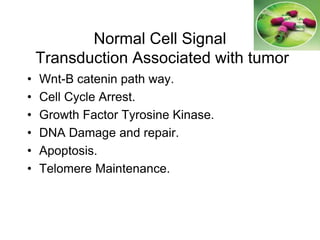
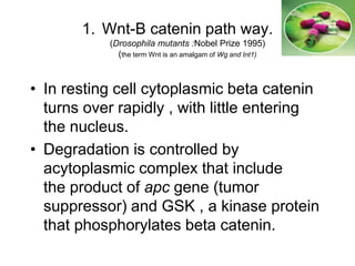
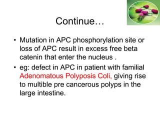
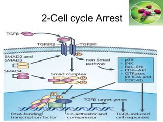
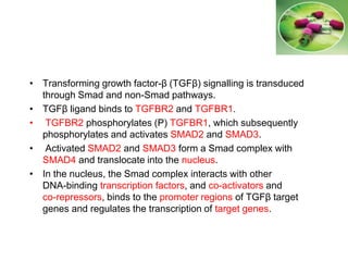
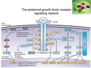
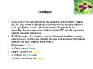
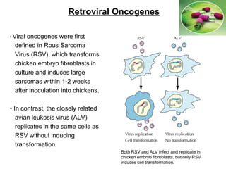
Molecular Pharmacology course
lecture No.5
Mutagenic Signal Transduction
and apoptosis
Dr.Omer Yahia Elhussein
B.sc. Pharmacy.
M.sc. Molecular Medicine.
Normal Cell Signal
Transduction Associated with tumor
• Wnt-B catenin path way.
• Cell Cycle Arrest.
• Growth Factor Tyrosine Kinase.
• DNA Damage and repair.
• Apoptosis.
• Telomere Maintenance.
1. Wnt-B catenin path way.
(Drosophila mutants :Nobel Prize 1995)
(the term Wnt is an amalgam of Wg and Int1)
• In resting cell cytoplasmic beta catenin
turns over rapidly , with little entering
the nucleus.
• Degradation is controlled by
acytoplasmic complex that include
the product of apc gene (tumor
suppressor) and GSK , a kinase protein
that phosphorylates beta catenin.
Wnt-B catenin path way
Continue…
• Mutation in APC phosphorylation site or
loss of APC result in excess free beta
catenin that enter the nucleus .
• eg: defect in APC in patient with familial
Adenomatous Polyposis Coli, giving rise
to multible pre cancerous polyps in the
large intestine.
The gene rearrangements resulting from
chromosome translocations frequently
lead to the generation of oncogenes.
The first characterized example of
oncogene activation by chromosome
translocation was the involvement of the
c-myc oncogene in human Burkitt's
lymphomas and mouse plasmacytomas,
which are malignancies of antibody-
producing B lymphocytes
(immunoglobulins).
For example, virtually all Burkitt's
lymphomas have translocations of a
fragment of chromosome 8 to one of the
immunoglobulin gene loci, which reside
on chromosomes 2, (k light chain), 14
(heavy chain), and 22 (g light chain).
Chromosomal abnormalities and cancer
The c-myc proto-oncogene is translocated from chromosome 8 to the
immunoglobulin heavy-chain locus (IgH) on chromosome 14 in Burkitt's
lymphomas, resulting in abnormal c-myc expression.
Cancer therapeutics that target
components of
the canonical Wnt pathway
APC, adenomatous polyposis coli ; TCF, T-cell factor
BCL9, B-cell lymphoma 9. P, phosphorylation
CBP, CREB-binding protein; PS1, presenilin 1
CK1α, casein kinase;1α; Pygo, Pygopus
DKK, Dickkopf; RTKs, receptor tyrosine kinases
DVL, Dishevelled; WIF,Wnt inhibitory factor 1
GSK3β, glycogen synthase kinase 3β LEF, lymphoid enhancer factor;
LRP5, LDL-receptor related protein 5MAPK,mitogen-activated protein kinase;
NRTKs, non-receptor tyrosine kinases; SFRP, secreted Frizzled-related protein
Cancer therapeutics that target
components of
the canonical Wnt pathway
Continue …..
• The non-steroidal anti-inflammatory drug (NSAID) celecoxib,
which targets the Wnt target gene COX2 (cyclooxygenase 2), is
already approved for the treatment of patients with familial
adenomatous polyposis (FAP)
• Therapeutic proteins, antibodies and low-molecular-weight
products that interfere with aberrant activity of the canonical Wnt
pathway are currently being developed or might be future
candidates.
• Imatinib mesylate (Gleevec), a tyrosine kinase inhibitor and an
approved therapeutic for chronic myeloid leukaemia (CML),
shows promise for the treatment of Wnt-induced gastrointestinal
tumours.
• Interference with members of other signalling
pathways, which directly or indirectly control
β-catenin stability (such as γ-secretase or ADAM, a
disintegrin and metalloproteinase ) might also provide
therapeutics against disease
Note :
• ADAM and γ-secretase are
• These metalloproteinase activities result in the
enhancement of migratory and invasive properties of
endothelial cells, which are required for tumour
angiogenesis.
2-Cell cycle Arrest
• Transforming growth factor-β (TGFβ) signalling is transduced
through Smad and non-Smad pathways.
• TGFβ ligand binds to TGFBR2 and TGFBR1.
• TGFBR2 phosphorylates (P) TGFBR1, which subsequently
phosphorylates and activates SMAD2 and SMAD3.
• Activated SMAD2 and SMAD3 form a Smad complex with
SMAD4 and translocate into the nucleus.
• In the nucleus, the Smad complex interacts with other
DNA-binding transcription factors, and co-activators and
co-repressors, binds to the promoter regions of TGFβ target
genes and regulates the transcription of target genes.
• TGFβ also regulates non-Smad pathways.
• TGFβ receptors activate p38, JNK, Ras–Erk, PI3K–Akt, and
small GTPases such as RHOA and CDC42.
• TGFβ stimulation inhibits cancer cell proliferation in some
cellular contexts and promotes it in others.
• Numerous factors are involved in TGFβ-regulated
cell proliferation and keep its signalling pathways balanced
1. It inhibit the proliferation of most adult cells (eg: TGF-beta null
mutation in mice causing death of inflammation in multiple
organs caused by excessive proliferation of lymphocytes )
2. TGF-beta stimulate production extra cellular matrix, including
collagen,proteoglycans and adhesion glycoprotein
• In addition to perturbation of TGFβ signalling, disruption or
mutation of regulators of TGFβ signalling can lead to a loss of
balanced TGFβ signalling, resulting in the generation and
progression of tumours.
• TGFβ signalling in cancer cells has dual roles in the regulation
of cell death and proliferation.
• TGFβ signalling has crucial roles in the maintenance of
self-renewal and tumorigenic activity of glioma-initiating cells
and leukaemia-initiating cells , whereas the function of TGFβ
signalling in breast cancer-initiating cells is controversial.
• TGFβ signalling is involved in several cell responses during
cancer cell metastasis, and cell type-dependent and
context-dependent factors contribute to the regulation of tumour
metastasis.
• Mutation in gene of TGF-beta or SMADS appear in up to 50% of
some types of eg: heridetary hemorrhagic telangiecta
• The TGFβ pathway has been targeted for cancer therapy using
multiple strategies. Some of them are currently in clinical trials
• Reserverotral (a potent antitumor factor) inhibit invasiveness of
human SCC cell at least in part , through Down regulation of
TGF-beta2 (Kopelovich L et al 2011)
3- FGFr signalling network.
Continue…
• Following ligand binding and receptor dimerization, the kinase domains
transphosphorylate each other, leading to the docking of adaptor proteins and
the activation of four key downstream pathways:
o RAS–RAF–MAPK signal transducer .
o PI3K(inositol triphosphate kinase)–AKT signal transducer.
o activator of transcription (STAT).
o phospholipase Cγ (PLCγ) .
• FGFRs have also been shown to bind and directly phosphorylate ribosomal S6
kinase17 .
• Signalling can be negatively regulated at several levels by receptor
internalization or the induction of negative regulators, including
o FGFR-like 1 (FGFRL1),
o SEF, Sprouty (SPRY),
o CBL,
o MAPK phosphatase 1 (MKP1) and MKP3 (brown).
Continue…
• These regulators may modulate ligand binding
(FGFRL1 and SEF) or interfere with intracellular
signalling, principally through modulation of the
MAPK pathway.
o DAG, diacylglycerol;
o FRS2α, FGFR substrate 2α;
o GRB2, growth factor receptor-bound 2;
o IP3, inositol triphosphate;
o P, phosphorylation;
o PIP2, phosphatidylinositol-4,5 biphosphate;
o PKC, protein kinase C;
o Sos, son of sevenless.
The epidermal growth factor receptor
signalling network
Continue…
• On activation (by ligand binding), the epidermal growth factor receptor
(EGFR, also known as ERBB1) autophosphorylates tyrosine residues
in its cytoplasmic domain, which serve as docking sites for the
assembly of protein complexes that transduce EGF signals to generate
specific biological responses.
• Scaffold protein : A protein that can simultaneously bind two or more
other proteins, and thereby facilitate physical and functional interactions
between the client proteins that bind to it.
o Kinases are light blue,
o scaffolds are dark blue,
o adaptor proteins are yellow,
o G proteins are green and
o transcription factors are orange.
Factor Principal
Source
Primary Activity Comments
PDGF platelets,
endothelial
cells, placenta
promotes proliferation of connective
tissue, glial and smooth muscle cells
two different protein chains form 3 distinct
dimer forms; AA, AB and BB
EGF submaxillary
gland, Brunners
gland
promotes proliferation of
mesenchymal, glial and epithelial
cells
TGF- common in
transformed
cells
may be important for normal wound
healing
related to EGF
FGF wide range of
cells; protein is
associated with
the ECM
promotes proliferation of many cells;
inhibits some stem cells; induces
mesoderm to form in early embryos
at least 19 family members, 4 distinct
receptors
NGF promotes neurite outgrowth and
neural cell survival
several related proteins first identified as
proto-oncogenes; trkA (trackA), trkB, trkC
Erythropoietin kidney promotes proliferation and
differentiation of erythrocytes
TGF- activated TH1
cells (T-helper)
and natural killer
(NK) cells
anti-inflammatory (suppresses
cytokine production and class II
MHC expression), promotes wound
healing, inhibits macrophage and
lymphocyte proliferation
at least 100 different family members
IGF-I primarily liver promotes proliferation of many cell
types
related to IGF-II and proinsulin, also called
Somatomedin C
IGF-II variety of cells promotes proliferation of many cell
types primarily of fetal origin
related to IGF-I and proinsulin
Growth Factors
4- DNA damage and repair
(Retinoblastoma , P53 , Mdm, E2F , Waf-1 , Bax)
• pRB primary function is to block cell cycle
progression and it is in activated in response
to mitogenic stimulation .
• So,loss of Prb can lead to in appropriate cycle
progression and cancer
• Rare individuals who inherit one inactivated
rb gene are predisposed to develop
Retinoblastoma as children and
Osteosarcomas as adult
• Homozygous loss of p RB is lethal during
embryogenesis, as the protein is essential for
promoting differentiation in various organs
and tissues.
• E2F is a tumor suppressor because it
opposes the cell cycle progression prior to
the restriction point when partnered with p RB
; However it can act also as an Oncogene as
it promotes the cell cycle when p RB in
activated
Retroviral Oncogenes
• Viral oncogenes were first
defined in Rous Sarcoma
Virus (RSV), which transforms
chicken embryo fibroblasts in
culture and induces large
sarcomas within 1-2 weeks
after inoculation into chickens.
• In contrast, the closely related
avian leukosis virus (ALV)
replicates in the same cells as
RSV without inducing
transformation.
Both RSV and ALV infect and replicate in
chicken embryo fibroblasts, but only RSV
induces cell transformation.
5- Apoptosis and cancer
• Cell dies because of :
Old and defective - incur some damage - surplus to the requirement of
tissues ( like finger formation )
• Cell dies in different ways:
1. Necrosis.
2. Apoptosis.
3. Autophagy.
Apoptosis :
controlled process leading to cell death that is triggered by intracellular
damage (e.g., DNA lesions) or by external signals from neighboring
cells. Also called programmed cell death.
Continue …..
Extrinsic path way
T cell induced death
Intrinsic path way
5-Telomer Maintenance
• Telomerase is the enzyme for maintaining the
end of the chromosome
• While the Telomerase enzyme is not active
in most normal human cells , the enzyme is
active in all types of cancer , but enables
cancer cells that have telomerase too short
for chromosome replication to divide
indefinitely by lengthening the telomeres and
stabilizing the chromosomes
• During successive cell cycles the telomeres shorten
because the primer needed by DNA Polymerase for
new DNA synthesis is removed.
• This action is counteracted by the action of
telomerase, which adds new telomere repeat
sequences to the ends of chromosomes.
• Eventually ,the telomeres become so short that the
complex between sequences and the proteins that
bind to them disrupted , and DNA damage occur.
Thanks
Mutagenic signal transduction (5)

Mutagenic signal transduction (5)

  • 1.
    Molecular Pharmacology course lectureNo.5 Mutagenic Signal Transduction and apoptosis Dr.Omer Yahia Elhussein B.sc. Pharmacy. M.sc. Molecular Medicine.
  • 3.
    Normal Cell Signal TransductionAssociated with tumor • Wnt-B catenin path way. • Cell Cycle Arrest. • Growth Factor Tyrosine Kinase. • DNA Damage and repair. • Apoptosis. • Telomere Maintenance.
  • 4.
    1. Wnt-B cateninpath way. (Drosophila mutants :Nobel Prize 1995) (the term Wnt is an amalgam of Wg and Int1) • In resting cell cytoplasmic beta catenin turns over rapidly , with little entering the nucleus. • Degradation is controlled by acytoplasmic complex that include the product of apc gene (tumor suppressor) and GSK , a kinase protein that phosphorylates beta catenin.
  • 5.
  • 6.
    Continue… • Mutation inAPC phosphorylation site or loss of APC result in excess free beta catenin that enter the nucleus . • eg: defect in APC in patient with familial Adenomatous Polyposis Coli, giving rise to multible pre cancerous polyps in the large intestine.
  • 8.
    The gene rearrangementsresulting from chromosome translocations frequently lead to the generation of oncogenes. The first characterized example of oncogene activation by chromosome translocation was the involvement of the c-myc oncogene in human Burkitt's lymphomas and mouse plasmacytomas, which are malignancies of antibody- producing B lymphocytes (immunoglobulins). For example, virtually all Burkitt's lymphomas have translocations of a fragment of chromosome 8 to one of the immunoglobulin gene loci, which reside on chromosomes 2, (k light chain), 14 (heavy chain), and 22 (g light chain). Chromosomal abnormalities and cancer The c-myc proto-oncogene is translocated from chromosome 8 to the immunoglobulin heavy-chain locus (IgH) on chromosome 14 in Burkitt's lymphomas, resulting in abnormal c-myc expression.
  • 9.
    Cancer therapeutics thattarget components of the canonical Wnt pathway APC, adenomatous polyposis coli ; TCF, T-cell factor BCL9, B-cell lymphoma 9. P, phosphorylation CBP, CREB-binding protein; PS1, presenilin 1 CK1α, casein kinase;1α; Pygo, Pygopus DKK, Dickkopf; RTKs, receptor tyrosine kinases DVL, Dishevelled; WIF,Wnt inhibitory factor 1 GSK3β, glycogen synthase kinase 3β LEF, lymphoid enhancer factor; LRP5, LDL-receptor related protein 5MAPK,mitogen-activated protein kinase; NRTKs, non-receptor tyrosine kinases; SFRP, secreted Frizzled-related protein
  • 10.
    Cancer therapeutics thattarget components of the canonical Wnt pathway
  • 11.
    Continue ….. • Thenon-steroidal anti-inflammatory drug (NSAID) celecoxib, which targets the Wnt target gene COX2 (cyclooxygenase 2), is already approved for the treatment of patients with familial adenomatous polyposis (FAP) • Therapeutic proteins, antibodies and low-molecular-weight products that interfere with aberrant activity of the canonical Wnt pathway are currently being developed or might be future candidates. • Imatinib mesylate (Gleevec), a tyrosine kinase inhibitor and an approved therapeutic for chronic myeloid leukaemia (CML), shows promise for the treatment of Wnt-induced gastrointestinal tumours.
  • 12.
    • Interference withmembers of other signalling pathways, which directly or indirectly control β-catenin stability (such as γ-secretase or ADAM, a disintegrin and metalloproteinase ) might also provide therapeutics against disease Note : • ADAM and γ-secretase are • These metalloproteinase activities result in the enhancement of migratory and invasive properties of endothelial cells, which are required for tumour angiogenesis.
  • 14.
  • 15.
    • Transforming growthfactor-β (TGFβ) signalling is transduced through Smad and non-Smad pathways. • TGFβ ligand binds to TGFBR2 and TGFBR1. • TGFBR2 phosphorylates (P) TGFBR1, which subsequently phosphorylates and activates SMAD2 and SMAD3. • Activated SMAD2 and SMAD3 form a Smad complex with SMAD4 and translocate into the nucleus. • In the nucleus, the Smad complex interacts with other DNA-binding transcription factors, and co-activators and co-repressors, binds to the promoter regions of TGFβ target genes and regulates the transcription of target genes.
  • 16.
    • TGFβ alsoregulates non-Smad pathways. • TGFβ receptors activate p38, JNK, Ras–Erk, PI3K–Akt, and small GTPases such as RHOA and CDC42. • TGFβ stimulation inhibits cancer cell proliferation in some cellular contexts and promotes it in others. • Numerous factors are involved in TGFβ-regulated cell proliferation and keep its signalling pathways balanced 1. It inhibit the proliferation of most adult cells (eg: TGF-beta null mutation in mice causing death of inflammation in multiple organs caused by excessive proliferation of lymphocytes ) 2. TGF-beta stimulate production extra cellular matrix, including collagen,proteoglycans and adhesion glycoprotein
  • 17.
    • In additionto perturbation of TGFβ signalling, disruption or mutation of regulators of TGFβ signalling can lead to a loss of balanced TGFβ signalling, resulting in the generation and progression of tumours. • TGFβ signalling in cancer cells has dual roles in the regulation of cell death and proliferation. • TGFβ signalling has crucial roles in the maintenance of self-renewal and tumorigenic activity of glioma-initiating cells and leukaemia-initiating cells , whereas the function of TGFβ signalling in breast cancer-initiating cells is controversial.
  • 19.
    • TGFβ signallingis involved in several cell responses during cancer cell metastasis, and cell type-dependent and context-dependent factors contribute to the regulation of tumour metastasis. • Mutation in gene of TGF-beta or SMADS appear in up to 50% of some types of eg: heridetary hemorrhagic telangiecta • The TGFβ pathway has been targeted for cancer therapy using multiple strategies. Some of them are currently in clinical trials • Reserverotral (a potent antitumor factor) inhibit invasiveness of human SCC cell at least in part , through Down regulation of TGF-beta2 (Kopelovich L et al 2011)
  • 21.
  • 22.
    Continue… • Following ligandbinding and receptor dimerization, the kinase domains transphosphorylate each other, leading to the docking of adaptor proteins and the activation of four key downstream pathways: o RAS–RAF–MAPK signal transducer . o PI3K(inositol triphosphate kinase)–AKT signal transducer. o activator of transcription (STAT). o phospholipase Cγ (PLCγ) . • FGFRs have also been shown to bind and directly phosphorylate ribosomal S6 kinase17 . • Signalling can be negatively regulated at several levels by receptor internalization or the induction of negative regulators, including o FGFR-like 1 (FGFRL1), o SEF, Sprouty (SPRY), o CBL, o MAPK phosphatase 1 (MKP1) and MKP3 (brown).
  • 23.
    Continue… • These regulatorsmay modulate ligand binding (FGFRL1 and SEF) or interfere with intracellular signalling, principally through modulation of the MAPK pathway. o DAG, diacylglycerol; o FRS2α, FGFR substrate 2α; o GRB2, growth factor receptor-bound 2; o IP3, inositol triphosphate; o P, phosphorylation; o PIP2, phosphatidylinositol-4,5 biphosphate; o PKC, protein kinase C; o Sos, son of sevenless.
  • 25.
    The epidermal growthfactor receptor signalling network
  • 26.
    Continue… • On activation(by ligand binding), the epidermal growth factor receptor (EGFR, also known as ERBB1) autophosphorylates tyrosine residues in its cytoplasmic domain, which serve as docking sites for the assembly of protein complexes that transduce EGF signals to generate specific biological responses. • Scaffold protein : A protein that can simultaneously bind two or more other proteins, and thereby facilitate physical and functional interactions between the client proteins that bind to it. o Kinases are light blue, o scaffolds are dark blue, o adaptor proteins are yellow, o G proteins are green and o transcription factors are orange.
  • 27.
    Factor Principal Source Primary ActivityComments PDGF platelets, endothelial cells, placenta promotes proliferation of connective tissue, glial and smooth muscle cells two different protein chains form 3 distinct dimer forms; AA, AB and BB EGF submaxillary gland, Brunners gland promotes proliferation of mesenchymal, glial and epithelial cells TGF- common in transformed cells may be important for normal wound healing related to EGF FGF wide range of cells; protein is associated with the ECM promotes proliferation of many cells; inhibits some stem cells; induces mesoderm to form in early embryos at least 19 family members, 4 distinct receptors NGF promotes neurite outgrowth and neural cell survival several related proteins first identified as proto-oncogenes; trkA (trackA), trkB, trkC Erythropoietin kidney promotes proliferation and differentiation of erythrocytes TGF- activated TH1 cells (T-helper) and natural killer (NK) cells anti-inflammatory (suppresses cytokine production and class II MHC expression), promotes wound healing, inhibits macrophage and lymphocyte proliferation at least 100 different family members IGF-I primarily liver promotes proliferation of many cell types related to IGF-II and proinsulin, also called Somatomedin C IGF-II variety of cells promotes proliferation of many cell types primarily of fetal origin related to IGF-I and proinsulin Growth Factors
  • 29.
    4- DNA damageand repair (Retinoblastoma , P53 , Mdm, E2F , Waf-1 , Bax) • pRB primary function is to block cell cycle progression and it is in activated in response to mitogenic stimulation . • So,loss of Prb can lead to in appropriate cycle progression and cancer • Rare individuals who inherit one inactivated rb gene are predisposed to develop Retinoblastoma as children and Osteosarcomas as adult
  • 30.
    • Homozygous lossof p RB is lethal during embryogenesis, as the protein is essential for promoting differentiation in various organs and tissues. • E2F is a tumor suppressor because it opposes the cell cycle progression prior to the restriction point when partnered with p RB ; However it can act also as an Oncogene as it promotes the cell cycle when p RB in activated
  • 34.
    Retroviral Oncogenes • Viraloncogenes were first defined in Rous Sarcoma Virus (RSV), which transforms chicken embryo fibroblasts in culture and induces large sarcomas within 1-2 weeks after inoculation into chickens. • In contrast, the closely related avian leukosis virus (ALV) replicates in the same cells as RSV without inducing transformation. Both RSV and ALV infect and replicate in chicken embryo fibroblasts, but only RSV induces cell transformation.
  • 36.
    5- Apoptosis andcancer • Cell dies because of : Old and defective - incur some damage - surplus to the requirement of tissues ( like finger formation ) • Cell dies in different ways: 1. Necrosis. 2. Apoptosis. 3. Autophagy. Apoptosis : controlled process leading to cell death that is triggered by intracellular damage (e.g., DNA lesions) or by external signals from neighboring cells. Also called programmed cell death.
  • 37.
    Continue ….. Extrinsic pathway T cell induced death Intrinsic path way
  • 39.
    5-Telomer Maintenance • Telomeraseis the enzyme for maintaining the end of the chromosome • While the Telomerase enzyme is not active in most normal human cells , the enzyme is active in all types of cancer , but enables cancer cells that have telomerase too short for chromosome replication to divide indefinitely by lengthening the telomeres and stabilizing the chromosomes
  • 41.
    • During successivecell cycles the telomeres shorten because the primer needed by DNA Polymerase for new DNA synthesis is removed. • This action is counteracted by the action of telomerase, which adds new telomere repeat sequences to the ends of chromosomes. • Eventually ,the telomeres become so short that the complex between sequences and the proteins that bind to them disrupted , and DNA damage occur.
  • 43.