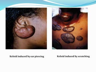
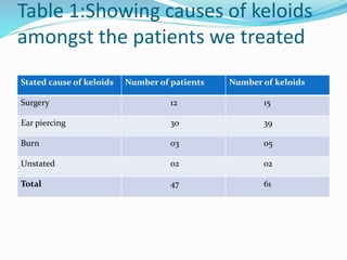
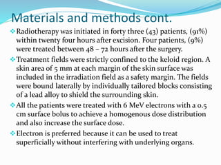
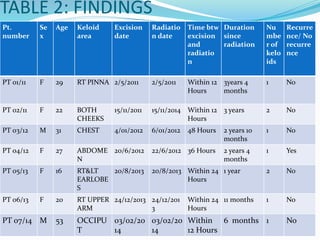
PRESENTED BY LOREH PETER
INTRODUCTION 
Keloid is a benign tumor, which arises from scar tissue is 
response to trauma. 
Although keloids have varying growth patterns, they are 
considered benign tumors, because the development of 
malignant cells in keloids is very rare. In fact, most 
patients seek treatment for cosmetic concerns. 
Most keloids are hard, raised and sometimes irritating 
or painful. 
Keloids have a tendency of recurring after surgical 
excision; the recurrence is usually seen within six 
months after excision. (Principles and practice of 
Radiation Oncology by Perez and Brady’s) 
As such, simple removal or plastic repair alone for keloid 
treatment is not always sufficient.
What are the causes of keloids? 
Cuts 
Scratches 
Insect bites 
Iatrogenic trauma as in vaccinations or surgical 
procedures 
Thermal or chemical burns 
And skin eruptions such as chicken pox
Keloid induced by ear piercing Keloid induced by scratching
Who is at risk? 
The risk of developing a keloid or keloids is equal for males 
and females. 
The most consistent risk factor for keloid development is a 
previous keloid. 
Hereditary: Analysis of patients history show that majority 
of people with keloids also had family members with them. 
Skin color/ethnicity: Keloids reportedly develop more 
often in people with darker skin pigmentations while they 
are NEVER seen in people without skin pigment (albinos). 
Blood type: People with blood type A have been linked 
with higher rates of keloid development. 
Age: Younger people (ages between 10 and 30) are more 
prone to keloids than older adults and the elderly. 
Endocrine factors: Pregnant women are more prone to 
keloid formation or worsening keloids whereas post-menopausal 
women rarely develop keloids.
How to prevent keloids 
People who are at risk of developing keloids should 
not get body piercing or tattoos. 
If you need surgery, you need to let your doctor know 
that you may develop keloids. 
Starting certain keloids treatments such as 
corticosteroid shots and pressure dressing 
immediately after surgery may help prevent keloids.
Materials and methods 
Between Jan, 2011 to date, forty seven (47) patients with a 
total of sixty one (61) keloids have been treated at Cancer 
Care Kenya. 
Out of these, forty (40) patients (85.25 %) were female 
between the ages of sixteen (16) and thirty four (34) years, 
three (3) patients (6.5 %) were male adults and four (4) 
patients (8.25 %) children below the age of ten. 
 For the purpose of this study, keloids that occurred at 
different sites in the same patient were considered as 
different keloids. 
The keloids were surgically removed and patients treated 
post-operatively with 9 Gy electron beam irradiation in a 
single fraction.
Table 1:Showing causes of keloids 
amongst the patients we treated 
Stated cause of keloids Number of patients Number of keloids 
Surgery 12 15 
Ear piercing 30 39 
Burn 03 05 
Unstated 02 02 
Total 47 61
Materials and methods cont. 
Radiotherapy was initiated in forty three (43) patients, (91%) 
within twenty four hours after excision. Four patients, (9%) 
were treated between 48 – 72 hours after the surgery. 
Treatment fields were strictly confined to the keloid region. A 
skin area of 5 mm at each margin of the skin surface was 
included in the irradiation field as a safety margin. The fields 
were bound laterally by individually tailored blocks consisting 
of a lead alloy to shield the surrounding skin. 
All the patients were treated with 6 MeV electrons with a 0.5 
cm surface bolus to achieve a homogenous dose distribution 
and also increase the surface dose. 
Electron is preferred because it can be used to treat 
superficially without interfering with underlying organs.
Pt. 
number 
Se 
x 
Age Keloid 
area 
Excision 
date 
Radiatio 
n date 
Time btw 
excision 
and 
radiatio 
n 
Duration 
since 
radiation 
Nu 
mbe 
r of 
kelo 
ids 
Recurre 
nce/ No 
recurre 
nce 
PT 01/11 F 29 RT PINNA 2/5/2011 2/5/2011 Within 12 
Hours 
3years 4 
months 
1 No 
PT 02/11 F 22 BOTH 
CHEEKS 
15/11/2011 15/11/2014 Within 12 
Hours 
3 years 2 No 
PT 03/12 M 31 CHEST 4/01/2012 6/01/2012 48 Hours 2 years 10 
months 
1 No 
PT 04/12 F 27 ABDOME 
N 
20/6/2012 22/6/2012 36 Hours 2 years 4 
months 
1 Yes 
PT 05/13 F 16 RT&LT 
EARLOBE 
S 
20/8/2013 20/8/2013 Within 24 
Hours 
1 year 2 No 
PT 06/13 F 20 RT UPPER 
ARM 
24/12/2013 24/12/201 
3 
Within 24 
Hours 
11 months 1 No 
PT 07/14 M 53 OCCIPU 
T 
03/02/20 
14 
03/02/20 
14 
Within 
12 Hours 
6 months 1 No 
TABLE 2: FINDINGS
Results 
All the patients came for review by their respective 
oncologists one month after the radiation. 
None of them reported any recurrence within this time. 
Phone call follow up six months later however revealed 
that two (2) keloids had recurred. Four patients (8.5%) 
with a total of five (5) keloids could not be reached for 
follow up while 54 keloids (88.7%) were reported to have 
healed completely. 
The two recurrences were observed in patients who had 
their radiation more than 24 hours after excision. 
The keloids related symptoms like itching and pain also 
improved in majority of the patients.
Conclusion and recommendations 
CONCLUSION 
Surgical excision of a keloid followed by immediate, single-fraction, 
high-dose radiotherapy is both safe and effective in 
preventing recurrence of therapy-resistant keloids. 
RECOMMENDATIONS 
The results above show that all the recurrences were observed 
in patients who showed up for radiation more than 24 hours 
after excision. 
This points to increase in recurrence as the time increases. We 
could however not make concrete conclusion on this due to 
limited sample size. 
I therefore recommend that more research be carried out to 
determine if delaying post operative radiotherapy would 
increase recurrence rate for keloids.
References 
Principles and practice of radiation oncology by Perez and 
Brady’s 
Keloid research foundation 
Thank you!!!

Post operative electron beam therapy for keloids by loreh peter

  • 2.
  • 3.
    INTRODUCTION Keloid isa benign tumor, which arises from scar tissue is response to trauma. Although keloids have varying growth patterns, they are considered benign tumors, because the development of malignant cells in keloids is very rare. In fact, most patients seek treatment for cosmetic concerns. Most keloids are hard, raised and sometimes irritating or painful. Keloids have a tendency of recurring after surgical excision; the recurrence is usually seen within six months after excision. (Principles and practice of Radiation Oncology by Perez and Brady’s) As such, simple removal or plastic repair alone for keloid treatment is not always sufficient.
  • 4.
    What are thecauses of keloids? Cuts Scratches Insect bites Iatrogenic trauma as in vaccinations or surgical procedures Thermal or chemical burns And skin eruptions such as chicken pox
  • 5.
    Keloid induced byear piercing Keloid induced by scratching
  • 6.
    Who is atrisk? The risk of developing a keloid or keloids is equal for males and females. The most consistent risk factor for keloid development is a previous keloid. Hereditary: Analysis of patients history show that majority of people with keloids also had family members with them. Skin color/ethnicity: Keloids reportedly develop more often in people with darker skin pigmentations while they are NEVER seen in people without skin pigment (albinos). Blood type: People with blood type A have been linked with higher rates of keloid development. Age: Younger people (ages between 10 and 30) are more prone to keloids than older adults and the elderly. Endocrine factors: Pregnant women are more prone to keloid formation or worsening keloids whereas post-menopausal women rarely develop keloids.
  • 7.
    How to preventkeloids People who are at risk of developing keloids should not get body piercing or tattoos. If you need surgery, you need to let your doctor know that you may develop keloids. Starting certain keloids treatments such as corticosteroid shots and pressure dressing immediately after surgery may help prevent keloids.
  • 8.
    Materials and methods Between Jan, 2011 to date, forty seven (47) patients with a total of sixty one (61) keloids have been treated at Cancer Care Kenya. Out of these, forty (40) patients (85.25 %) were female between the ages of sixteen (16) and thirty four (34) years, three (3) patients (6.5 %) were male adults and four (4) patients (8.25 %) children below the age of ten.  For the purpose of this study, keloids that occurred at different sites in the same patient were considered as different keloids. The keloids were surgically removed and patients treated post-operatively with 9 Gy electron beam irradiation in a single fraction.
  • 9.
    Table 1:Showing causesof keloids amongst the patients we treated Stated cause of keloids Number of patients Number of keloids Surgery 12 15 Ear piercing 30 39 Burn 03 05 Unstated 02 02 Total 47 61
  • 10.
    Materials and methodscont. Radiotherapy was initiated in forty three (43) patients, (91%) within twenty four hours after excision. Four patients, (9%) were treated between 48 – 72 hours after the surgery. Treatment fields were strictly confined to the keloid region. A skin area of 5 mm at each margin of the skin surface was included in the irradiation field as a safety margin. The fields were bound laterally by individually tailored blocks consisting of a lead alloy to shield the surrounding skin. All the patients were treated with 6 MeV electrons with a 0.5 cm surface bolus to achieve a homogenous dose distribution and also increase the surface dose. Electron is preferred because it can be used to treat superficially without interfering with underlying organs.
  • 12.
    Pt. number Se x Age Keloid area Excision date Radiatio n date Time btw excision and radiatio n Duration since radiation Nu mbe r of kelo ids Recurre nce/ No recurre nce PT 01/11 F 29 RT PINNA 2/5/2011 2/5/2011 Within 12 Hours 3years 4 months 1 No PT 02/11 F 22 BOTH CHEEKS 15/11/2011 15/11/2014 Within 12 Hours 3 years 2 No PT 03/12 M 31 CHEST 4/01/2012 6/01/2012 48 Hours 2 years 10 months 1 No PT 04/12 F 27 ABDOME N 20/6/2012 22/6/2012 36 Hours 2 years 4 months 1 Yes PT 05/13 F 16 RT&LT EARLOBE S 20/8/2013 20/8/2013 Within 24 Hours 1 year 2 No PT 06/13 F 20 RT UPPER ARM 24/12/2013 24/12/201 3 Within 24 Hours 11 months 1 No PT 07/14 M 53 OCCIPU T 03/02/20 14 03/02/20 14 Within 12 Hours 6 months 1 No TABLE 2: FINDINGS
  • 13.
    Results All thepatients came for review by their respective oncologists one month after the radiation. None of them reported any recurrence within this time. Phone call follow up six months later however revealed that two (2) keloids had recurred. Four patients (8.5%) with a total of five (5) keloids could not be reached for follow up while 54 keloids (88.7%) were reported to have healed completely. The two recurrences were observed in patients who had their radiation more than 24 hours after excision. The keloids related symptoms like itching and pain also improved in majority of the patients.
  • 14.
    Conclusion and recommendations CONCLUSION Surgical excision of a keloid followed by immediate, single-fraction, high-dose radiotherapy is both safe and effective in preventing recurrence of therapy-resistant keloids. RECOMMENDATIONS The results above show that all the recurrences were observed in patients who showed up for radiation more than 24 hours after excision. This points to increase in recurrence as the time increases. We could however not make concrete conclusion on this due to limited sample size. I therefore recommend that more research be carried out to determine if delaying post operative radiotherapy would increase recurrence rate for keloids.
  • 15.
    References Principles andpractice of radiation oncology by Perez and Brady’s Keloid research foundation Thank you!!!