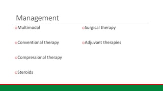
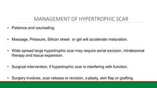
MAKERERE UNIVERSITY SCHOOL OF MEDICINE
DEPARTMENT OF SURGERY
Plastic and Reconstructive Surgery_ clinical rotation
Keloids and Hypertrophic Scars
Supervisor:
MR Kazibwe Simon
Plastics and Reconstructive Surgery
Fellow
Presenter:
Paul Ssempebwa
General surgery
Resident
outline
◦ Objectives
◦ Definitions
◦ epidemiology
◦ Aetiology and Pathogenesis
◦ Clinical presentation
◦ Diagnosis
◦ Management Options & prevention
Objectives
oTo define keloids and hypertrophic scar
oTo understand the aetiology and pathophysiology of keloids and HTS formation
oTo understand the different risk factors to formation of keloids & Hypertrophic scars
oTo understand the differences between keloids and hypertrophic scars and how to diagnose
them.
oTo understand the different management options for keloids, complications and follow up care.
Definitions
• Keloids and hypertrophic scars are fibroproliferative disorders that are due to
aberrant wound healing of susceptible individuals following tissue insult due to
trauma/ surgery/inflammation.
•Keloid grow beyond the boundaries of the original site of skin injury.
•HTS: widened or unsightly scar that does not extend beyond the original
boundaries of the wound
Scar classification
• Immature Scar: erythema, edema, collagen. Time is 6 months but can take up to years
• Mature Scar: no erythema, disorganized collagen as compared to adjacent skin
• Atrophic scar: due to reduced proliferation/inflammation. E.g striae following steroids/cuhings
• Linear hypertrophic Scar: HTS following a linear insult on skin
• Widespread hypertrophic scar: HTS that follows a wide spread insult like burns
• Minor keloid: grow beyond the margins of the insult but are self limiting. E.g the ones on the ear lobes
• Major keloid: continuous growth of the keloids giving no chance to treatment. Give a butterfly pattern no
quiescent stage
Epidemiology
Incidence 0.15% (Taiwanese database).
Prevalence of keloids 3.5% - 16% in black
communities. 8.3% in Kenya. (Kiprono et
al,2015)
African, Hispanic and Asian ancestry 2 to
3.5times > white Americans
Age 20 – 30 years more risk to trauma/
pubertal hormones
Familial; isolated genes in Japanese 2q23,
7p11 in African families , and 18q21 in
Chinese associated with Keloids.(Glass et
al,2017)
Occurrence with syndromes:
Rubenstein Taybi syndrome
Females > male; role of estrogen.
Hypertension; vascular endothelial
damage
aetiopathogensis
Exact mechanism is unclear,
understanding the pathogenesis is needed
to optimize the multimodal management
insult (trauma/burns/infection) to the
reticular dermis of a susceptible individual
Abnormal dysregulation of one or more of
the wound healing processes;
Inflammation, proliferation, Remodeling.
Role of : Pro inflammatory, IL 6 & 8, and
anti inflammatory IL 10.
Macrophages
TGF B 1 ,2 and 3
amplified inflammatory response,
overexpression of growth factor signals,
and increased activation of fibroblasts and
collagen production
• Hypertrophic scar contains islands composed of aggregates
of fibroblasts, small vessels and well organized type 3
collagen throughout the dermis.
• Keloids contains disorganized type 1 and 3 hyalinized
collagen bundles that form acellular node like structures in the
deep dermis with paucity of cells centrally.
Clinical Features
Clinical presentation
HEAD AND NECK KELOIDS
Adapted from Dr Achebe U lecture
note26
EAR LOBE KELOIDS
Adapted from Dr Achebe U lecture
note
27
TRUNCAL KELOIDS
28
Adapted from Dr Achebe U lecture
note
ABDOMINAL/PUBIC KELOIDS
Adapted from Dr Achebe U lecture
note
16
KELOIDS OF FOOT
Adapted from Dr Achebe U lecture
note
17
DDx
• Dermatofibrosarcoma protuberance
• Neurofibromatosis
• Lobomycosis
• Nodular scleroderma
Management
oMultimodal
oConventional therapy
oCompressional therapy
oSteroids
oSurgical therapy
oAdjuvant therapies
21
MANAGEMENT OF HYPERTROPHIC SCAR
• Patience and counseling.
• Massage, Pressure, Silicon sheet or gel will accelerate maturation.
• Wide spread large hypertrophic scar may require serial excision, intralesional
therapy and tissue expansion.
• Surgical intervention, if hypertrophic scar is interfering with function.
• Surgery involves, scar release or revision, z-plasty, skin flap or grafting.
Adapted from Dr Achebe U lecture
note
22
2
3
Prevention of Keloids
• First rule in keloid therapy.
• Avoid nonessential surgery in keloid formers.
• Close all surgical wounds with minimal tension.
• Incisions should not cross joint space.
• Avoid making midchest incisions.
• Incisions follow skin crease where possible.
• Control of inflammatory acne.
• Prophylactic pressure garment.
Conventional therapies
Occlusive dressing
Compression dressing : button compression,
pressure earrings
Steroids:
intralesional used as independent or in
combination
◦ TAC, administered at a concentration of
10 to 40 mg/mL, over intervals of 4 to 6
weeks.
◦ Response rates range from 50% to
100% clinical efficacy, with recurrence
rates of 9% to 50%.
◦ side effects :
telangiectasia, atrophy, and pigmentary
changes including hyper- and hypo-
pigmentation
Surgical therapy
Surgical excision:
◦ Usually used with adjuncts.
◦ Recurrency rates range from 45% to 100%
Prevention: gentle tissue handling, closure within
relaxed skin tension lines, and use of buried sutures
when needed to reduce tension on the wound closure.
Postoperative care should include pressure dressings to
prevent recurrence
Cryotherapy: contact, spray or intralesional
ranging from -4 to -7C for melanocytes, to colder
temperatures of -30 to -35C
remission rates of 68% to 81% with rare recurrence
(2%), but are limited by the
Disadvantages: large number of multiple monthly
sessions often required.
Side effects: blisters, local pain, and hypo- or hyper-
pigmentation.
Adjuvant therapies
•Radiotherapy:
•Interferon: alpha 2B
•5 fluoro Uracil
•Imiquimod
•Tacrolimus
•bleomycin
others
•Silcon based camouflage
•Hydrogel scaffold
•Skin tension offloading device
References
1. Kassi, Komenan et al. “Quality of life in black African patients with keloid
scars.” Dermatology reports vol. 12,2 8312. 22 Oct. 2020, doi:10.4081/dr.2020.8312
2. Davis, Scott A et al. “Management of keloids in the United States, 1990-2009: an analysis
of the National Ambulatory Medical Care Survey.” Dermatologic surgery : official publication
for American Society for Dermatologic Surgery [et al.] vol. 39,7 (2013): 988-94.
doi:10.1111/dsu.12182
3. Glass, Donald A 2nd. “Current Understanding of the Genetic Causes of Keloid
Formation.” The journal of investigative dermatology. Symposium proceedings vol. 18,2
(2017): S50-S53. doi:10.1016/j.jisp.2016.10.024

keloids and Hypertrophic Scars. Presentation and management options

  • 1.
    MAKERERE UNIVERSITY SCHOOLOF MEDICINE DEPARTMENT OF SURGERY Plastic and Reconstructive Surgery_ clinical rotation Keloids and Hypertrophic Scars Supervisor: MR Kazibwe Simon Plastics and Reconstructive Surgery Fellow Presenter: Paul Ssempebwa General surgery Resident
  • 2.
    outline ◦ Objectives ◦ Definitions ◦epidemiology ◦ Aetiology and Pathogenesis ◦ Clinical presentation ◦ Diagnosis ◦ Management Options & prevention
  • 3.
    Objectives oTo define keloidsand hypertrophic scar oTo understand the aetiology and pathophysiology of keloids and HTS formation oTo understand the different risk factors to formation of keloids & Hypertrophic scars oTo understand the differences between keloids and hypertrophic scars and how to diagnose them. oTo understand the different management options for keloids, complications and follow up care.
  • 4.
    Definitions • Keloids andhypertrophic scars are fibroproliferative disorders that are due to aberrant wound healing of susceptible individuals following tissue insult due to trauma/ surgery/inflammation. •Keloid grow beyond the boundaries of the original site of skin injury. •HTS: widened or unsightly scar that does not extend beyond the original boundaries of the wound
  • 5.
    Scar classification • ImmatureScar: erythema, edema, collagen. Time is 6 months but can take up to years • Mature Scar: no erythema, disorganized collagen as compared to adjacent skin • Atrophic scar: due to reduced proliferation/inflammation. E.g striae following steroids/cuhings • Linear hypertrophic Scar: HTS following a linear insult on skin • Widespread hypertrophic scar: HTS that follows a wide spread insult like burns • Minor keloid: grow beyond the margins of the insult but are self limiting. E.g the ones on the ear lobes • Major keloid: continuous growth of the keloids giving no chance to treatment. Give a butterfly pattern no quiescent stage
  • 6.
    Epidemiology Incidence 0.15% (Taiwanesedatabase). Prevalence of keloids 3.5% - 16% in black communities. 8.3% in Kenya. (Kiprono et al,2015) African, Hispanic and Asian ancestry 2 to 3.5times > white Americans Age 20 – 30 years more risk to trauma/ pubertal hormones Familial; isolated genes in Japanese 2q23, 7p11 in African families , and 18q21 in Chinese associated with Keloids.(Glass et al,2017) Occurrence with syndromes: Rubenstein Taybi syndrome Females > male; role of estrogen. Hypertension; vascular endothelial damage
  • 7.
    aetiopathogensis Exact mechanism isunclear, understanding the pathogenesis is needed to optimize the multimodal management insult (trauma/burns/infection) to the reticular dermis of a susceptible individual Abnormal dysregulation of one or more of the wound healing processes; Inflammation, proliferation, Remodeling. Role of : Pro inflammatory, IL 6 & 8, and anti inflammatory IL 10. Macrophages TGF B 1 ,2 and 3 amplified inflammatory response, overexpression of growth factor signals, and increased activation of fibroblasts and collagen production
  • 8.
    • Hypertrophic scarcontains islands composed of aggregates of fibroblasts, small vessels and well organized type 3 collagen throughout the dermis. • Keloids contains disorganized type 1 and 3 hyalinized collagen bundles that form acellular node like structures in the deep dermis with paucity of cells centrally.
  • 10.
  • 11.
  • 13.
    HEAD AND NECKKELOIDS Adapted from Dr Achebe U lecture note26
  • 14.
    EAR LOBE KELOIDS Adaptedfrom Dr Achebe U lecture note 27
  • 15.
    TRUNCAL KELOIDS 28 Adapted fromDr Achebe U lecture note
  • 16.
    ABDOMINAL/PUBIC KELOIDS Adapted fromDr Achebe U lecture note 16
  • 17.
    KELOIDS OF FOOT Adaptedfrom Dr Achebe U lecture note 17
  • 18.
    DDx • Dermatofibrosarcoma protuberance •Neurofibromatosis • Lobomycosis • Nodular scleroderma
  • 20.
  • 21.
    21 MANAGEMENT OF HYPERTROPHICSCAR • Patience and counseling. • Massage, Pressure, Silicon sheet or gel will accelerate maturation. • Wide spread large hypertrophic scar may require serial excision, intralesional therapy and tissue expansion. • Surgical intervention, if hypertrophic scar is interfering with function. • Surgery involves, scar release or revision, z-plasty, skin flap or grafting.
  • 22.
    Adapted from DrAchebe U lecture note 22
  • 23.
    2 3 Prevention of Keloids •First rule in keloid therapy. • Avoid nonessential surgery in keloid formers. • Close all surgical wounds with minimal tension. • Incisions should not cross joint space. • Avoid making midchest incisions. • Incisions follow skin crease where possible. • Control of inflammatory acne. • Prophylactic pressure garment.
  • 24.
    Conventional therapies Occlusive dressing Compressiondressing : button compression, pressure earrings Steroids: intralesional used as independent or in combination ◦ TAC, administered at a concentration of 10 to 40 mg/mL, over intervals of 4 to 6 weeks. ◦ Response rates range from 50% to 100% clinical efficacy, with recurrence rates of 9% to 50%. ◦ side effects : telangiectasia, atrophy, and pigmentary changes including hyper- and hypo- pigmentation
  • 25.
    Surgical therapy Surgical excision: ◦Usually used with adjuncts. ◦ Recurrency rates range from 45% to 100% Prevention: gentle tissue handling, closure within relaxed skin tension lines, and use of buried sutures when needed to reduce tension on the wound closure. Postoperative care should include pressure dressings to prevent recurrence Cryotherapy: contact, spray or intralesional ranging from -4 to -7C for melanocytes, to colder temperatures of -30 to -35C remission rates of 68% to 81% with rare recurrence (2%), but are limited by the Disadvantages: large number of multiple monthly sessions often required. Side effects: blisters, local pain, and hypo- or hyper- pigmentation.
  • 26.
    Adjuvant therapies •Radiotherapy: •Interferon: alpha2B •5 fluoro Uracil •Imiquimod •Tacrolimus •bleomycin
  • 27.
    others •Silcon based camouflage •Hydrogelscaffold •Skin tension offloading device
  • 29.
    References 1. Kassi, Komenanet al. “Quality of life in black African patients with keloid scars.” Dermatology reports vol. 12,2 8312. 22 Oct. 2020, doi:10.4081/dr.2020.8312 2. Davis, Scott A et al. “Management of keloids in the United States, 1990-2009: an analysis of the National Ambulatory Medical Care Survey.” Dermatologic surgery : official publication for American Society for Dermatologic Surgery [et al.] vol. 39,7 (2013): 988-94. doi:10.1111/dsu.12182 3. Glass, Donald A 2nd. “Current Understanding of the Genetic Causes of Keloid Formation.” The journal of investigative dermatology. Symposium proceedings vol. 18,2 (2017): S50-S53. doi:10.1016/j.jisp.2016.10.024

Editor's Notes

  • #7 Darker skin tone most common, Fitzpatrick phototype 3 (hotchman et al) Trauma physical:(ear lobe piercing, surgery) pathological: (chicken pox, acne, folliculitis, insect bites,Herpes zooster infection, burns) Syndromes associated with keloids: Rubinstein-Taybi syndrome, Goeminne syndrome, Noonan syndrome, Ehler-Danlos syndrome,Leigh necrotising encephalopathy, Dubowitz syndrome,Ullrich congenital muscular dystrophy,Conjuctivocorneal dystrophy
  • #8 Why keloids are not tumors: fibroproliferative driven by chronic inflammation not cellular proliferation. 2. no cellular atypia, IL 6, 8 Liechty and colleagues showed an increase in scarring to these wounds after administration of IL-6. Van den Broek and colleagues found a decrease in IL-10 .
  • #10 Kelloid evolution after 7 years. Major kelloids
  • #11 Adapted from SRB’s Manual of General Surgery
  • #12 BAD SCAR
  • #14 Earlobes Head and Neck Pre-sternal Breasts Deltoid/upper back Upper extremities Abdomen/pubis Lower extremities Feet Genitalia
  • #19 Dermatofibrosarcoma protuberance: rare skin d/o arising from the dermal connective tissue. Looks like a pimple. Lobomycosis: keloidal deep cutaneos blastomycosis Nodular scleroderma:
  • #20 Dermatofibrosarcoma protuberance, Neurofibromatosis, Lobomycosis,Nodular scleroderma
  • #25 Occlusive dressing due to compression and hydration. First line for prevention and treatment of scars Compression dressing activates the mechanoreceptors which activate apoptosis Steroids mediate their effects by reducing the synthesis of collagen, altering extracellular matrix components such as glycosaminoglycans, and reducing proinflammatory mediators.