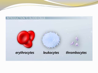
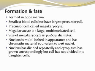
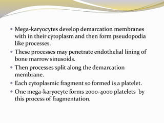
 George Gulliver in 1841 drew pictures of platelets using
the twin lens (compound) microscope invented in 1830
by Joseph Jackson Lister.
 William Addison in 1842 drew pictures of a platelet-fibrin
clot.
 Max Schultze in 1865 described what he called "spherules“.
 James Wright examined blood smears using the stain
named for him, and used the term plates in his 1906
publicationbut changed to platelets in his 1910 publication
which has become the universally accepted term.
 The term thrombocyte (clot cell) came into use in the early
1900s.
 Literally means, a small plate.
 Also k/as Thrombocytes ( Thrombus= lump or clot:
kytos= cell).
 Size 2-4µ.
 Small irregularly shaped blood cells.
 No nucleus but mitochondria present.
 Extensive canalicular system present.
 Microtubular system composed of contractile proteins.
 Besides contractile proteins, the metabolic pathways
are also remarkably similar to skeletal muscle.
 Normal count= 150,000 to 400,000/mm3.
 Circulating platelets are 2/3rd of platelet pool of body.
 Remaining 1/3rd reside in the spleen.
Bleeding
Acutely Stressful condition to the body
Activation of sympathoadrenal system
Release of epinephrine
Action on spleen
Release of platelets as well as erythrocytes
Help in haemostasis
 Structurally the platelet can be divided into four zones,
from peripheral to innermost:
 Peripheral zone – is rich in glycoproteins required for
platelet adhesion, activation, and aggregation.
 Sol-gel zone – is rich in microtubules and
microfilaments, allowing the platelets to maintain
their discoid shape.
 Organelle zone – is rich in platelet granules.
 Membranous zone – contains membranes derived
from megakaryocytic smooth endoplasmic reticulum .
 Organelle zone – is rich in platelet granules.
 Alpha granules contain clotting mediators such as factor V,
factor VIII, fibrinogen, fibronectin, platelet-derived growth
factor, and chemotactic agents.
 Delta granules, or dense bodies, contain ADP, calcium,
serotonin, which are platelet-activating mediators.
 γ granules (gamma granules) – similar to lysosomes and
contain several hydrolytic enzymes.
 λ granules (lambda granules) – contents involved in clot
resorption during later stages of vessel repair.
 Membranous zone –is responsible for thromboxane A2
synthesis. This dense tubular system is connected to the
surface platelet membrane to aid thromboxane A2 release.
Formation & fate
 Formed in bone marrow.
 Smallest blood cells but have largest precursor cell.
 Precursor cell, called megakaryocyte.
 Megakaryocyte is a large, multinucleated cell.
 Size of megakaryocyte is 35-160 µ diameter.
 Nucleus is multi-loabed in appearance and has
chromatin material equivalent to 4-16 nuclei.
 Nucleus has divided repeatedly and cytoplasm has
grown correspondingly but cell has not divided into
daughter cells.
 Mega-karyocytes develop demarcation membranes
with in their cytoplasm and then form pseudopodia
like processes.
 These processes may penetrate endothelial lining of
bone marrow sinusoids.
 Then processes split along the demarcation
membrane.
 Each cytoplasmic fragment so formed is a platelet.
 One mega-karyocyte forms 2000-4000 platelets by
this process of fragmentation.
 Life span of throbocytes is 9-12 days.
 They are destroyed in the spleen.
 Spleen serves as both graveyard and reservoir.
 Death of a platelet may be due to senescence or
random destruction.
 Thrombopoiesis and destruction is balanced process.
 Number of platelets fluctuates within narrow range.
 Thrombopoiesis seems to be regulated by a group of
humoral factors collectively called thrombopoietin.
Functions
 The functions of platelets are primarily related to
haemostasis.
 Serotonin released by platelets contributes to the
vasoconstriction observed immediately after vascular
injury.
 Platelets aggregates to plug the vascular injury.
 Platelets provide platelet factor 3 which accelerate the
clotting process.
 Contractile proteins of the platelets bring about clot
retraction.
 It has growth factors which stimulate mitosis in vascular
wall thus repair the damage of vessel wall.
Laboratory tests for platelet function
Applied
 Low platelet concentration is thrombocytopenia and is
due to either decreased production or increased
destruction.
 Elevated platelet concentration is thrombocytosis and
is either congenital, reactive (to cytokines), or due
to unregulated production: one of the
myeloproliferative neoplasms or certain other myeloid
neoplasms.
 Thrombocytopenia
 Immune thrombocytopenias (ITP) – formerly known
as immune thrombocytopenic purpura and idiopathic
thrombocytopenic purpura
 Splenomegaly
 Gaucher's disease
 Familial thrombocytopenia
 Chemotherapy
 Babesiosis
 Dengue
Altered platelet function
 Congenital
 Gray platelet syndrome
 ADP Receptor defect
 Decreased cyclooxygenase activity
 Storage pool defects, acquired or congenital
 Acquired
 Paroxysmal nocturnal hemoglobinuria
 Asthma
 Cancer
 Malaria
 Decreased cyclooxygenase activity
Thrombocytosis and thrombocythemia
 Chronic infection
 Chronic inflammation
 Malignancy
 Hyposplenism (post-splenectomy)
 Iron deficiency
 Acute blood loss
 Myeloproliferative neoplasms – platelets are both elevated and
activated
 Essential thrombocytosis
 Polycythemia vera
 Associated with other myeloid neoplasms
 Congenital
Platelets

Platelets

  • 3.
     George Gulliverin 1841 drew pictures of platelets using the twin lens (compound) microscope invented in 1830 by Joseph Jackson Lister.  William Addison in 1842 drew pictures of a platelet-fibrin clot.  Max Schultze in 1865 described what he called "spherules“.  James Wright examined blood smears using the stain named for him, and used the term plates in his 1906 publicationbut changed to platelets in his 1910 publication which has become the universally accepted term.  The term thrombocyte (clot cell) came into use in the early 1900s.
  • 4.
     Literally means,a small plate.  Also k/as Thrombocytes ( Thrombus= lump or clot: kytos= cell).  Size 2-4µ.  Small irregularly shaped blood cells.  No nucleus but mitochondria present.  Extensive canalicular system present.
  • 5.
     Microtubular systemcomposed of contractile proteins.  Besides contractile proteins, the metabolic pathways are also remarkably similar to skeletal muscle.  Normal count= 150,000 to 400,000/mm3.  Circulating platelets are 2/3rd of platelet pool of body.  Remaining 1/3rd reside in the spleen.
  • 8.
    Bleeding Acutely Stressful conditionto the body Activation of sympathoadrenal system Release of epinephrine Action on spleen Release of platelets as well as erythrocytes Help in haemostasis
  • 9.
     Structurally theplatelet can be divided into four zones, from peripheral to innermost:  Peripheral zone – is rich in glycoproteins required for platelet adhesion, activation, and aggregation.  Sol-gel zone – is rich in microtubules and microfilaments, allowing the platelets to maintain their discoid shape.  Organelle zone – is rich in platelet granules.  Membranous zone – contains membranes derived from megakaryocytic smooth endoplasmic reticulum .
  • 10.
     Organelle zone– is rich in platelet granules.  Alpha granules contain clotting mediators such as factor V, factor VIII, fibrinogen, fibronectin, platelet-derived growth factor, and chemotactic agents.  Delta granules, or dense bodies, contain ADP, calcium, serotonin, which are platelet-activating mediators.  γ granules (gamma granules) – similar to lysosomes and contain several hydrolytic enzymes.  λ granules (lambda granules) – contents involved in clot resorption during later stages of vessel repair.  Membranous zone –is responsible for thromboxane A2 synthesis. This dense tubular system is connected to the surface platelet membrane to aid thromboxane A2 release.
  • 12.
    Formation & fate Formed in bone marrow.  Smallest blood cells but have largest precursor cell.  Precursor cell, called megakaryocyte.  Megakaryocyte is a large, multinucleated cell.  Size of megakaryocyte is 35-160 µ diameter.  Nucleus is multi-loabed in appearance and has chromatin material equivalent to 4-16 nuclei.  Nucleus has divided repeatedly and cytoplasm has grown correspondingly but cell has not divided into daughter cells.
  • 13.
     Mega-karyocytes developdemarcation membranes with in their cytoplasm and then form pseudopodia like processes.  These processes may penetrate endothelial lining of bone marrow sinusoids.  Then processes split along the demarcation membrane.  Each cytoplasmic fragment so formed is a platelet.  One mega-karyocyte forms 2000-4000 platelets by this process of fragmentation.
  • 14.
     Life spanof throbocytes is 9-12 days.  They are destroyed in the spleen.  Spleen serves as both graveyard and reservoir.  Death of a platelet may be due to senescence or random destruction.  Thrombopoiesis and destruction is balanced process.  Number of platelets fluctuates within narrow range.  Thrombopoiesis seems to be regulated by a group of humoral factors collectively called thrombopoietin.
  • 15.
    Functions  The functionsof platelets are primarily related to haemostasis.  Serotonin released by platelets contributes to the vasoconstriction observed immediately after vascular injury.  Platelets aggregates to plug the vascular injury.  Platelets provide platelet factor 3 which accelerate the clotting process.  Contractile proteins of the platelets bring about clot retraction.  It has growth factors which stimulate mitosis in vascular wall thus repair the damage of vessel wall.
  • 17.
    Laboratory tests forplatelet function
  • 18.
    Applied  Low plateletconcentration is thrombocytopenia and is due to either decreased production or increased destruction.  Elevated platelet concentration is thrombocytosis and is either congenital, reactive (to cytokines), or due to unregulated production: one of the myeloproliferative neoplasms or certain other myeloid neoplasms.
  • 19.
     Thrombocytopenia  Immunethrombocytopenias (ITP) – formerly known as immune thrombocytopenic purpura and idiopathic thrombocytopenic purpura  Splenomegaly  Gaucher's disease  Familial thrombocytopenia  Chemotherapy  Babesiosis  Dengue
  • 20.
    Altered platelet function Congenital  Gray platelet syndrome  ADP Receptor defect  Decreased cyclooxygenase activity  Storage pool defects, acquired or congenital  Acquired  Paroxysmal nocturnal hemoglobinuria  Asthma  Cancer  Malaria  Decreased cyclooxygenase activity
  • 21.
    Thrombocytosis and thrombocythemia Chronic infection  Chronic inflammation  Malignancy  Hyposplenism (post-splenectomy)  Iron deficiency  Acute blood loss  Myeloproliferative neoplasms – platelets are both elevated and activated  Essential thrombocytosis  Polycythemia vera  Associated with other myeloid neoplasms  Congenital