The document discusses the structure and types of plant cell walls and tissues. It begins by describing the four layers of the plant cell wall: middle lamella, primary wall, secondary wall, and tertiary wall. It then discusses the ultrastructure of cell walls under electron microscopes. The document also covers the structure and functions of the plasma membrane, pits, plasmodesmata, and the four main types of plant tissues: meristematic tissues, parenchyma tissues, collenchyma tissues, and sclerenchyma tissues. It provides detailed information on each tissue type.
economic importance of gymnosperms.Gymnosperms are simple and primitive seed-bearing plants without flowers.
The plant body is sporophytic and is differentiated into root,stem and leaves.
All gymnosperms are usually wind-pollinated.
Leaves have thick cuticle and sunken stomata.
Gymnosperms are heterosporous.magasporangia and microsporangia occur on mega and microsporophylls respectively.
the top three theories of root apical meristem in plants. The theories are: 1. Apical Cell Theory 2. Histogen Theory 3. Korper-Kappe Theory.The root apical meristem, or root apex, is a small region at the tip of a root in which all cells are capable of repeated division and from which all primary root tissues are derived. The root apical meristem is protected as it passes through the soil by an outer region of living parenchyma cells called the root cap.
The Shoot apex is also known as the terminal bud of plants that grows from 0.1-1.0 mm and consists of the apical meristem, developing leaves and the immediate surrounding leaf primordial. The shoot apex is present in both dicot and monocot plants.
This is a detailed presentation on Morphology, anatomy and reproduction of Marchantia spp. with high quality pics and eye capturing transitions and animations
Vascular Cambium & Seasonal activity & its Role in Stem & RootFatima Ramay
Vascular Cambium & Seasonal activity & its Role in Stem & Root:
The vascular cambium (pl. cambia or cambiums) is a lateral meristem in the vascular tissue of plants.
The vascular cambium is a cylindrical layer of cambium that runs through the stem of a plant that undergoes secondary growth.
In Dicots:
The vascular cambium is in dicot stems and roots, located between the xylem and the phloem in the stem and root of a vascular plant, and is the source of both the secondary xylem growth (inwards, towards the pith) and the secondary phloem growth (outwards).
In Monocots:
Monocot stems, such as corn, palms and bamboos, do not have a vascular cambium and do not exhibit secondary growth by the production of concentric annual rings. They cannot increase in girth by adding lateral layers of cells as in conifers and woody dicots.
Cambium of some plants remains active for the entire period of their life, i.e., cambial cells divide and resulting cells mature to form xylem and phloem elements.
This type of seasonal activity usually found in the plants present in the tropical regions, and not all plants show cambial activity.
Percentage of ringless trees in the rain forests of; India : 75% Amazon : 43% Malaysia : 15%
In regions with definite seasonal climate; seasonal activity of cambium ceased with onset of unfavorable conditions; In Autumn, it enters the dormant state and lasts for the end of summer; In Spring, cambium again becomes active.
Duration of cambial activity is also affected by day-length, e.g., In Robinia pseudoacacia, cambium is dormant under short-day condition.
The cambium cells formed in circular in cross section from the beginning onwards.
The cambial ring is partially primary (fascicular cambium) and partially secondary (interfascicular cambium).
Periderm originates from the cortical cells (extra stelar in origin).
In Dicot stem, for mechanical support xylem is with comparatively smaller vessels, greater fibers and less parenchyma.
More amount of cork is produces for protection.
Lenticels on periderm are very prominent.
The cambial ring formed is wavy in the beginning and later becomes circular.
The cambium ring is completely secondary in origin.
Periderm originates from the pericycle (intra stelar in origin).
In Dicot root, xylem is with big thin walled vessels with few fibers and more parenchyma.
Less amount of cork is produced as root is underground.
Lenticels on periderm are not very prominent.
Equisetum popularly known a the ‘horse-tail’ or ‘scouring rush’.
It is now represented by nearly 30 species which are seen world wide except in Australia and New Zealand.
Some species prefer damp and shady places while others grow in marshes, ponds or stream banks
Some are found in xerophytic habitats
Plant systems: Extracellular matrix components of plants-cell wall, cellulose and hemicelluloses, extensins, WAKs, secondary wall structure, pits-primary and secondary pits and their development, plasmodesmota-structure and functions, pectins, cutins, lignins, turnover of cell wall components
Embracing GenAI - A Strategic ImperativePeter Windle
Artificial Intelligence (AI) technologies such as Generative AI, Image Generators and Large Language Models have had a dramatic impact on teaching, learning and assessment over the past 18 months. The most immediate threat AI posed was to Academic Integrity with Higher Education Institutes (HEIs) focusing their efforts on combating the use of GenAI in assessment. Guidelines were developed for staff and students, policies put in place too. Innovative educators have forged paths in the use of Generative AI for teaching, learning and assessments leading to pockets of transformation springing up across HEIs, often with little or no top-down guidance, support or direction.
This Gasta posits a strategic approach to integrating AI into HEIs to prepare staff, students and the curriculum for an evolving world and workplace. We will highlight the advantages of working with these technologies beyond the realm of teaching, learning and assessment by considering prompt engineering skills, industry impact, curriculum changes, and the need for staff upskilling. In contrast, not engaging strategically with Generative AI poses risks, including falling behind peers, missed opportunities and failing to ensure our graduates remain employable. The rapid evolution of AI technologies necessitates a proactive and strategic approach if we are to remain relevant.
How to Make a Field invisible in Odoo 17Celine George
It is possible to hide or invisible some fields in odoo. Commonly using “invisible” attribute in the field definition to invisible the fields. This slide will show how to make a field invisible in odoo 17.
Unit 8 - Information and Communication Technology (Paper I).pdfThiyagu K
This slides describes the basic concepts of ICT, basics of Email, Emerging Technology and Digital Initiatives in Education. This presentations aligns with the UGC Paper I syllabus.
Biological screening of herbal drugs: Introduction and Need for
Phyto-Pharmacological Screening, New Strategies for evaluating
Natural Products, In vitro evaluation techniques for Antioxidants, Antimicrobial and Anticancer drugs. In vivo evaluation techniques
for Anti-inflammatory, Antiulcer, Anticancer, Wound healing, Antidiabetic, Hepatoprotective, Cardio protective, Diuretics and
Antifertility, Toxicity studies as per OECD guidelines
Macroeconomics- Movie Location
This will be used as part of your Personal Professional Portfolio once graded.
Objective:
Prepare a presentation or a paper using research, basic comparative analysis, data organization and application of economic information. You will make an informed assessment of an economic climate outside of the United States to accomplish an entertainment industry objective.
Palestine last event orientationfvgnh .pptxRaedMohamed3
An EFL lesson about the current events in Palestine. It is intended to be for intermediate students who wish to increase their listening skills through a short lesson in power point.
Honest Reviews of Tim Han LMA Course Program.pptxtimhan337
Personal development courses are widely available today, with each one promising life-changing outcomes. Tim Han’s Life Mastery Achievers (LMA) Course has drawn a lot of interest. In addition to offering my frank assessment of Success Insider’s LMA Course, this piece examines the course’s effects via a variety of Tim Han LMA course reviews and Success Insider comments.
A Strategic Approach: GenAI in EducationPeter Windle
Artificial Intelligence (AI) technologies such as Generative AI, Image Generators and Large Language Models have had a dramatic impact on teaching, learning and assessment over the past 18 months. The most immediate threat AI posed was to Academic Integrity with Higher Education Institutes (HEIs) focusing their efforts on combating the use of GenAI in assessment. Guidelines were developed for staff and students, policies put in place too. Innovative educators have forged paths in the use of Generative AI for teaching, learning and assessments leading to pockets of transformation springing up across HEIs, often with little or no top-down guidance, support or direction.
This Gasta posits a strategic approach to integrating AI into HEIs to prepare staff, students and the curriculum for an evolving world and workplace. We will highlight the advantages of working with these technologies beyond the realm of teaching, learning and assessment by considering prompt engineering skills, industry impact, curriculum changes, and the need for staff upskilling. In contrast, not engaging strategically with Generative AI poses risks, including falling behind peers, missed opportunities and failing to ensure our graduates remain employable. The rapid evolution of AI technologies necessitates a proactive and strategic approach if we are to remain relevant.
Acetabularia Information For Class 9 .docxvaibhavrinwa19
Acetabularia acetabulum is a single-celled green alga that in its vegetative state is morphologically differentiated into a basal rhizoid and an axially elongated stalk, which bears whorls of branching hairs. The single diploid nucleus resides in the rhizoid.
1. PLANT ANATOMY AND MICROTECHNIQUES
Unit 1
Structure of plant cell wall
Plant cell is distinguished from the animal cells
Cell wall is a outermost covering layer of plant cell
It is absent in animal cell
Each cell has own cell wall
Thickness in relation to age and type of cells
Generally, young cells have thin, elastic, transparent and
colourless
The Cell wall itself is taken as the non-living part of the
cells
It maintain the shape of cell
Protects the protoplasm form external injuries
Permeable to water
Cell wall is made up of cellulose, hemicelluloses and
pectin
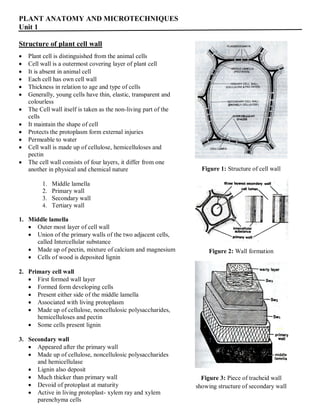
The cell wall consists of four layers, it differ from one
another in physical and chemical nature
1. Middle lamella
2. Primary wall
3. Secondary wall
4. Tertiary wall
1. Middle lamella
Outer most layer of cell wall
Union of the primary walls of the two adjacent cells,
called Intercellular substance
Made up of pectin, mixture of calcium and magnesium
Cells of wood is deposited lignin
2. Primary cell wall
First formed wall layer
Formed form developing cells
Present either side of the middle lamella
Associated with living protoplasm
Made up of cellulose, noncellulosic polysaccharides,
hemicelluloses and pectin
Some cells present lignin
3. Secondary wall
Appeared after the primary wall
Made up of cellulose, noncellulosic polysaccharides
and hemicellulase
Lignin also deposit
Much thicker than primary wall
Devoid of protoplast at maturity
Active in living protoplast- xylem ray and xylem
parenchyma cells
Figure 1: Structure of cell wall
Figure 2: Wall formation
Figure 3: Piece of tracheid wall
showing structure of secondary wall
2. It consist of three layers
a. Outer – SW1
b. Middle – SW2
c. Inner - SW3
It vary on orientation of microfibrils
Maintain the cells shape and mechanical strength
4. Tertiary wall
Present innerside of secondary wall
dried residue of degenerating plasmamembrane
Rarely present
Ultrastructure of cell wall
The cell walls under the electron microscope shows
two parts
Matrix- non cellulosic part
Fibrils- embedded in the matrix and made
up of cellulose
The largest fibril could be seen by a light microscope
and is called a macrofibril
The smaller ones are seen under the electron
microscope and are called microfibrils
With the increase in resolving power smaller and
smaller fibrils are visualized as subunits of microfibrils
These are called micells which in turn are further divided to form cellulose molecules
The microfibrils display a dense textile like pattern in electron microscope preparation
Plasma membrane or plasmalemma
There is present a living membrane called plasma membrane or plasmalemma in all the plant and animal
cells around the cytoplasm
Like other membranes it is also a unit membrane or 100 Aº thickness
The unit membrane contains about 40% lipid and 60% protein
It consists of a bi-molecular layer of phospholipids (40-60 Aº dia) with their not polar oriented inwardly
perpendicular to the plane of the membrane
The external surface of the double layer is made up of phospholipids on the both side of the polar moieties
and is covered by a layer of protein
Functions
Controls cellular semipermeability, resorption and excretion acting in a selective pathway manner
Protective function
The material inside or outside the plasma membrane could be transported pinocytosis or phagocytosis.
The plamma membrane invaginates to form vacuole which passes sinto the cell to form food vacuole or
phagocytic vacuole
By reverse process material is thrown out of plasma membrane
Figure: Ultra structure of cell wall
3. Chemical nature of cell wall
1. The cell wall is composed of carbohydrate rich
materials
2. The major component of cells are
cellulose, hemicelluloses, pectins, proteins
and phenolics
a. Cellulose
Provides shape and strength of cell wall
Hydrophilic crystalline compound
Long chain of linked glucose residues, more
than 100
Molecules are Chain or ribbon like structure
General formula is (C6H10O5)n
Cellulose is an unbranched β 1,4-glucan
b. Non cellulosic polysaccharides / hemicellulose
Closely allied to cellulose
Built up of a variety of different sugars
Examples:
Xylan:
Linked with xylose and arabinose
Primary and secondary wall of dicot plants
Glucomannan:
Secondary wall of gymnosperm and
angiospermous cells
Glucose and mannose in the ratio of 1:3
Mannan and Galactomannan:
Found in wall of endosperm
Reserves food
Glucuronomannan:
Low proportion in the cell walls
Contain mannose, glucuronic acid, xylose
and arabinose
Xyloglucan:
Storage polysaccharides
Xyloses are major components
It also contain glucose
Thick storage wall in some seeds, eg.
Nasturtium
Present in primary wall of dicot and grasses
Absent in secondary wall
Backbone of cellulose
c. Pectin
Derivative of polyglacturonic acid
Rich in galacturonic acid
Linked to cellulose, proteins and phenols
Present in primary and secondary walls
High concentration in middle lamella
Highly hydrophilic polysaccharides
Necessary for wall expansion
d. Lignin and other phenolic compounds
Organic compound of high carbon content
Component of scelerenchyma, fibres,
sclereids
Also found in tracheids and vessels of
xylem
The process of conversion of cellulose into
lignin is called as lignification
e. Cutin, suberin and waxes
These are fatty substances
Cutin found in walls of roots, stem and
leaves
Suberin deposited in seed coats, cork and
casparian strips
Wax occurs in surface of leaves and fruits
f. Gum and mucilages
Compounds of carbohydrates related pectin
Posses the property of swelling in water
Gums appear in breakdown of walls
Mucilages found in seed coat, increase the
water holding capacity
g. Mineral substances
Like silica, calcium carbonate, calcium
oxalate and several organic compounds
may deposit on cell wall
Silica found in Equisetum
Calcium carbonate noted in Ficus
4. Plasmodesmata
1. The cytoplasm of the two neighbouring cells is interconnected through fine protoplasm threads called
Plasmodesmata (singular: Plasmadesma)
2. Plasmodesma is thin irregular cylinder of cytoplasm lined by plasmolemma, forming fine pores in the cell
wall
3. Plasmodesma originates during cytokinesis when cell plate is formed
4. These strands bind together to protoplast of the neighbouring cells into a large called symplast and
facilitate the movement of food and information, form once cell to another cell
5. The plasmodesma have been seen in red algae, liverworts, mosses, vascular cryptogams, gymnosperms
and angiosperms
6. They found throughout all living tissues of a plant including the meristematic tissue
7. Plasmodesma either occur in groups or are distributed throughout a wall
8. In groups are mostly found in the primary pit fields
9. Plasmodesma exists in thick cell also, eg. endosperm of the seeds Phoenix, Coffea
10. The plasmodesma are also regarded as channels permitting the movement of viruses from cell to cell
11. Plant hormones move through plasmodesma
12. The movement through palsmodesma is bi-directional, desmotubules act as a valve and regulate the
direction of flow
13. small molecules and ions pass readily through plasmodesma
Figure: Plasmodesma between adjoining cell walls
5. Pit
The entire inner surface of the cell wall is thickened, leaving small unthickened areas or depressions here
and there, called as the pits
The pits are formed in pairs lying against each other on the opposite sides of the wall, and morphologically
more correct they are called pit pairs
Structure
The space found inside the pit is called the pit cavity or pit chamber.
The separating membrane which separates the two chambers or cavities of a pit pair, is called the pit
membrane, or pit aperture
The pit cavity opens internally in the lumen of the cell and is closed by the closing or pit membrane along
the line of junction of two contiguous cells
A pit pair has two pit cavities, two pit apertures, and one pit or closing membrane
The pit membrane is common to both pits of a pit pair and consists of two primary walls and a middle
lamella or intercellular substance
Two types of pits are met with in the cells of various plants viz
Simple pits
Bordered pits
Two bordered pits make up a bordered pit pair
Two simple pits form a simple pit pair
A bordered pit and a simple pit lying opposite to each other in contiguous cells, constitute a half bordered
pit pair
A pit occurs opposite an intercellular space has no complementary pit and is known as blind pit
a. Simple pits
Simple pit pairs occur in parenchyma cells, in medullary rays, in phloem fibres, campanion cells, and in
tracheids of several flowering plants
In simple pits, the pit cavity remains of the same diameter and the pit or closing membrane also remains
same and uniform in its structure
The simple pit may be circular, oval, polygonal, elongated or somewhat irregular in its facial view
b. Bordered pits
They are abundantly found in the vessels of many angiospers and in the tracheids of many conifers
They are more complex and variable in their structure than simple pits
The overarching secondary wall which encloses a part of the pit cavity is called the pit border
The opens outside by a small rounded mouth known as pit aperture
The overarching rim forms a border around the aperture and thus named bordered pits
Figure: Pits. A Surface view of simple pits, B Simple pit pairs in section
C Surface view of bordered pits, D Bordered pit pairs in section
6. Tissue system
A. Meristematic tissues
Meristematic tissue consists of thin-walled, tightly packed living cells
Occurs frequent cell division
Undergo cell division and wall formation followed by differential cell expansion
Classification of meristems
Basis of different characters like
Origin
Stage of development
Position in plant body and
Functions
1. Meristems based on origin
Two types
a) Primary meristems
b) Secondary meristems
a) Primary meristems
Present from embryonic stage
Persist throughout the life of a plant
Known as primary mersitems
Forms primary or fundamental part of the plant body
Present in the apices of stem and root, primordial of leaves and similar organs
b) Secondary meristems
Arise from non-meristematic or permanent tissue
Cork cambium forms the typical example of secondary meristem
Developed from mature cells of epidermis, cortex or pericycle
Vascular cambium in stem is partly a secondary meristem
Fsicular cambium it developes form the cells of ground tissue (medulary ray cells)
2. Meristems based on plan of cell division
There types
a) Mass meristem
b) Plate meristem
c) Rib meristem
a) Mass meristem
Exhibits division in all planes
Results increased in volume
Example- development of pith, cortex, endosperm etc
b) Plat meristem
Cells divide in two planes
Increase in area of the organ
7. Results in the formation of flat structures
Examples- epidermal growth and leaf
formation
c) Rib meristem
Cell division is in one plane
Results in the formation of row of cells
Example- formation of young roots, pith and
cortex of young stems
3. Meristems based on position in plant body
Three types
a) Apical meristem
b) Lateral meristem
c) Intercalary meristem
a) Apical meristem
Existent at the growing tips or apical of stems
and roots
Apical meristem upsurges the length of the
plant
b) Lateral meristem
Existent in the radial portion of the stem or
root
Lateral meristem upsurges the thickness of the
plant
c) Intercalary meristem
Found at the internodes or at the base of the
leaves
Intercalary meristem upsurges the size of the
internode
4. Meristems based on function
The promeristem, the first formed meristem
Promeristem differentiated into three regions
a) Protoderm- outermost and forms the epidermis
b) Procambium- rise to the primary vascular tissues (phloem, cambium and xylem)
c) Fundamental or ground meristem- primary cortex, pericycle, primary medullary rays and pith
These layers have distinct functions
Figure: Longitudinal (L.S.) and transverse
section (T.S.) of meristems
8. B. Permanent tissues
Cells which have lost their ability to distribute but are specialized to offer elasticity, flexibility and
strength to the plant
The cells of these tissues may be living or dead and thin walled or thick walled.
These tissues can be categorized into
1. Simple Permanent Tissue
Simple tissue classified into
a) Parenchyma
b) Collenchyma and
c) Sclerenchyma
2. Complex Permanent Tissue
Complex tissues include phloem and xylem.
Xylem is valuable for the transportation of water and solvable constituents.
Xylem is made up of
a) Xylem parenchyma
b) Fibres
c) Vessels and
d) Tracheids
Phloem is valuable in the transportation of food particles
Phloem consists of
a) Phloem parenchyma
b) Phloem fibres
c) Companion cells and
d) Sieve tubes
1. Simple tissue
A. Parenchyma
These are alive
Thin-walled, polyhedral or otherwise variously shaped, sometimes lobed
Present intercellular spaces
Parenchymatous cells create ground tissue and pith
Further categorized into
a. Chlorenchyma
Parenchyma comprising of chloroplasts are termed as chlorenchyma
The chlorenchyma helps in photosynthesis
Figure: Types of parenchyma cells
9. b. Aerenchyma
Aerenchyma is a specialized parenchymatous tissue
Occurs in aquatic plants (hydrophytes).
It possesses a regular, well developed system of large intercellular air spaces
It facilitates internal diffusion of gases.
Associated with a system of transverse septa or diaphragms that provide mechanical resistance
Some parenchymatous cells perform as storage chambers for starch in vegetable and fruits
C. Collenchyma
Collenchyma consists of axially elongated, tightly packed cells with unevenly thickened walls.
This tissue has a strengthening function
Collenchyma cells differ from fibres in that they often retain
Their cell walls are made up of pectin and cellulose
Collenchyma is found in the marginal regions of leaves and stems
Offers flexibility with the structural framework and mechanical support in plants
Figure: Types of collenchyma cells
D. Sclerenchyma
Sclerenchyma, also a supporting or protective tissue
Consists of cells with thickened, often lignified, walls, which usually lack contents at maturity
Sclerenchyma cells occur in primary or secondary tissue
They have no intercellular gaps
Sclerenchyma is found in the covering of seeds and nuts
Present around the vascular tissues in stems and the veins of leaves
Sclerenchyma provides strength to the plant
They are categorized as either fibres or sclereids
a. Fibres
Fires are very much elongated and narrow cells with pointed ends
walls are very thick with reduced cavity and cells are dead at maturity
Thickening is due to the thick secondary wall
Imparts strength to the fibre bundles
Based on position the fibres are
i) Wood fibres or xylem fibres- long cells with lignified secondary walls
ii) Phloem fibres or bast fibres or extraxylary fibres- found in stems, originate in the early
part of phloem tissue
b. Sclereids
Known as stone of cells or sclerotic cells
These are widely distributed in the plant body
10. Shape and size are variable
Very thick secondary walls, strongly lignified
Lumen is very much reduced
Classified into
i) Brachysclereids- stone cells or scelerids are short and more or less isodimetric
ii) Macrosclerids- rod shaped or columnar
iii) Osteosclereids- bone shaped, columnar with enlarged ends
iv) Astrosclerids- star shaped
v) Trichosclereids- branched hair like
Figure: L.S. and T.S. of Sclerenchyma fibres Figure: Types of sclereids
2. Complex tissue
Vascular tissues have been treated as complex tissues
a. Xylem and
b. Phloem
A. Xylem
Xylem is a conducting tissue
It helps in the transport of dissolved substances and water all through the plant
Xylem is composed of different kinds of elements
They are
i. Tracheids
ii. Vessels
iii. Wood fibres and
iv. Wood parenchyma
i) Tracheids
Elongated cell with tapering ends (one side)
Dead cells and lacking protoplast at maturity (like empty lumen)
Walls are hard and lignified
Angular, may be rounded
Thickening may be annular, spiral, scalariform in primary xylem
Pitted in secondary or metaxylem
Passage of water from cell to cell is facilitated by pit pairs
The function is conduction and storage
Important role in supporting of an organ
11. ii) Vessels
Cylindrical tube-like structures found in wood of angiosperms
Formed by absorption of the end walls from row of procambium cells placed end to end
The mature cells have no protoplasm and the wall are lignified
The cell walls have various types- annular, spiral, scalariform, reticulate and pitted
The function is water conduction and mechanical support
iii) Wood fibres (xylem fibres)
Integrate part of the xylem
Consist of long, slender, pointed, dead and sclerenchymatous cells
Fibres are usually very long and narrow cells with tapered or branched ends
Two types based on wall thickness type and amount of pits
a. Libriform fibres- extremely thick walls and simple pits
b. Fibre tracheids- medium thickness, bordered pits
The function of wood fibres provide mechanical strength to specified parts
Commercial uses- textile industry, cordage industry, brush fibres and filling fibres
iv) Wood parenchyma (xylem parenchyma)
More are less elongated, placed end to end and may be thick or thin walled
Usually present in the secondary xylem is thick walled due to lignifications
Xylem remain alive as long tissue
The function is conduction of water upward directly or indirectly
Helps in storage of food materials
Figure: Xylem tissue
B. Phloem
This tissue helps in the transportation of food all through the plant.
The diverse elements of phloem include
i. Sieve elements
ii. Companion cells
iii. Phloem fibres and
iv. Phloem parenchyma
The basic cell type is sieve elements
i) Sieve elements
Conducting elements of phloem are known as sieve elements
Segregated into less specialized sieve cells and more specialized sieve tubes or sieve tube elements
Sieve areas each connecting strand enclosed in cylinder of substance called callose
12. The wall is double structure consisting of two layers of primary wall
Wall parts bearing highly specialized sieve areas are called sieve plats
The conduction of food materials takes place through cytoplasmic strands
ii) Companion cells
Specialized type of parenchyma cell, closely associated in origin, position and function with sieve tube
elements
Small, triangular, rounded or rectangular cell beside a sieve tube element
Cells are living, abundant granular cytoplasm, prominent elongated nucleus
This is retained throughout the life of cell, do not contain
starch
iii) Phloem fibres
Form a prominent part of both primary and secondary
phloem in flowering plants
Rarely found or absent in phloem of living pteridophytes,
also in gymnosperms
Walls may be lignified or non-lignified
Associated with sclerenchyma and phloem
Among four phloem elements, phloem fibres are only
dead tissue
iv) Phloem parenchyma
Contains parenchyma cells
Concerned with many activities such as storage of starch,
fat and other organic substances
Found tannins and resins
Elongated and are oriented, like the sieve elements
Two systems of parenchyma found in the secondary
phloem- vertical and horizontal
Horizontal parenchyma is composed of phloem rays
Figure: Phloem tissue
13. C. Secretory tissues
The substances separated from the protoplast are deposited
in the non-living cells,
in vacuoles of the living cells,
in cavities or in canals
This phenomenon is called secretion
Secretory tissues are the cells or glands concerned with the secretion of
Gums,
Resins,
Terpenes,
Tannins,
Essential oils,
Nectors or
Hormones etc.
These are further subdivided into two groups
a. Laticiferous tissues
b. Glandular tissues
a. Laticiferous Tissues
Latex is present in the family of many flowering plants
Substance like sugars, proteins, gums, alkaloids, enzymes, rubber etc.
Substance may be white, yellow or pinkish in colour
Viscous fluid and established to the colloidal in nature
These are of two kinds
i) Latex vessels and
ii) Latex cells
i) Latex vessels
Formed by the fusion of walls of many cells and the dissolution of septa
Living and possess lateral branches, coenocytic structure
Called as articulated laticifers
Found in Papaveraceae, Cactaceae, Musaceae and Aroideae etc.
ii) Latex cells
Similar in shape to the vessels
Elongated and branched individual cells
Contain numerous nuclei and secrete latex
The branches in this case do not anastomose
Called non-articulated laticifer
Found in Urticaceae, Asclepidaceae, Moraceae, Euphorbiaceae and Apocynaceae
b. Glandular Tissues
Special structures contain some secretory or excretory products
Consist of isolated cells or small group of cells with or without central cavity
Various kinds and more common types are secrete digestive enzymes called digestive glands
Secrete nectar, known as nectarines
They may be
Internal- lying embedded in inferior tissues are oil glands, mucilage, gums, resin and tannin
digestive glands secreting enzymes, water secreting glands, known as hydathodes
External- occur on the epidermis are glandular epidermal hairs, nectarines
14. i) Digestive glands
Secrete digestive enzymes in certain insectivorous plants
Secrete protein digesting enzyme and viscid substances to hold insects
ii) Oil glands
Secrete essential oils in leaves and fruits of orange and lemeon
These oils are volatile and odoriferous
iii) Mucilage secreting glands- Betel leaf
iv) Glands secreting resins, gums etc- Gymnosperms and many angiosperms family
v) Water secreting glands (Hydathodes)
Special organs through which exudation of water mixed salts takes place
Present at the tip or margins of leaves of plants that grow in moist places
Known as hydathodes or water stomata
vi) Nectaries
Insect-pollinated plants produce nectar
Which is attracts insects
Substance is secreted by special cellular structures
Figure: Secretory tissues. A Hydathodes, B Glandular hair, C Stinging hair, D-F Nectary, G Digestive gland
15. Trichomes or hairs
The epidermal cells of most plant, grow out in the form of hairs or trichomes
Found singly or less frequently in groups
Unicellular or multicellular and occur in various forms
Vary from small protuberances of epidermal cells to complex branched or stellate multicellular structures
Hairs may be dead or living
Loss their protoplasm in their cells
The hairs may be several types
Stinging hairs
Laticiferous hairs
Bladder like hairs
Mucilage hairs
Arachnoid hairs
Calcified or silicified hairs
Non-glandular shaggy hairs
Glandular shaggy hairs
Non-glandular tufted hairs
Two-armed non-glandular hairs
Stellate glandular hairs
Branched non-glandular hairs
Branched glandular hairs
Capitate sessile hairs
Glandular capitates stalked hairs
Non-glandular peltate hairs
Glandular peltate hairs and
Uniseriate hairs
Trichomes may be classified into different morphological categories
Common type is referred to as hair
Hairs may be subdivided into
Unicellular- branched or unbranched
Multicellular-consist single row of cells or several layers
branched in dendroid (tree-like) or branches oriented largely in one plane (stellate hairs)
Important types are
a. Stinging hairs
Most interesting types of trichomes
contains poisonous liquid and consists of a basal bulb like portion
Stiff, slender and tapering structure
Tapering structure ends in a small knob like or sharp point
b. Glandular hairs
Secrete oil, resin or mucilage
Possesses a stalk and enlarged terminal portion, referred to as gland
Uni- or multicellular hairs
Dense protoplast and elaborate various substances like volatile oils, resins, mucilages and gums
Substances are excreted and accumulate between the walls and cuticle
c. Scale or pelate hair
Common type of trichome is the scale, also called pellate hair
Scale consists of discoid plate of cells
Borne on a stalk or attached directly to the foot
16. Figure: Different types of trichomes
Cell wall
Trichomes are commonly or cellulose and covered with a cuticle
May be lignified, produce thick secondary walls
Sometimes impregnated with silica or calcium carbonate
Cystoliths and other crystals may develop in hairs
Functions of trichomes
Dense covering of wooly trichomes control the rate of transpiration
Reduce the heating effect of sunlight
Protection of plant body from outer injurious agencies
Insectivours plants give enzymes
Present on seeds these are helpful in dispersal
17. Unit 2
Anatomy of dicot stem
Epidermis
1. Outermost layer of the stem
2. Surrounded by a well-defined cuticle
3. Single-layered
4. Develop multicellular epidermal hair
5. Epidermal layer is broken by certain stomata
Cortex
6. Consists of outer collenchyma, some parenchyma
7. Collenchyma region is 3 to 6 layered, cells are thickened due to the deposition of pectin and cellulose
8. Parenchyma is present inner to the collenchymas, contains many intercellular spaces
9. Innermost layer of cortex is is endodermis contain casparian strips and starch grains
Pericycle
10. Sclerenchymatous, outside the phloem of each vascular bundle
Vascular Bundles
11. Conjoint, collateral, open and endarch
12. Arranged in a ring, and each consists of phloem, cambium and xylem
13. Phloem consists, of sieve tubes, companion cells and phloem parenchyma
14. Cambium is present in between xylem and phloem
15. Xylem consists of vessels, tracheids, wood fibres and woody parenchyma
16. Big xylem vessels represent metaxylem and smaller is protoxylem
Pith
17. Present in the centre consists of thin walled, rounded or polygonal, parenchymatous cells
Figure: Transverse section of Dicot Stem
18. Anatomy of monocot stem
Epidermis
1. Outermost layer
2. Made up of single layer of tightly packed parenchymatous cells
3. Present thick cuticle
Hypodermis
4. Few layers of sclerenchymatous cells lying below the epidermis
5. Gives mechanical strength
Ground tissue
6. It is not differentiated into cortex, endodermis, pericycle and pith
7. Loosely arranged parenchyma cells
8. Present intercellular spaces
9. Storage layer of food
Vascular bundles
10. Scattered in the ground tissue
11. Vascular bundles are numerous, small and closely arranged in the peripheral portion
12. Towards the centre, the bundles are large in size and loosely arranged
13. Each vascular bundle is surrounded by a sheath of sclerenchymatous fibres called bundle sheath
14. The vascular bundles are conjoint, collateral, endarch and closed
15. Phloem consists of sieve tubes and companion cells
16. Phloem parenchyma and phloem fibres are absent.
17. Xylem are two metaxylem vessels are located at the upper two arms
18. The one or two protoxylem vessels at the base. (Y shaped)
Figure: T.S. of Monocot stem
19. Anatomy of dicot root
Rhizodermis or epiblema (Epidermis)
1. Outermost layer
2. Made up of single layered
3. Parenchymatous cells
4. Wthout intercellular spaces
5. Stomata and cuticle are absent
6. Root hairs are always single celled
Cortex
7. Oval or rounded and loosely arranged cells
8. Parenchymatous
9. Cells are stored food
Endodermis
10. Made up of single layer of barrel shaped
parenchymatous cells
11. Radial and the inner tangential walls are
thickened with suberin
12. Thickenings are known as casparian strips
13. Casparian strips are absent in opposite to the
protoxylem
Stele
14. All the tissues present inside endodermis
comprise the stele
Pericycle
15. Single layered
16. Parenchymatous cells
17. Found inner to the endodermis
18. Lateral roots originate from the pericycle
Vascular system
19. Radial arrangement
20. Xylem and phloem are separated by
conjunctive tissue
21. Xylem is exarch and tetrarch condition
22. Metaxylem vessels are generally polygonal
in shape
Pith
23. Absent
Figure: Transverse section of Dicot root
20. Anatomy of monocot root
Rhizodermis or epiblema (Epidermis)
1. Outermost layer
2. Parenchymatous cells
3. Without intercellular spaces
4. Stomata and cuticle are absent
5. Root hairs are always single celled
Cortex
6. The cortex is homogenous
7. Consists of oval or rounded and loosely
arranged cells
8. Parenchymatous
9. Function is storage.
Endodermis
10. Single layer of barrel shaped
11. Parenchymatous cells
12. Radial and inner tangential walls are thickened
with suberin,
13. Thickenings are known as casparian strips.
14. Casparian strips are absent in opposite to the
protoxylem
Stele
15. All the tissues present inside endodermis
comprise the stele
Pericycle
16. A single layer of parenchymatous cells
17. Found inner to the endodermis
18. Lateral roots originate from the pericycle
Vascular system
19. Radial arrangement.
20. Xylem and phloem are separated by
sclerenchymatous conjunctive tissue
21. Xylem is exarch and polyarch condition
22. Metaxylem vessels are generally circular in
shape
Pith
23. The central portion is occupied by a large pith
24. It consist of thin walled parenchyma cells with intercellular spaces
25. Cells are filled with starch grain
Figure: Transverse section of Monocot root
21. Unit 3
Normal secondary thickening in dicot stem
Secondary growth is the formation of secondary
tissues
It increases the diameter of the stem
They take part in providing protection,
Support and
Conduction of water and nutrients
Secondary tissues are formed by two types of
lateral meristems
a. vascular cambium and
b. cork cambium or phellogen
I. Formation of Secondary Vascular Tissues
They are formed by the vascular cambium.
Vascular cambium is produced by two types of
meristems
i. Fascicular or intra-fascicular occurs as
strips in vascular bundles
ii. Inter-fascicular cambium arises secondarily
from the cells of medullary rays
These two types of meristematic tissues get
connected to form a ring of vascular cambium.
Vascular cambium divide periclinally both on
the outer and inner sides (bipolar divisions) to
form secondary permanent tissues
The cells of vascular cambium are of two types
Elongated spindle-shaped fusiform initials and
Shorter isodiametric ray initials
Fusiform initials divide to form secondary
phloem on the outer side and secondary xylem
on the inner side
The formation of secondary xylem on the inner
side
The vascular cambium moves gradually to the
outside by adding new cells.
The phenomenon is called dilation. New ray
cells are also added
a. Vascular Rays
The vascular rays or secondary medullary
rays are rows of radially arranged cells
Which are formed in the secondary vascular
tissues.
b. Secondary Phloem (Bast)
It forms a narrow circle on the outer side of
vascular cambium.
Secondary phloem does not grow in
thickness
c. Secondary Xylem:
It forms the bulk of the stem and is commonly called wood.
Secondary xylem does not show distinction into protoxylem and meta-xylem
Figure: Normal secondary thickening in dicot stem
22. d. Annual Rings (Growth Rings)
Annual rings are formed due to sequence of rapid growth (favourable season, e.g., spring), slow growth
(before the onset of un-favourable period, e.g., autumn) and no growth (un-favourable season, e.g.,
winter)
e. Spring wood and Autumn wood
It is the wood formed in a single year.
It consists of two types of wood, spring wood and
autumn wood.
Spring wood
The spring or early wood is much wider than the
autumn or late wood.
It is lighter in colour and of lower density.
Spring wood consists of larger and wider xylem
elements.
Autumn wood
The autumn or late wood is dark coloured and of
higher density.
It contains compactly arranged smaller and
narrower elements which have comparatively
thicker walls.
Sapwood and Heartwood
The wood of the older stems (Dalbergia, Acacia) gets differentiated into two zones,
The outer light coloured and functional sapwood or alburnum
The inner darker and nonfunctional heartwood or duramen
II. Formation of Periderm
Increase in girth and prevent harm on the rupturing of the outer ground tissues due to the formation of
secondary vascular tissues
a. Cork cambium or phellogen
Dicot stems produce a cork cambium or phellogen in the outer cortical cells
Phellogen cells divide on both the outer side as well as the inner side
b. Secondary cortex or phelloderm
Secondary tissue produced on the inner side of the phellogen is parenchymatous or collenchymatous
It is called secondary cortex or phelloderm.
Its cells show radial arrangement.
c. Cork or phellem
Phellogen produces cork or phellem on the
outer side.
It consists of dead and compactly arranged
rectangular cells
Possess suberised cell walls.
The cork cells contain tannins
They appear brown or dark brown in colour
d. Lenticels:
Lenticels are aerating pores in the bark of
plants.
They appear on the surface of the bark
Containing oval, rounded or oblong depressions
They are facilitating gas exchange.
e. Bark
All the dead cells lying outside phellogen are collectively called bark.
Figure: Extra-stelar growth in dicot stem
23. Normal secondary thickening in dicot root
The roots of some herbaceous and woody dicotyledons show secondary increase in thickness
Dicotyledonous roots show secondary growth in thickness
Similar to that of dicotyledonous stems
The roots have limited number of radially arranged vascular bundles with exarch xylem.
Pith is usually absent.
A few parenchyma cells beneath each phloem, meristematic and thus form strips of cambium
The number of strips being equal to the number of phloem present.
Cambial cells divid and produce secondary tissues.
The cells of pericycle against the protoxylem
The first- formed cambium now extends both ways
Cambium reaches the innermost xylem.
The cambial cells produce more xylem than phloem
The first-formed cambium produce secondary xylem more rapidly
The cambium cylinder is circular.
The secondary vascular tissues are fundamentally similar to those of the stem.
The root structure is radially arranged, exarch primary xylem located at the central region
The strands of secondary vascular tissues being collaterally arranged like stem
The sieve elements of the primary phloem often get crushed.
The cambial cells originating from the pericycle against protoxylem
It produces broad bands of vascular rays.
These rays running between xylem and phloem through the cambium
They are also called main medullary rays.
Periderm is formed in the outer region.
Phellogen arises in the outer cells of the pericycle
It produces phellem or cork cells on the outer side, and phelloderm on the inner.
The pressure caused by formation of secondary tissues inside the cortex with endodermis
Lenticels may be formed.
Figure: Normal secondary thickening in dicot root
24. Anomalous secondary growth in
Boerhaavia stem
Epidermis:
1. Single layered, small, radially elongated cells.
2. Multicellular epidermal hairs
3. A thick cuticle is present on the epidermis.
Cortex:
4. Well differentiated and consists of few layered
collenchymatous hypodermis followed by
chlorenchyma.
5. Collenchyma is 3 to 4 cells.
6. Chlorenchyma is present inner to collenchyma
in the form of 3 to 7 layers.
7. Chlorenchymatous cells are thin walled, oval,
full of chloroplasts and many intercellular
spaces.
8. Endodermis is clearly developed and made up
of many, tubular, thick-walled cells.
Pericycle:
9. Inner to the endodermis
10. Present parenchymatous pericycle
11. Some places present sclerenchyma cells.
Vascular System:
12. Vascular bundles are present in three rings.
Innermost ring are present two large
bundles
Middle ring the number ranges from 6 to 14
Outermost ring consists of 15 to 20
vascular bundles.
13. Vascular bundles of innermost and middle
rings are medullary bundles.
14. Conjoint, collateral and endarch.
15. Two vascular bundles of the innermost ring arc
large, oval and lie opposite to each other with
their xylem facing towards centre and phloem
outwards.
16. Inner and middle rings may show a little
secondary growth.
17. Phloem consists of sieve tubes, companion cells and phloem parenchyma
18. Xylem consists of vessels, tracheids and xylem parenchyma.
19. Outermost ring of the vascular bundles contain inter-fascicular cambium
20. Inter-facicular cambium is absent in other two rings.
21. Cambium develops secondarily from the pericycle and becomes active.
22. It cuts secondary phloem towards outer side and secondary xylem towards inner side.
23. The primary phloem present next to pericycle.
24. Primary xylem is situated near the pith.
25. Interfascicular cambium also soon becomes active and cuts internally the row of cells which become
thick walled and lignified and are known as conjunctive tissue.
Pith:
26. It is well developed,
27. Parenchymatous and present in the centre.
Figure: Anomalous secondary growth in
Boerhaavia stem
25. Anomalous secondary growth in Dracaena stem
Epidermis
1. The outer most, single layerd, parenchyatous cells
Hypodermis
2. Situated blow the epidermis
3. composed of sclerenchymatous cells
Ground tissue
4. Undifferentiated parenchymatous cells
Vascular bundles
5. Closed type, scattered in the ground tissue, xylem is endarch
6. The secondary thickening is initiated by the
formation of special meristematic zone in the
innter cortical region
7. Multilayered cambial ring is differentiated from
the inner cortical cells
8. Lying outside the vascular bundles
9. The shape of cambial cells varies and activities
are abnormal
10. Large number of cells to the inner side and few of
outside
11. The cells produced external to the cambium are
parenchymatous
12. Inner side is partially parenchymatous and
partially vascular in nature
13. These parenchymatous cells develop into lignified
conjunctive tissue
14. Cambium differentiated into secondary vascular
bundles
15. Secondary vascular bundles are concentric-
amphivassal type
16. Consist centrally placed phloem and surrounded
by the xylem
17. Initially divide anticlinal division and these cells
undergo a periclinal division
18. Later division to form a mass of cells
19. Centrally placed cells form phloem cells
20. Peripheral cells differentiated into xylem elements
Figure: Anomalous secondary growth in
Dracaena stem
26. Unit 4
Anatomy of dicot leaf
Epidermis:
1. Present on the upper and lower surfaces.
2. It consist one-celled thick layers of barrel-shaped, compactly arranged cells.
3. Thick cuticle is present on the upper epidermis than lower epidermis.
4. Stomata are present only on the lower epidermis.
Mesophyil:
5. Differentiated into palisade and spongy parenchyma.
Palisade parenchyma
6. Palisade lies just inner to the upper epidermis.
7. It is composed, elongated cells arranged in two layers.
8. The cells of palisade region are compactly arranged and filled with chloroplasts.
9. Palisade cells are arranged at a plane at right angle to the upper epidermis
10. The chloroplasts in them are arranged along their radial walls.
11. Parenchymatous cells are present above and below the large vascular bundles.
Spongy parenchyma
12. Spongy parenchyma region is present just below the palisade
13. It extends upto the lower epidermis
14. The cells of spongy parenchyma are loosely arranged, filled with many chloroplasts
15. They leave big intercellular spaces.
Vascular Region:
16. Many large and small vascular bundles are present.
17. Vascular bundles are conjoint, collateral and closed.
18. Each vascular bundle is surrounded by a bundle sheath.
19. Bundle sheath is parenchymatous
20. Large bundles it extends upto the epidermis with the help of thin-walled parenchymatous cells.
21. The xylem is present towards the upper epidermis
22. Xylem consists of vessels and xylem parenchyma.
23. Protoxylem is present towards upper epidermis
24. Metaxylem is present towards the lower epidermis.
25. Phloem is situated is present towards the lower epidermis
26. Phloem consists of sieve tubes, companion cells and phloem parenchyma
Figure: Transverse section of Dicot Leaf
27. Lateral root formation
Lateral roots are branches of the tap root
They are initiated in relatively mature tissues some distance from the apex, often in acropetal sequence
The most recently-formed lateral roots are usually those nearest to the root apical meristem
In angiosperms, lateral roots have a deep-seated (endogenous) origin
Root formation is usually initiated in groups of ‘‘founder cells’’ in the pericycle, xylem poles
The position of lateral root initiation in the pericycle is usually at a point adjacent to a protoxylem pole
The root is diarch, initiation is opposite a phloem pole
In monocots lateral root initiation can be opposite either protoxylem or phloem poles
The founder cells undergo a series of periclinal and anticlinal divisions to form a lateral root
primordium
In many species some subsequent cell divisions occur in the endodermis
Both the pericycle and the endodermis contribute to the tissues of the lateral root
The growing lateral root pushes its way through the cortex and epidermis of the parent root, either by
mechanical or enzymatic action
Adventitious roots are formed in other parts of the plant, primarily stem tissue
They have various sites of origin, from deep-seated (endogenous), to (more rarely) exogenous
Arise from superficial tissues such as the epidermis (e.g. in surface-rooting Begonia leaves)
In most monocots adventitious roots arise from cell divisions in the pericyclic region of the stem
The primary thickening meristem contributes to adventitious root formation
Adventitious roots are often formed at nodes on the stem, which is why in horticulture cuttings are most
commonly taken from just below a node
Adventitious roots may also form from callus tissue at the site of a wound.
Figure: Formation of lateral roots
28. Nodal anatomy
A shoot bears ‘nodes’ and ‘internodes’
The vascular system of the leaf is in continuation with the vasculature of stem
This connection is found in the nodal region of stems
This part the strand from the stem bundles move towards the leaf
This bundle extending from the base of leaf to the point of its junction with a strand in the stem is termed leaf trace
Leaf trace is a vascular bundle that connects the vascular system of the leaf with that of the stem
The leaf trace may thus be described as the cauline part of the vascular supply of the leaf
At the point in the node form where the leaf trace begins, a region of parenchyma occurs in the vascular cylinder
of stem and is known as leaf gap or lacunae
The leaf gap is formed due to the origin of leaf trace
The term lacuna is used in place of gap and the nodes are called
Unilacunar
Trilacunar and
Multilacunar
a) Unilacunar
The node with a single gap and a single trace to the leaf
b) Trilacunar
The node with three gaps and three trace to the leaf (one
median and two lateral)
c) Multilacunar
The node with several to many gaps and traces to a leaf
Figure: Cross section of stems with different types of nodal anatomy
Figure: Unilacunar and Trilacunar node
29. Unit 5
Microtechniques
Microtechnique is an aggregate of methods used to prepare micro-objects for studying.
It is currently being employed in many fields in life science.
Two well-known branches of microtechnique are
(i) Botanical (plant) microtechnique and
(ii) Zoological (animal) microtechnique.
E. Fixation of plant materials
Fixation is the preservation of biological tissues from decay
Decay due to autolysis or putrefaction
It terminates biochemical reactions
Increase the treated tissues' mechanical strength or stability
Tissue fixation is a critical step in the preparation of histological sections
Its broad objective being to preserve cells and tissue components
To allow for the preparation of thin, stained sections
Allows the investigation of the tissues' structure, shapes and sizes of such macromolecules
(proteins and nucleic acids)
F. Sectioning
Sections are known as thin slices
It needs to be tested cellular structures
This technique used for the preparation of tissue of animals and plants
For using under optical microscopy, the thickness of the material should be between above 2 and 25
micrometers
Observing under electron microscopy, sections should be from 20 to 30 nanometers
Microtome used in sectioning of sufficiently thin slices
Objects cannot satisfy the requirement of thickness, materials are required to be dehydrated using
alcohol before section
Three commonly used sectioning method are
(i) Freehand section technique
(ii) Paraffin method, and
(iii)Celloidin method
30. Freehand section
This method requires simple laboratory supplies
Freehand slicing is a method of making thin slices of fresh or fixed experimental materials with a hand-
held blade (double-edged razor blades)
Freehand slicing refers to the method of directly cutting fresh or fixed materials (generally plants with a
low degree of lignification) into thin slices without special instruments or special chemical reagents
G. Staining
Plant tissues have a color, there is little chromatically difference between plant tissues makes it difficult
to differentiate botanical structure
Material is usually dyed before installation
The process is called staining, which can be used to prepare botanical specimens so that it is possible to
distinguish one part of the sample from another in terms of color.
Proper selection of staining methods can provide specific histochemical information and define cellular
components clearly
Acid dyes can be used when staining micro slides
Example, acid dyes are in use when coloring nuclei
Other cellular components are stained using alkaline
There are also staining machine used for staining, which allows tissue to be stained automatically
H. Mounting
Before you start building your slides, make sure you have everything you will need, including slides,
cover slips, droppers or pipets and any chemicals or stains you plan to use.
Two main types of slides
Well slides have a small well, or indentation, in the center to hold a drop of water or liquid substance
(glycerol)
(i) Dry Mount
In a dry mount, the specimen is placed directly on the slide.
A cover slip may be used to keep the specimen in place and to help protect the objective lens.
Dry mounts are suitable for specimens such as samples of pollen, hair, feathers or plant materials
(ii) Wet Mount
In wet mount, a drop of water or glycerol is used to suspend the specimen between the slide and cover
slip
Place a sample on the slide.
31. Using a pipette, place a drop of water on the specimen.
Then place on edge of the cover slip over the sample and carefully lower the cover slip into place using
a toothpick or equivalent.
This method will help prevent air bubbles from being trapped under the cover slip
(iii) Whole Mount Preparation
The whole mount method is superior to the sectioning method because the sample is able to be viewed
globally and 3-D reconstruction is possible.