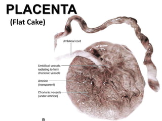
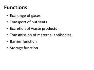
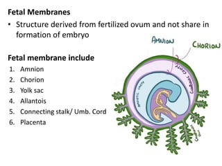
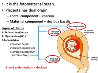
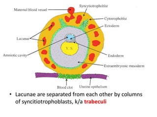
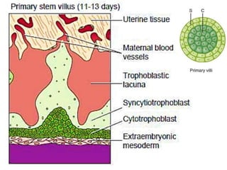
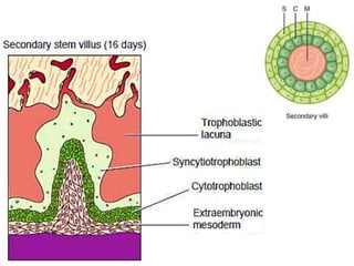
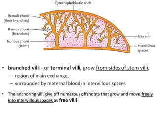
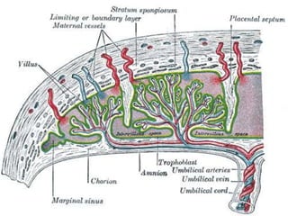
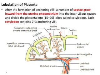
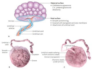
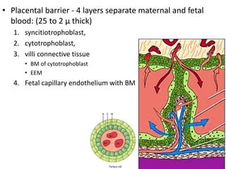
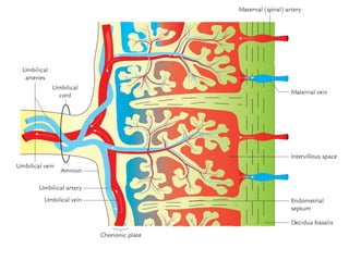
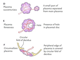
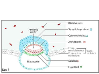
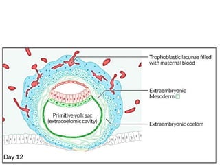
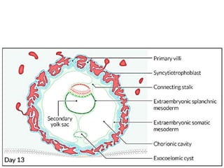
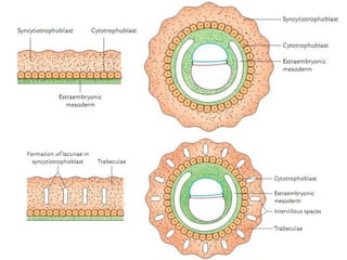
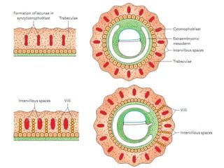
PLACENTA
(Flat Cake)
Functions:
• Exchange of gases
• Transport of nutrients
• Excretion of waste products
• Transmission of maternal antibodies
• Barrier function
• Storage function
Fetal Membranes
• Structure derived from fertilized ovum and not share in
formation of embryo
Fetal membrane include
1. Amnion
2. Chorion
3. Yolk sac
4. Allantois
5. Connecting stalk/ Umb. Cord
6. Placenta
• It is the fetomaternal organ.
• Placenta has dual origin
– Foetal component – chorion
– Maternal component – decidua basalis
Layers of Uterus
1. Perimetrium/Serosa
2. Myometrium (LCL)
3.Endometrium
i. Stratum basalis
ii.stratum spongiosum
iii.stratum compactum
(decidual layer)
Gravid endometrium = decidua
Interstitial type of implantation
Syncitiotrophoblast start secreting hCG
After implantation endometrium
undergoes changes k/a Decidual
Reaction
• Finger like chorionic villi formed
all around the chorionic sac
• Formed by trophoblast and EEM
• As the chorionic sac enlarges
– Chorionic villi in region of D. capsularis
get compressed and degenrate. This
smooth chorion is k/a chorionic leavae
– Chorionic villi in region of D. basalis
grow extensively. This part of chorion is
k/a chorionic frondosum
• Lacunae are separated from each other by columns
of syncitiotrophoblasts, k/a trabeculi
• Cells of cytotrophoblast invade the trabiculae in center.
– trabiculae has two layers – k/a Primary Villi
• Outer layer - syncitiotrophoblasts
• Inner core - cytotrophoblast
• Somatopleuric EEM deep to cytotrophoblast invade inner
core of Primary Villi – k/a Secondary Villi
• Blood vessels develops in EEM – k/a Tertiary villi
Tertiary villi continuous between chorion and decidua
these villi are also called STEM VILLI - or ANCHORING VILLI
(cytotrophoblast cells attached to maternal tissue)
• branched villi - or terminal villi, grow from sides of stem villi,
– region of main exchange,
– surrounded by maternal blood in intervillous spaces
• The anchoring villi give off numerous offshoots that grow and move freely
into intervillous spaces as free villi
Lobulation of Placenta
• After the formation of anchoring villi, a number of septae grow
inward from the uterine endometrium into the inter-villous spaces
and divide the placenta into (15–20) lobes called cotyledons. Each
cotyledon contains 2–3 anchoring villi.
Features of a full-term placenta
• It is like a flat circular disc (i.e., discoid).
• Haemochorial placenta - where the chorion comes in
direct contact with maternal blood (e.g. human)
• It has a diameter of 15–20 cm.
• It weighs about 500 g.
• It presents two surfaces: maternal and fetal.
a) Its maternal surface presents 15–20 lobes/cotyledons.
b) Its fetal surface presents a smooth shining surface, at
the approximate center of which the umbilical cord is
attached
• Placental barrier - 4 layers separate maternal and fetal
blood: (25 to 2 µ thick)
1. syncitiotrophoblast,
2. cytotrophoblast,
3. villi connective tissue
• BM of cytotrophoblast
• EEM
4. Fetal capillary endothelium with BM
Placental circulation
• Maternal placental circulation
– About 80–100 spiral arteries & veins open into the
intervillous spaces.
– under pressure of blood in arteries, the blood reaches
right up to the chorionic plate.
• Fetal placental circulation
– fetal blood comes to placenta through the umbilical
arteries.
– These arteries after entering the placenta ramify freely in
the chorion and their branches enter the chorionic villi.
– The veins from the chorionic villi drain into the umbilical
vein that carries blood rich in oxygen (O2) and nutrients
to the fetus from placenta.
Congenital Anomalies of the Placenta
• Anomalies Due to Abnormal Shape
• Anomalies Due to Abnormal Site of Attachment of UC
Abnormal Site of Attachment of UC
Abnormal Shape
THANK YOU
@~PLACENTA.ppt
@~PLACENTA.ppt
@~PLACENTA.ppt
@~PLACENTA.ppt
@~PLACENTA.ppt
@~PLACENTA.ppt

@~PLACENTA.ppt

  • 1.
  • 2.
    Functions: • Exchange ofgases • Transport of nutrients • Excretion of waste products • Transmission of maternal antibodies • Barrier function • Storage function
  • 3.
    Fetal Membranes • Structurederived from fertilized ovum and not share in formation of embryo Fetal membrane include 1. Amnion 2. Chorion 3. Yolk sac 4. Allantois 5. Connecting stalk/ Umb. Cord 6. Placenta
  • 4.
    • It isthe fetomaternal organ. • Placenta has dual origin – Foetal component – chorion – Maternal component – decidua basalis Layers of Uterus 1. Perimetrium/Serosa 2. Myometrium (LCL) 3.Endometrium i. Stratum basalis ii.stratum spongiosum iii.stratum compactum (decidual layer) Gravid endometrium = decidua
  • 6.
  • 8.
    Syncitiotrophoblast start secretinghCG After implantation endometrium undergoes changes k/a Decidual Reaction
  • 11.
    • Finger likechorionic villi formed all around the chorionic sac • Formed by trophoblast and EEM • As the chorionic sac enlarges – Chorionic villi in region of D. capsularis get compressed and degenrate. This smooth chorion is k/a chorionic leavae – Chorionic villi in region of D. basalis grow extensively. This part of chorion is k/a chorionic frondosum
  • 12.
    • Lacunae areseparated from each other by columns of syncitiotrophoblasts, k/a trabeculi
  • 13.
    • Cells ofcytotrophoblast invade the trabiculae in center. – trabiculae has two layers – k/a Primary Villi • Outer layer - syncitiotrophoblasts • Inner core - cytotrophoblast • Somatopleuric EEM deep to cytotrophoblast invade inner core of Primary Villi – k/a Secondary Villi • Blood vessels develops in EEM – k/a Tertiary villi
  • 19.
    Tertiary villi continuousbetween chorion and decidua these villi are also called STEM VILLI - or ANCHORING VILLI (cytotrophoblast cells attached to maternal tissue)
  • 20.
    • branched villi- or terminal villi, grow from sides of stem villi, – region of main exchange, – surrounded by maternal blood in intervillous spaces • The anchoring villi give off numerous offshoots that grow and move freely into intervillous spaces as free villi
  • 22.
    Lobulation of Placenta •After the formation of anchoring villi, a number of septae grow inward from the uterine endometrium into the inter-villous spaces and divide the placenta into (15–20) lobes called cotyledons. Each cotyledon contains 2–3 anchoring villi.
  • 24.
    Features of afull-term placenta • It is like a flat circular disc (i.e., discoid). • Haemochorial placenta - where the chorion comes in direct contact with maternal blood (e.g. human) • It has a diameter of 15–20 cm. • It weighs about 500 g. • It presents two surfaces: maternal and fetal. a) Its maternal surface presents 15–20 lobes/cotyledons. b) Its fetal surface presents a smooth shining surface, at the approximate center of which the umbilical cord is attached
  • 25.
    • Placental barrier- 4 layers separate maternal and fetal blood: (25 to 2 µ thick) 1. syncitiotrophoblast, 2. cytotrophoblast, 3. villi connective tissue • BM of cytotrophoblast • EEM 4. Fetal capillary endothelium with BM
  • 26.
    Placental circulation • Maternalplacental circulation – About 80–100 spiral arteries & veins open into the intervillous spaces. – under pressure of blood in arteries, the blood reaches right up to the chorionic plate. • Fetal placental circulation – fetal blood comes to placenta through the umbilical arteries. – These arteries after entering the placenta ramify freely in the chorion and their branches enter the chorionic villi. – The veins from the chorionic villi drain into the umbilical vein that carries blood rich in oxygen (O2) and nutrients to the fetus from placenta.
  • 28.
    Congenital Anomalies ofthe Placenta • Anomalies Due to Abnormal Shape • Anomalies Due to Abnormal Site of Attachment of UC Abnormal Site of Attachment of UC
  • 29.
  • 31.

Editor's Notes

  • #26 villi connective tissue – mesenchymal core