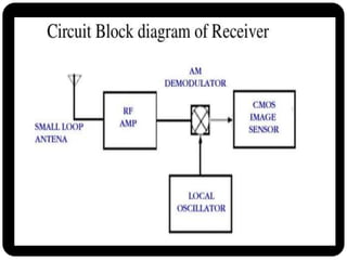
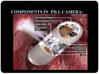
2. ABSTRACT…
Advancement in science & technology and
nanotechnology results in the development of the
pill camera.
Until recently, only the proximal (esophagus,
stomach and duodenum) and the distal (colon)
portions of the gastrointestinal tract were easily
visible using available technology.
But pill camera technology is capable of viewing
unreachable portions of small intestine.
3. INTRODUCTION…
A pill camera is a miniature camera, along with a light,
transmitter, and batteries, called Capsule Cam, is housed in
a capsule, of the size of vitamin pill.
It is used in a process called capsule endoscopy, which is an
easiest way of viewing the esophagus and the small
intestine.
Once swallowed, the capsule is propelled through the
small intestine by peristalsis, and acquires and
transmits digital images.
The pill camera can detect any abnormalities in the
stomach and also it is biocompatible.
7. WORKING:
The device, called the Given Diagnostic Imaging System,
comes in capsule form and contains a camera, lights,
transmitter & batteries.
The capsule has a clear end that allows the camera to
view the lining of the small intestine & large intestine too.
The pill camera is sized at 26*11 mm & is capable of
transmitting 50,000 color Images during its traversal
through the digestive system of patient.
Video chip consists of the IC CMOS image sensor which is
used to take pictures of intestine .
The LED is used for proper illumination in the intestine for
taking photos.
8. The patient swallows the capsule, and the natural muscular
waves of the digestive tract propel it forward through the
stomach, into the small intestine and large intestine, and then
out through the stool.
It takes snaps as it glides through digestive tract twice a
second.
The capsule transmits the images to a data recorder, which is
worn on a belt around the patient's waist, while the physician
then transfers the stored data to a computer for processing and
analysis.
The complete traversal takes around eight hours after it has
completed taking pictures it comes out of body as excreta.
9. ADVANTAGES OVER ENDOSCOPY…
Diseases of the gastrointestinal tract (such as esophageal
cancer) are currently diagnosed by this technology.
In endoscopy, the doctor passes a thin, flexible tube with a
camera on the end (endoscope) down your throat to your
stomach looking for abnormalities along the way.
The doctor can also pass instruments down the centre of the
tube to extract a small amount of tissue from areas that
appear abnormal which would be painful.
These cells are then analyzed in a laboratory to see if they are
cancerous.
10.
11. DIAGONISING CANCER:
This new investigative technique may help spot early signs of
cancers of the digestive system, such as oesophagal
cancer (also known as cancer of the gullet).
The researchers suggest the pill camera is a quick, simple
and pain free imaging method that patients may prefer
where a thin tube with a camera and a light source at the
end is passed down into the oesophagus.
Endoscopies have a number of practical disadvantages,
including they are often performed under sedation, so they
can be time-consuming.
12. • All the visual information is fed back to a processing console
that builds the information into a detailed 3D image.
• So that a doctor can review for signs of disease such as
oesophageal or stomach cancer.
13. EMERGING TECHNOLOGY:
Previous pill cameras place the camera at one end, facing
forward, so the tissue walls are visible only in the periphery
of their photos.
Sayaka is the first that gets a clearer picture with 32 mega
pixel resolution by mounting the camera facing the side and
spinning 360 degrees so that it shoots directly at the tissue
walls.
15. PILLCAM COLON:
With an imaging device on both ends of the capsule,
PillCam COLON provides a near 360˚ view of the colon.
Automatic light control provides optimal illumination
for improved visualization throughout the 30 mm
depth of field.
With bi-directional communication between the
capsule and the data recorder (which is worn around
the patient’s waist), real-time adjustments can be
made to maximize colon tissue coverage.
Images, captured at a rate from 4 to 35 frames per
second, are displayed as a smooth, continuous video.
Proprietary RAPID software optimizes reading
efficiency while aiding in diagnostic interpretation.
16. USING MOTOR FOR DRIVING THE
CAPSULE:
Even though the natural muscular movement fails to
drive the capsule. Using a grain sized motor attached,
the capsule can be driven through the intestine.
Any obstruction in the intestine can also be distorted
by the driving pill.
Even it has the ability to rupture the kidney stones on
its way which cannot be done by techniques like
endoscopy.
Thus the camera will drive the pill inspite of any
obstruction around it.
17. ADVANTAGES…
This pill camera makes us free from taking appointment and
waiting for doctors for hours and hours.
Painful treatments can be avoided.
Using the pill camera does not require highly trained staff.
The patient who has swallowed the capsule can go to his
work as usual with complete comfortability.
The size is smaller and so the cost is also reduced.
The patient does not need any sedation.
The process is relatively very quick.
18. CONCLUSION…
Gallstones form when liquid stored in the gallbladder hardens
into pieces of stone-like material.
Due to the controlled movement of pill camera it gradually
moves to its destination and in case any stone is detected, it
will rupture it.