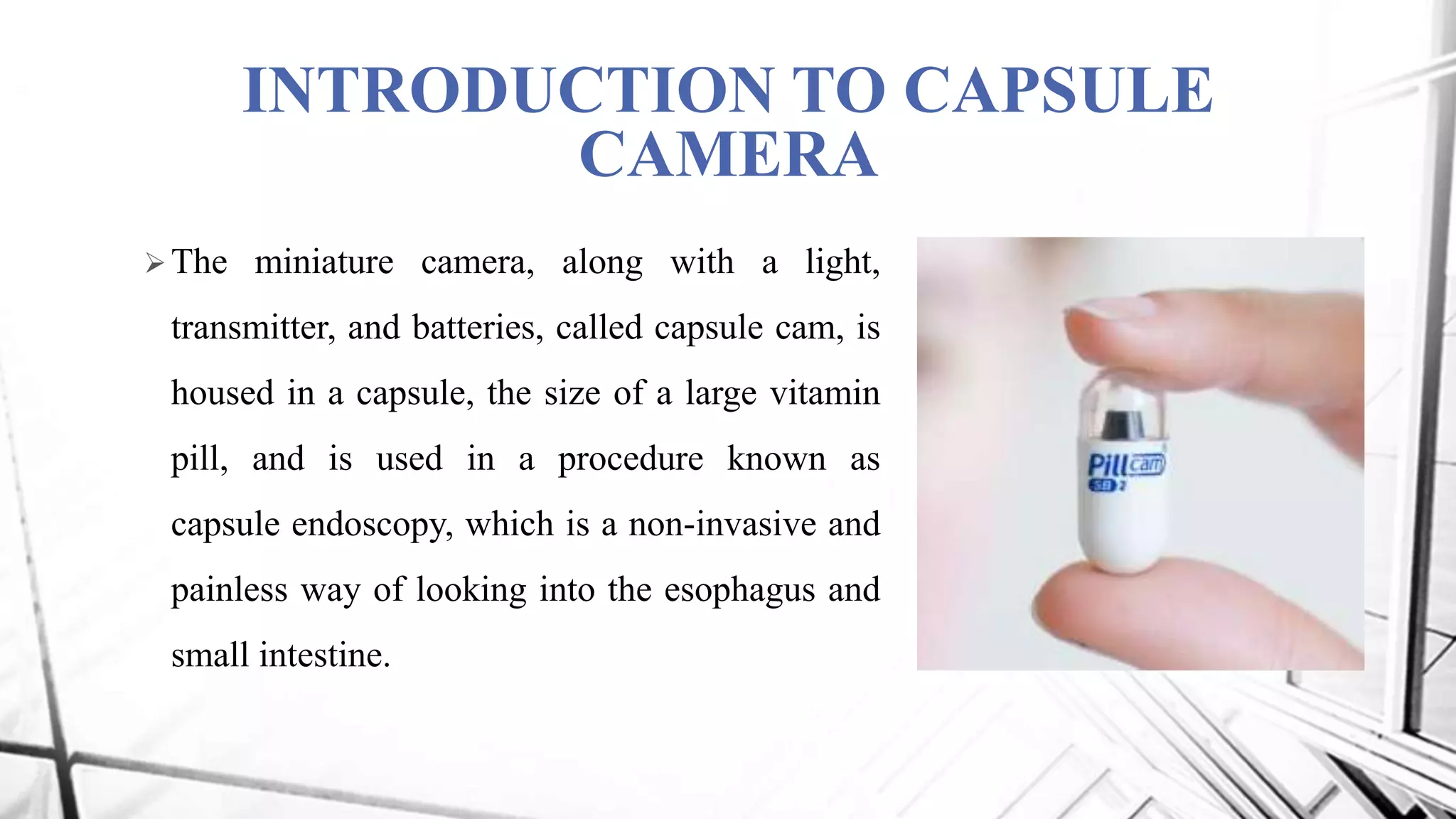
CAPSULE CAMERA
- Well known as Capsule Endoscopy
PRESENTED BY:
Ch. Bhavani 15R91A0208
D. Mounika 15R91A0209
CONTENTS
History
Introduction to capsule camera
Objectives
Components Required
Block Diagram
Description
Inside a Capsule Camera
Operation
Results
Advantages
Applications
Future Scope
Conclusion
Reference
HISTORY
 Endoscopic Ultrasound (EUS) endoscopes are unique
because they offer ultrasound guided needle biopsy,
colour Doppler and advanced image.
 Basic endoscopy was introduced in the late 1960s, and
about 20 years later, ultrasound was added, enabling
us to look at internal GI structures as never before.
 Pill Camera was invented by GAVRIEL LDDAN in
the year 2000 and it was approved by U.S.Food and
Drug Administration in 2001.
INTRODUCTION TO CAPSULE
CAMERA
The miniature camera, along with a light,
transmitter, and batteries, called capsule cam, is
housed in a capsule, the size of a large vitamin
pill, and is used in a procedure known as
capsule endoscopy, which is a non-invasive and
painless way of looking into the esophagus and
small intestine.
Continues….
Imagine a vitamin pill-sized camera that could travel through your
body taking pictures, helping to diagnose a problem which doctor
previously would have found only through EUS(Endoscopic
Ultrasound) Endoscopy and Colonoscopy.
Capsule Camera is developed on the basis of NANO
TECHNOLOGY.
OBJECTIVES
To evaluate the effectiveness of capsule endoscopy in the management of
patients with obscure gastrointestinal bleeding.
COMPONENTS REQUIRED
TRANSMITTER:
 CMOS Image Sensor
 AM Modulation
 RF Amplifier
 Oscillator
 Antenna
RECIEVER:
 LNA
 AM Demodulator
 IF Amplifier
 Oscillator
 OOK Demodulator
 Decoder
 High current Buffer
 CMOS Image Sensor
 LEDS
 Antenna
BLOCK DIAGRAM
CMOS
Image
Sensor
Local
Oscillator
(315MHZ)
RF
Amplifier
AM Modulation Small Loop
Antenna
VIDEO SIGNAL TRANSMITTER OF CAPSULE INSIDE
In this block diagram, one SMD type transistor amplifies the video signal for
efficient modulation using a 3 biasing resistor and 1 inductor.
In the bottom block, a tiny SAW resonator oscillates at 315 MHZ for
modulation of the video signal.
This modulated signal is then radiated from inside the body to outside the
body.
RECIEVER CIRCUIT INSIDE CAPSULE
Local
Oscillator
LNA
IF
Amplifier
OOK
Demodulator
Decoder
High
Current
Buffer
CMOS
Image
Sensor
Small Loop
Antenna
AM
Demodulator
4-LED’s
 This block diagram a commercialized ASK/OOK (ON/OFF Keyed) super heterodyne receiver with
an 8-pin SMD was used.
 This single chip receiver for remote wireless communications, which includes an internal local
oscillator fixed at a single frequency, is based on an external reference crystal or clock.
 The decoder IC receives the serial stream and interprets the serial information as 4 bits of binary data.
Each bit is used for channel recognition of the control signal from outside the body.
 lightning LED’s also use significant amount of power, the individual ON/OFF control of each LED is
equally necessary.
 As such the control system is divided into 4 channels in the current study. A high output current
amplifier with a single supply is utilized to drive loads in capsule.
DESCRIPTION
• The device, called the Diagnostic Imaging System, comes in capsule form and
contains a camera, lights, transmitter and batteries.
• The latest pill camera is sized at 26*11 mm and is capable of transmitting 50,000
color images during its traversal through the digestive system of patient.
INSIDE A CAPSULE CAMERA
Optical Dome
Lens Holder
Lens
Illuminating LED’s
CMOS Image Sensor
Battery
ASIC Transmitter
Antenna
OPTICAL DOME :It is the front part of the capsule and it is bullet shaped. Optical
dome is the light receiving window of the capsule and it is a non- conductor material. It
prevents the filtration of digestive fluids inside the capsule.
LENS HOLDER : This accommodates the lens. Lenses are tightly fixed in the capsule
to avoid dislocation of lens.
LENS : It is the integral component of pill camera. This lens is placed behind the
Optical Dome. The light through window falls on the lens.
ILLUMINATING LED’S :Illuminating LEDs illuminate an object. Non reflection
coating is placed on the light receiving window to prevent the reflection. Light
irradiated from the LED’s pass through the light receiving window.
CMOS IMAGE SENSOR :It has 140 degree field of view and detect object as small as
0.1mm. It have high precise.
BATTERY : Battery used in the pill camera is button shaped and two in number and
silver oxide primary batteries are used. It is disposable and harmless material.
ASIC TRANSMITTER : It is application specific integrated circuit and is placed
behind the batteries. Two transmitting electrodes are connected to this transmitter and
these electrodes are electrically isolated
ANTENNA :Parylene coated on to polyethylene or polypropylene antennas are used.
Antenna receives data from transmitter and then sends to data recorder.
OPERATION
 Capsule is swallowed by the patient like a
conventional pill.
 It takes images as it is propelled forward by
peristalsis.
 A wireless recorder, worn on a belt, receives the
images transmitted by the pill.
 A computer workstation processes the data and
produces a continuous still images.
MOVEMENT OF CAPSULE THROUGH THE DIGESTIVE SYSTEM :
Produces two images per second approximately 2600
DATA
RECORDER
COMPUTER
DATAACQUISITION AND STORAGE OF DATA
ON COMPUTER
RESULTS
Images Obtained From Capsule Camera
ADVANTAGES
Painless, no side effects.
Miniature size.
Accurate, precise (view of 150 degree).
High quality images.
Harmless material.
Simple procedure.
High sensitivity and specificity.
Avoids risk in sedation.
Efficient than X-ray CT-scan, normal endoscopy.
APPLICATIONS
Biggest impact in the medical industry.
Nano robots perform delicate surgeries.
Pill cam ESO can detect esophageal diseases,
gastrointestinal reflex diseases, and barreff’s esophagus.
Pill cam SB can detect Crohn’s disease, small bowel
tumours, small bowel injury, celiac disease, ulcerative
colitis etc.
FUTURE SCOPE
 The image quality is not bad but needs to be improved if it is to become a
realistic substitute for flexible upper and lower gastrointestinal endoscopy.
 An increase in the frame rate, angle of view, depth of field, image numbers,
duration of the procedure and improvements in illumination seem likely.
 Colonic, esophageal and gastric capsules will improve in quality, eroding the
supremacy of flexible endoscopy, and become embedded into screening
programs.
CONCLUSION
 The Given Endoscopy capsule is a pioneering
concept for Medical Technology of the 21st
century.
 The endoscopy system is the first of its kind to be
able to provide non-invasive imaging of the entire
small intestine.
 It has revolutionized the field of diagnostic
imaging to a great extent and has proved to be of
great help to physicians all over the world.
REFERENCES
Biomedical Circuits and Systems Conference, 2009. BioCAS 2009. IEEE.
Intelligent Systems, 2006 3rd International IEEE Conference on capsule
endoscopy.
Medical Imaging, IEEE Transactions on Dec. 2008.
Sidhu, Reena, etal. " Gastrointestinal capsule endoscopy: from tertiary centers
to primary care". BMJ, March 4 2006. 332:528-531.
doi:10.1136/bmj.332.7540.528.
"Capsule Endoscopy in Gastroenterology". Mayo Clinic. Accessed October 5
2007.
Capsule camera

Capsule camera

  • 1.
    CAPSULE CAMERA - Wellknown as Capsule Endoscopy PRESENTED BY: Ch. Bhavani 15R91A0208 D. Mounika 15R91A0209
  • 2.
    CONTENTS History Introduction to capsulecamera Objectives Components Required Block Diagram Description Inside a Capsule Camera Operation Results Advantages Applications Future Scope Conclusion Reference
  • 3.
    HISTORY  Endoscopic Ultrasound(EUS) endoscopes are unique because they offer ultrasound guided needle biopsy, colour Doppler and advanced image.  Basic endoscopy was introduced in the late 1960s, and about 20 years later, ultrasound was added, enabling us to look at internal GI structures as never before.  Pill Camera was invented by GAVRIEL LDDAN in the year 2000 and it was approved by U.S.Food and Drug Administration in 2001.
  • 4.
    INTRODUCTION TO CAPSULE CAMERA Theminiature camera, along with a light, transmitter, and batteries, called capsule cam, is housed in a capsule, the size of a large vitamin pill, and is used in a procedure known as capsule endoscopy, which is a non-invasive and painless way of looking into the esophagus and small intestine.
  • 5.
    Continues…. Imagine a vitaminpill-sized camera that could travel through your body taking pictures, helping to diagnose a problem which doctor previously would have found only through EUS(Endoscopic Ultrasound) Endoscopy and Colonoscopy. Capsule Camera is developed on the basis of NANO TECHNOLOGY.
  • 6.
    OBJECTIVES To evaluate theeffectiveness of capsule endoscopy in the management of patients with obscure gastrointestinal bleeding.
  • 7.
    COMPONENTS REQUIRED TRANSMITTER:  CMOSImage Sensor  AM Modulation  RF Amplifier  Oscillator  Antenna
  • 8.
    RECIEVER:  LNA  AMDemodulator  IF Amplifier  Oscillator  OOK Demodulator  Decoder  High current Buffer  CMOS Image Sensor  LEDS  Antenna
  • 9.
    BLOCK DIAGRAM CMOS Image Sensor Local Oscillator (315MHZ) RF Amplifier AM ModulationSmall Loop Antenna VIDEO SIGNAL TRANSMITTER OF CAPSULE INSIDE
  • 10.
    In this blockdiagram, one SMD type transistor amplifies the video signal for efficient modulation using a 3 biasing resistor and 1 inductor. In the bottom block, a tiny SAW resonator oscillates at 315 MHZ for modulation of the video signal. This modulated signal is then radiated from inside the body to outside the body.
  • 11.
    RECIEVER CIRCUIT INSIDECAPSULE Local Oscillator LNA IF Amplifier OOK Demodulator Decoder High Current Buffer CMOS Image Sensor Small Loop Antenna AM Demodulator 4-LED’s
  • 12.
     This blockdiagram a commercialized ASK/OOK (ON/OFF Keyed) super heterodyne receiver with an 8-pin SMD was used.  This single chip receiver for remote wireless communications, which includes an internal local oscillator fixed at a single frequency, is based on an external reference crystal or clock.  The decoder IC receives the serial stream and interprets the serial information as 4 bits of binary data. Each bit is used for channel recognition of the control signal from outside the body.  lightning LED’s also use significant amount of power, the individual ON/OFF control of each LED is equally necessary.  As such the control system is divided into 4 channels in the current study. A high output current amplifier with a single supply is utilized to drive loads in capsule.
  • 13.
    DESCRIPTION • The device,called the Diagnostic Imaging System, comes in capsule form and contains a camera, lights, transmitter and batteries. • The latest pill camera is sized at 26*11 mm and is capable of transmitting 50,000 color images during its traversal through the digestive system of patient.
  • 14.
    INSIDE A CAPSULECAMERA Optical Dome Lens Holder Lens Illuminating LED’s CMOS Image Sensor Battery ASIC Transmitter Antenna
  • 15.
    OPTICAL DOME :Itis the front part of the capsule and it is bullet shaped. Optical dome is the light receiving window of the capsule and it is a non- conductor material. It prevents the filtration of digestive fluids inside the capsule. LENS HOLDER : This accommodates the lens. Lenses are tightly fixed in the capsule to avoid dislocation of lens. LENS : It is the integral component of pill camera. This lens is placed behind the Optical Dome. The light through window falls on the lens. ILLUMINATING LED’S :Illuminating LEDs illuminate an object. Non reflection coating is placed on the light receiving window to prevent the reflection. Light irradiated from the LED’s pass through the light receiving window.
  • 16.
    CMOS IMAGE SENSOR:It has 140 degree field of view and detect object as small as 0.1mm. It have high precise. BATTERY : Battery used in the pill camera is button shaped and two in number and silver oxide primary batteries are used. It is disposable and harmless material. ASIC TRANSMITTER : It is application specific integrated circuit and is placed behind the batteries. Two transmitting electrodes are connected to this transmitter and these electrodes are electrically isolated ANTENNA :Parylene coated on to polyethylene or polypropylene antennas are used. Antenna receives data from transmitter and then sends to data recorder.
  • 17.
    OPERATION  Capsule isswallowed by the patient like a conventional pill.  It takes images as it is propelled forward by peristalsis.  A wireless recorder, worn on a belt, receives the images transmitted by the pill.  A computer workstation processes the data and produces a continuous still images.
  • 18.
    MOVEMENT OF CAPSULETHROUGH THE DIGESTIVE SYSTEM : Produces two images per second approximately 2600 DATA RECORDER COMPUTER DATAACQUISITION AND STORAGE OF DATA ON COMPUTER
  • 19.
  • 20.
    ADVANTAGES Painless, no sideeffects. Miniature size. Accurate, precise (view of 150 degree). High quality images. Harmless material. Simple procedure. High sensitivity and specificity. Avoids risk in sedation. Efficient than X-ray CT-scan, normal endoscopy.
  • 21.
    APPLICATIONS Biggest impact inthe medical industry. Nano robots perform delicate surgeries. Pill cam ESO can detect esophageal diseases, gastrointestinal reflex diseases, and barreff’s esophagus. Pill cam SB can detect Crohn’s disease, small bowel tumours, small bowel injury, celiac disease, ulcerative colitis etc.
  • 22.
    FUTURE SCOPE  Theimage quality is not bad but needs to be improved if it is to become a realistic substitute for flexible upper and lower gastrointestinal endoscopy.  An increase in the frame rate, angle of view, depth of field, image numbers, duration of the procedure and improvements in illumination seem likely.  Colonic, esophageal and gastric capsules will improve in quality, eroding the supremacy of flexible endoscopy, and become embedded into screening programs.
  • 23.
    CONCLUSION  The GivenEndoscopy capsule is a pioneering concept for Medical Technology of the 21st century.  The endoscopy system is the first of its kind to be able to provide non-invasive imaging of the entire small intestine.  It has revolutionized the field of diagnostic imaging to a great extent and has proved to be of great help to physicians all over the world.
  • 24.
    REFERENCES Biomedical Circuits andSystems Conference, 2009. BioCAS 2009. IEEE. Intelligent Systems, 2006 3rd International IEEE Conference on capsule endoscopy. Medical Imaging, IEEE Transactions on Dec. 2008. Sidhu, Reena, etal. " Gastrointestinal capsule endoscopy: from tertiary centers to primary care". BMJ, March 4 2006. 332:528-531. doi:10.1136/bmj.332.7540.528. "Capsule Endoscopy in Gastroenterology". Mayo Clinic. Accessed October 5 2007.