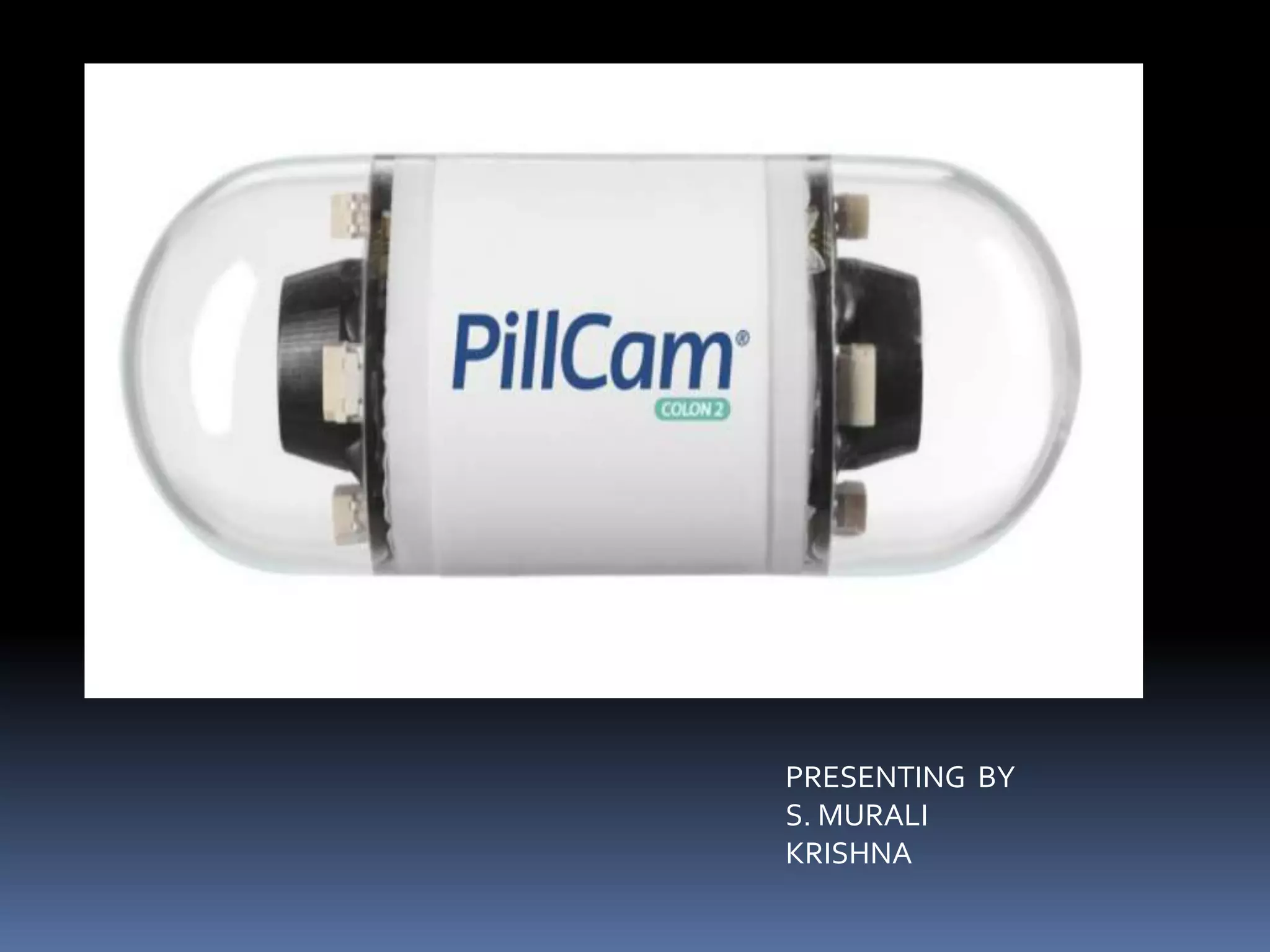
The document discusses a pill-sized camera called a capsule endoscopy or pill camera. Developed in 2000 using nanotechnology, the pill camera contains a camera, light, battery, and antenna to transmit images of the digestive tract as it passes through the body. Measuring around 25mm by 10mm, the pill camera provides a clear view of the intestines and allows for easier endoscopy exams compared to other methods. However, there are drawbacks for patients with gastrointestinal obstructions as the pill could get stuck.
The document discusses capsule cameras, which are pill-sized cameras that can be swallowed to take pictures as they pass through the digestive tract. Conventional methods like endoscopes are limited in the areas they can examine. Capsule cameras contain lenses, lights, batteries, and a transmitter to send over 50,000 high quality images to a data recorder. They help diagnose conditions like Crohn's disease and tumors. Capsule cameras are painless and accurate but have drawbacks like being unable to control behavior, though new technologies may help overcome limitations.
pill camera/ capsule endoscopy used inn medical field to find out the abnormalities of small intestine. This replaced the convectional endoscopy method.
The document describes a pill-sized camera called a pill camera that can be swallowed to take over 50,000 photos as it passes through the digestive tract. It consists of components like a lens, light source, and antenna to transmit photos. The pill camera allows physicians to non-invasively examine the small intestine for conditions like Crohn's disease and tumors. It provides advantages over endoscopy by being painless, having no side effects, and enabling high-quality internal images of areas endoscopy can't reach. However, it risks obstruction if partial blockages are present in the small intestine.
The document describes the components and operation of a pill camera. The pill camera is about 26x11 mm in size and can take over 50,000 color images as it passes through the digestive tract. It contains an optical dome with a light receiving window, lens, LED lights, CMOS image sensor, batteries, transmitter, and antenna. The capsule transmits images to an external recorder as it is propelled through the intestines by peristalsis, allowing physicians to non-invasively examine the small intestine.
The document discusses a pill camera, which is a capsule-sized camera that can be swallowed to take pictures of the digestive tract. It consists of an optical dome, lens, LED lights, CMOS image sensor, battery, transmitter, and antenna. The procedure involves swallowing the capsule, which then transmits over 50,000 color images as it passes through the digestive system. The capsule provides a painless and accurate way to diagnose gastrointestinal conditions without surgery. However, it is very expensive and cannot be reused. Future improvements could include zooming, auto-focus, smaller size, and longer battery life.
The document discusses the pill camera, which is a capsule-sized endoscopy device used to non-invasively examine the esophagus and small intestine. The pill camera contains a camera, light source, transmitter and batteries. It is swallowed and passes naturally through the digestive tract, transmitting over 50,000 images to an external recorder. Compared to conventional endoscopy, the pill camera offers a painless procedure without sedation that provides images of the entire small intestine. However, it cannot stop to examine areas of interest and cannot perform biopsies. The document reviews the capsule components, procedure, advantages of providing detailed images without risks of other methods, and applications in diagnosing gastrointestinal conditions.
The document describes a capsule camera, also known as capsule endoscopy. It provides a brief history of endoscopy and describes how the capsule camera was invented in 2000. The summary provides an overview of the key components of the capsule camera, including a CMOS image sensor, batteries, transmitter, and antenna. It also describes how the capsule camera works, taking images as it passes through the digestive tract that are transmitted to an external recorder. The summary concludes by mentioning some of the advantages of capsule cameras, such as being painless and providing accurate images of the small intestine.
The document discusses a pill camera, which is a capsule endoscopy device used to examine the small intestine. It consists of a pill-sized capsule containing a camera, light source, and transmitter. Patients swallow the capsule, which passes naturally through the digestive tract while transmitting thousands of images. These images are received by a sensor array worn by the patient and recorded for physicians to review on specialized software. The capsule does not require sedation and allows examination of the entire small intestine, providing a more complete evaluation than other endoscopic methods. Key advantages include it being non-invasive, comfortable for patients, and providing better imaging of hard to reach areas of the digestive tract.
Pill camera technology allows a miniature camera to be housed in a capsule and passed through the digestive tract to provide images of the small intestine. The capsule captures images as it is propelled through the tract and transmits them to an external recorder. This provides a painless alternative to endoscopy for diagnosing conditions like cancer by visualizing areas that standard techniques cannot reach. The capsule offers advantages over endoscopy like being non-invasive and allowing patients to avoid sedation and return to normal activities after swallowing.
The document describes a pill camera, which is a capsule-sized, swallowable camera that can capture over 50,000 images of the inside of the body as it passes through the gastrointestinal tract. It has a diameter of 11mm and length of 26mm, with components like an optical dome, lens, LED lights, CMOS image sensor, batteries, transmitter, and antenna that allow it to wirelessly transmit the captured images to an external data recorder. The pill camera provides a painless and effective way to examine the small intestine for conditions like tumors or ulcers.
The document discusses the "camera in a pill", or capsule endoscopy. It provides a high-level overview of the technology, including that a pill-sized camera is swallowed to take images of the small intestine as it passes through. The camera transmits over 56,000 images wirelessly to a sensor array and data recorder worn on the body. The images can later be reviewed by a doctor on specialized software to diagnose conditions of the small intestine.
A pill camera is a capsule-sized device used in endoscopy to record images of the digestive tract. It contains a tiny camera that takes pictures after being swallowed. The primary use is to examine the small intestine, which other endoscopy methods cannot access well. The capsule transmits images wirelessly to an external receiver as it passes through the tract. Images are then reviewed by doctors to diagnose problems, though the capsule cannot treat any issues found. While generally safe, there is a small risk of the capsule being retained in the body for an extended time. It works using electromagnetic waves and has protections to safely pass through the digestive system.
The document describes the pill camera, a vitamin-sized device invented in 2000 that can be swallowed to take pictures inside the digestive tract to help diagnose problems. It contains a camera, lights, transmitter and batteries. Images are transmitted to a recorder worn by the patient. The capsule passes naturally through the digestive system, providing a clear view of the intestines. It is an alternative to conventional endoscopy for examining parts of the intestine that cannot be seen with other methods.
The document discusses a pill camera, which is a capsule endoscopy device used to examine the digestive tract. It is about the size of a pill and contains a camera, lights, batteries, and transmitter. Patients swallow the capsule, which takes pictures as it passes through the digestive system. The images are transmitted to a data recorder and can be reviewed later by doctors. The capsule offers a non-invasive alternative to traditional endoscopy for examining the small intestine. Key benefits include increased patient comfort and ability to capture images of hard to reach areas of the digestive tract.
The technology used to achieve manufacturing the product at molecular level is “NANOTECHNOLOGY”.
Nanotechnology is the creation of useful materials, devices and system through manipulation of such miniscule matter (nanometer).
Trillions of assemblers will be needed to develop products in viable time
frame.
Capsule endoscopy provides a non-invasive method to examine the small intestine using a pill-sized camera that is swallowed. It takes and transmits images that are recorded for physicians to review. Previous endoscopy methods could not fully examine the small intestine. Research shows capsule endoscopy can diagnose certain gastrointestinal diseases. The document discusses the
The document describes a pill camera, which is a pill-sized device that can be swallowed to examine the gastrointestinal tract by taking pictures. It works by transmitting pictures wirelessly as it passes through the digestive system, helping doctors diagnose conditions. The pill camera provides a noninvasive alternative to surgery for visualization of the GI tract. However, image quality and lack of control are still limitations that need improvement.
Biomedical instrumentation involves using measuring instruments to monitor and control processes within the body. Pill cameras are a type of biomedical instrumentation that can take pictures inside the body without surgery. A pill camera is the size of a pill and contains a camera, lights, transmitter and batteries. As it passes through the digestive system, it takes over 50,000 color images that are transmitted to a recorder. Doctors can then view the continuous images on a computer to diagnose conditions like Crohn's disease or tumors in the small intestine.
The document discusses a pill camera, which is a small, capsule-sized camera that can be swallowed to examine the gastrointestinal tract. It describes how the pill camera works, including how it is administered and how images are transmitted. Key advantages are that it provides detailed images of the entire digestive tract without pain or sedation compared to other examination methods. Potential disadvantages are that it is expensive, can become stuck, and doctors have limited control over its movement. The conclusion states it is a pioneering medical technology that has revolutionized diagnostic imaging.
The document describes a pill camera, which is a capsule that contains a camera and travels through the digestive system to take pictures. It is about 26x11mm in size and can capture 50,000 color images. The camera helps doctors diagnose problems without surgery. The pill camera contains components like an optical dome, lens holder, LED lights, image sensor, battery, transmitter and antenna to capture and transmit images. It provides a painless and easy alternative to endoscopy for examining the small intestine. However, it cannot control camera behavior and may get stuck if an obstruction is present. The endoscopy capsule is a pioneering medical technology that has revolutionized diagnostic imaging.
Imagine a vitamin pill-sized camera that could travel through your body taking pictures, helping diagnose a problem which doctor previously would have found only through surgery.
The capsule camera is a pill-sized device that can be swallowed to take pictures of the digestive tract as it passes through. It contains a camera, lights, transmitter and batteries. Images are transmitted wirelessly to a recorder and over 2,600 high quality images can be captured. The capsule allows non-invasive imaging of the small intestine to diagnose conditions like Crohn's disease. It is painless for the patient but cannot be controlled and could get obstructed, though newer models aim to overcome these limitations. The capsule camera has revolutionized digestive imaging.
Pill Camera is a vitamin pill-sized capsule that travels through the human body taking pictures, helping diagnose a problem which previously would have been found only through surgery
The document discusses a pill camera, which is a capsule-sized camera that can be swallowed to take pictures as it passes through the digestive system. It has revolutionized medical diagnosis by allowing doctors to non-invasively examine the small intestine. The pill camera contains a lens, light source, image sensor and transmitter inside a biocompatible casing. It transmits over 5,000 images during its 8-hour operation to an external recorder for analysis by doctors. The pill camera provides a painless alternative to invasive endoscopy and has improved diagnosis of conditions like cancer and ulcers.
The pill camera was invented in the 1990s as a minimally invasive way to examine the digestive tract. It is a capsule-sized device containing a camera, lights, batteries, and transmitter. As it passes through the digestive system, it takes over 50,000 images that are transmitted to an external data recorder. The pill camera has revolutionized gastrointestinal diagnosis and detection of conditions like Crohn's disease with its painless, non-invasive procedure.
Imagine a vitamin pill-sized camera that could travel through your body taking pictures, helping diagnose a problem which doctor previously would have found only through surgery.
pill camera based on endoscopy and its procedureabdullaabrar61
The document describes a pill-sized camera called a pill camera that can be ingested to take pictures and help diagnose problems within the digestive system. The pill camera is about the size of a large vitamin and contains a camera, lights, transmitter and batteries. As it passes through the digestive tract, it takes 50,000 color images that are wirelessly transmitted to a data recorder. Doctors can then review the images to identify any abnormalities. The pill camera provides a painless and accurate way to examine the small intestine without surgery. It has advantages for diagnosing various digestive conditions but also has some drawbacks like inability to control movement, which newer technologies are seeking to address.
The document discusses capsule endoscopy, a non-invasive medical procedure using a pill-sized camera to examine the digestive tract. The capsule camera contains LED lights, lenses, batteries and an image sensor, transmitting over 50,000 images as it passes through the tract. Doctors can view the high-quality images to diagnose conditions like Crohn's disease. The procedure is painless but the capsule may get obstructed; newer models are being developed to overcome limitations. Capsule endoscopy has revolutionized digestive imaging and become a valuable diagnostic tool for physicians.
The document discusses capsule cameras, which are pill-sized cameras that can be ingested to take pictures of the digestive tract. It describes the components of capsule cameras, including lenses, lights, batteries, and transmitters. Capsule cameras travel through the digestive system taking pictures, which are transmitted to an external recorder. Their images help diagnose conditions like Crohn's disease and small bowel tumors. While early capsule cameras had limitations like getting stuck, newer models use bi-directional telemetry to help overcome issues. Capsule cameras provide a non-invasive alternative to endoscopy and have revolutionized digestive imaging.
The document describes a capsule camera that can be swallowed to take pictures inside the digestive system. It consists of a camera, lights, transmitter and batteries inside a capsule the size of a vitamin pill. It takes 50,000 color images as it passes through the digestive tract, transmitting them wirelessly to an external recorder. The capsule camera provides a non-invasive alternative to conventional endoscopy and helps diagnose conditions of the small intestine.
The document describes a capsule camera that can be swallowed to take pictures inside the digestive system. It consists of a camera, lights, transmitter and batteries inside a capsule the size of a vitamin pill. It takes 50,000 color images as it passes through the digestive tract, transmitting them wirelessly to an external recorder. The capsule camera provides a non-invasive alternative to conventional endoscopy and has applications in diagnosing various digestive conditions. Some early limitations have been addressed by newer bidirectional telemetry cameras.
The document summarizes a seminar presentation on pill cameras. Pill cameras are capsule-sized devices that can be swallowed to take pictures of the digestive tract as they pass through. They provide a non-invasive alternative to endoscopy. The capsule contains a camera, lights, batteries, and transmitter to send over 50,000 color images as it travels through the digestive system. It has advantages like being painless, allowing imaging of the entire small intestine, and providing high quality images. However, it could get stuck if there is an obstruction and is difficult to control. New technologies aim to overcome issues like using a rice-grain sized motor or bi-directional telemetry camera. In conclusion, pill cameras have revolutionized diagnostic imaging
The document discusses a pill camera, which is a capsule device used to take pictures of the inside of the digestive system. It contains a camera, lights, transmitter and batteries and transmits 50,000 images as it passes through the digestive tract. The images help doctors diagnose conditions of the small intestine. It provides a safer alternative to endoscopy for examining the small intestine. The capsule works by being swallowed and passing naturally through the digestive system, transmitting images to an external receiver worn by the patient.
The document describes a pill-sized camera that can be swallowed to take pictures inside the digestive tract. It contains a camera, lights, transmitter and batteries inside a capsule. Over 50,000 color images are transmitted as it passes through the tract. Components include an optical dome, lens, LED lights, image sensor, battery and transmitter. The capsule is swallowed and images are transmitted to a receiver and computer for processing. It can diagnose conditions like Crohn's disease without surgery. Advantages are it is painless and provides high quality images of the small intestine. Drawbacks are it may get stuck if obstructions are present, though new bi-directional cameras aim to overcome this.
1) The document describes a capsule camera, also known as a pill camera, which is a pill-sized device that can be swallowed to take pictures inside the digestive tract to diagnose conditions.
2) The capsule contains a camera, lights, transmitter and batteries and transmits up to 50,000 color images as it passes through the digestive system, with the images being received by an external recorder.
3) The capsule camera provides a non-invasive alternative to endoscopy and is useful for diagnosing various conditions of the small intestine like Crohn's disease, ulcers, and tumors. It allows detection of disorders at an earlier stage compared to other tests.
The document describes a microelectronic pill used for remote biomedical measurements in the gastrointestinal tract. The pill contains components like an optical dome, lens, LED lights, CMOS image sensor, battery, transmitter, and antenna. It takes images as it passes through the GI tract, transmitting data to a wireless recorder. Compared to endoscopy, the pill provides painless, high-quality imaging of the intestines without risks of sedation or radiation. Future improvements could include zooming, auto-focus, smaller sizes, and using lasers to identify and treat precancerous cells.
The document describes the components and functioning of a pill camera, which is a capsule-sized, swallowable camera that can capture over 50,000 images of the inside of the body as it passes through the gastrointestinal tract. It has a diameter of 11mm, length of 26mm, and weighs less than 4g. Key components include an optical dome, lens, LED lights, CMOS image sensor, batteries, transmitter, and antenna that wirelessly transmits the images to an external data recorder. The pill camera provides a painless and effective way to diagnose conditions like tumors or ulcers throughout the intestines.
The document describes a pill-sized camera that can be swallowed to take pictures of the digestive tract. It consists of a camera, lights, transmitters and batteries in a capsule sized at 26x11mm. As it travels through the digestive system, it takes 50,000 color images which are transmitted to an external recorder and computer. The capsule helps diagnose conditions like Crohn's disease and tumors of the small intestine in a non-invasive way. It provides high quality images and has advantages over conventional endoscopy, though early models had disadvantages of getting stuck or lack of control that newer technologies have helped overcome.
The document describes a capsule endoscopy procedure using a pill-sized camera to diagnose problems in the intestines. It discusses how conventional endoscopy requires inserting a scope through the mouth, while capsule endoscopy involves swallowing a small camera pill. The pill camera contains components like a lens, image sensor, batteries, and transmitter to take 50,000 color images as it passes through the digestive tract, transmitting the images wirelessly to an external recorder and computer for analysis. The procedure involves swallowing the capsule, which then takes images as it moves through the intestines via peristalsis before the images are processed to produce a continuous video stream, allowing diagnosis without invasive surgery.
The document discusses capsule endoscopy, which uses a small, pill-sized camera to take images of the digestive tract. It provides details on the history, components, and working of the capsule camera. Key points include that the camera pill transmits thousands of images as it passes through the digestive system, allowing doctors to examine the small intestine without invasive procedures. The capsule camera offers advantages like being non-invasive and painless, but challenges include risks of obstruction and high costs. Future applications may include additional diagnostic and treatment abilities.
Capsule endoscopy is a noninvasive diagnostic procedure to visualize the inside of your digestive tract. You swallow a capsule that contains a tiny camera, a transmitter and a light. As it passes through your stomach, intestines, colon and rectum, the capsule takes thousands of pictures and transmits them to a recorder that you wear outside of your body.
Your doctor uploads the data from the recorder using a system that combines pictures into a video. These visualizations help identify problems in the digestive tract and formulate a diagnosis.
The document summarizes the key components and functioning of a pill camera. The pill camera is a vitamin pill-sized camera that can travel through the digestive system, taking pictures to help diagnose problems. It has an optical dome containing a light receiving window and lens, LED lights, a CMOS image sensor, batteries, an ASIC transmitter, and antenna. Some advantages are that it is painless, miniature, accurate, and produces high-quality images. Drawbacks like getting stuck in obstructions can be overcome with newer technologies like a rice-grain sized motor or bi-directional telemetry camera. In conclusion, the pill camera is a pioneering medical technology that provides non-invasive imaging of the small intestine.
The document describes a pill camera, which is a miniature camera housed in a capsule the size of a vitamin pill. It is used in capsule endoscopy to view the esophagus and small intestine. The capsule captures and transmits images as it passes through the digestive tract, allowing doctors to examine the images later. Key components include an optical dome, lens, LED lights, image sensor, batteries, and antenna. It provides detailed images of the entire digestive tract without needing endoscopy or sedation. While expensive and unable to be controlled, pill cameras have revolutionized diagnosis of small intestine conditions and have bright future applications.
Pill Camera - An Application of Bio Medical TechnologyIshanMalpotra
This is the research paper I presented on "Pill Camera - An Application of Bio Medical Technology" at a national level conference held at T.K.M. Institute of Technology on March 15, 2013
Digital Marketing Trends in 2024 | Guide for Staying AheadWask
https://www.wask.co/ebooks/digital-marketing-trends-in-2024
Feeling lost in the digital marketing whirlwind of 2024? Technology is changing, consumer habits are evolving, and staying ahead of the curve feels like a never-ending pursuit. This e-book is your compass. Dive into actionable insights to handle the complexities of modern marketing. From hyper-personalization to the power of user-generated content, learn how to build long-term relationships with your audience and unlock the secrets to success in the ever-shifting digital landscape.
Your One-Stop Shop for Python Success: Top 10 US Python Development Providersakankshawande
Simplify your search for a reliable Python development partner! This list presents the top 10 trusted US providers offering comprehensive Python development services, ensuring your project's success from conception to completion.
Have you ever been confused by the myriad of choices offered by AWS for hosting a website or an API?
Lambda, Elastic Beanstalk, Lightsail, Amplify, S3 (and more!) can each host websites + APIs. But which one should we choose?
Which one is cheapest? Which one is fastest? Which one will scale to meet our needs?
Join me in this session as we dive into each AWS hosting service to determine which one is best for your scenario and explain why!
Taking AI to the Next Level in Manufacturing.pdfssuserfac0301
Read Taking AI to the Next Level in Manufacturing to gain insights on AI adoption in the manufacturing industry, such as:
1. How quickly AI is being implemented in manufacturing.
2. Which barriers stand in the way of AI adoption.
3. How data quality and governance form the backbone of AI.
4. Organizational processes and structures that may inhibit effective AI adoption.
6. Ideas and approaches to help build your organization's AI strategy.
zkStudyClub - LatticeFold: A Lattice-based Folding Scheme and its Application...Alex Pruden
Folding is a recent technique for building efficient recursive SNARKs. Several elegant folding protocols have been proposed, such as Nova, Supernova, Hypernova, Protostar, and others. However, all of them rely on an additively homomorphic commitment scheme based on discrete log, and are therefore not post-quantum secure. In this work we present LatticeFold, the first lattice-based folding protocol based on the Module SIS problem. This folding protocol naturally leads to an efficient recursive lattice-based SNARK and an efficient PCD scheme. LatticeFold supports folding low-degree relations, such as R1CS, as well as high-degree relations, such as CCS. The key challenge is to construct a secure folding protocol that works with the Ajtai commitment scheme. The difficulty, is ensuring that extracted witnesses are low norm through many rounds of folding. We present a novel technique using the sumcheck protocol to ensure that extracted witnesses are always low norm no matter how many rounds of folding are used. Our evaluation of the final proof system suggests that it is as performant as Hypernova, while providing post-quantum security.
Paper Link: https://eprint.iacr.org/2024/257
Dandelion Hashtable: beyond billion requests per second on a commodity serverAntonios Katsarakis
This slide deck presents DLHT, a concurrent in-memory hashtable. Despite efforts to optimize hashtables, that go as far as sacrificing core functionality, state-of-the-art designs still incur multiple memory accesses per request and block request processing in three cases. First, most hashtables block while waiting for data to be retrieved from memory. Second, open-addressing designs, which represent the current state-of-the-art, either cannot free index slots on deletes or must block all requests to do so. Third, index resizes block every request until all objects are copied to the new index. Defying folklore wisdom, DLHT forgoes open-addressing and adopts a fully-featured and memory-aware closed-addressing design based on bounded cache-line-chaining. This design offers lock-free index operations and deletes that free slots instantly, (2) completes most requests with a single memory access, (3) utilizes software prefetching to hide memory latencies, and (4) employs a novel non-blocking and parallel resizing. In a commodity server and a memory-resident workload, DLHT surpasses 1.6B requests per second and provides 3.5x (12x) the throughput of the state-of-the-art closed-addressing (open-addressing) resizable hashtable on Gets (Deletes).
Fueling AI with Great Data with Airbyte WebinarZilliz
This talk will focus on how to collect data from a variety of sources, leveraging this data for RAG and other GenAI use cases, and finally charting your course to productionalization.
Building Production Ready Search Pipelines with Spark and MilvusZilliz
Spark is the widely used ETL tool for processing, indexing and ingesting data to serving stack for search. Milvus is the production-ready open-source vector database. In this talk we will show how to use Spark to process unstructured data to extract vector representations, and push the vectors to Milvus vector database for search serving.
Ivanti’s Patch Tuesday breakdown goes beyond patching your applications and brings you the intelligence and guidance needed to prioritize where to focus your attention first. Catch early analysis on our Ivanti blog, then join industry expert Chris Goettl for the Patch Tuesday Webinar Event. There we’ll do a deep dive into each of the bulletins and give guidance on the risks associated with the newly-identified vulnerabilities.
Skybuffer AI: Advanced Conversational and Generative AI Solution on SAP Busin...Tatiana Kojar
Skybuffer AI, built on the robust SAP Business Technology Platform (SAP BTP), is the latest and most advanced version of our AI development, reaffirming our commitment to delivering top-tier AI solutions. Skybuffer AI harnesses all the innovative capabilities of the SAP BTP in the AI domain, from Conversational AI to cutting-edge Generative AI and Retrieval-Augmented Generation (RAG). It also helps SAP customers safeguard their investments into SAP Conversational AI and ensure a seamless, one-click transition to SAP Business AI.
With Skybuffer AI, various AI models can be integrated into a single communication channel such as Microsoft Teams. This integration empowers business users with insights drawn from SAP backend systems, enterprise documents, and the expansive knowledge of Generative AI. And the best part of it is that it is all managed through our intuitive no-code Action Server interface, requiring no extensive coding knowledge and making the advanced AI accessible to more users.
GraphRAG for Life Science to increase LLM accuracyTomaz Bratanic
GraphRAG for life science domain, where you retriever information from biomedical knowledge graphs using LLMs to increase the accuracy and performance of generated answers
2. CONTENTS
INTRODUCTION
TECHNOLOGY USED
DESCRIPTION
BODY OF THE PIN CAMERA
INSIDE OF THE CAPSULE CAMERA
WORKING
TRANSMITTER AND RECEIVER EXTERNAL
CONTROL UNIT
USES
ADVANTAGES
DISADVANTAGES
CONCLUSION
3. INTRODUCTION
An application of nanotechnology.
A vitamin pill sized camera.
Travels through body and taking pictures.
Helps in diagnosing abnormality in small
intestine.
4. TECHNOLOGIES USED
Pill Camera developed on the basis of
NANOTECHNOLOGY.
Nanotechnology deals with objects measured
in nanometers.
Nanometer can be visualized as billionth of a
meter or millionth of a millimeter or it is
1/80000 width of human hair.
5. DESCRIPTION
Comes in capsule form and contains a camera,
lights, transmitter and batteries.
6. •Video chip : consists of IC CMOS image sensor
which is used to take pictures of the intestine.
•LAMP : It is used for proper Illumination in the
intestine for taking photos.
•Micro actuator: Acts as memory to store the
software code that is the instructions.
•Antenna : Used to transmit the images to the
receiver.
9. Optical Dome : Contains the light receiving window.
Lens Holder : Accommodates the lens.
Lens : Arranged behind the light receiving window.
Illuminating LED: Around the lens and CMOS
image sensor.
CMOS [ Complementary metal oxide semiconductor
]Image Sensor:
Produces very high quality images. High field view
and detect small objects.
Battery : Two button shaped silver oxide primary
batteries are arranged behind CMOS.
ASIC Transistors: Arranged behind the batteries.
Antennae :At the end.
10. WORKING
Pill camera is also called as Capsule
Endoscopy.
The capsule contains a camera, an LED light,
a battery and a built-in antenna.
The antenna transmits the images to a small
recording unit that the patient wears on their
waist during the study.
15. USES
Crohn’s disease.
Mal absorption disease.
Tumors of the small intestine.
Vascular disorders.
Ulcerative colitis.
Medication related to small injury.
16. ADVANTAGES
Painless, no side effect and complications.
Miniature size.
Accurate, precise and effective.
Images taken are of high quality.
Made of bio compatible material.
17. DISADVANTAGES
Patients with narrowing are not good for this
procedure due to risk of obstruction.
Impossible to control camera behavior.
Very expensive.
Not reusable.
18. CONCLUSION
The given endoscopy capsule is a pioneering
concept for Medical Technology of the 21st
century.
The endoscopy system is the first of its kind to
be able to provide non-invasive of the entire
smallest intestine.
It has revolutionized the field of diagnostic
imaging to great extent and has proved to be
of great help to physicians all over the world.