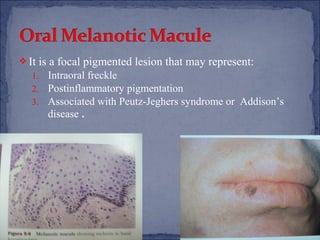
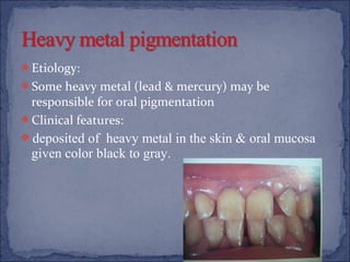
:Done by

Yousef alothman
Abdullah alsaad
Faheed al zoabi
Melanocytes have their embryologic orgin in the neural

crest.
It is migrate and reside among basal cells .
Melanocytes are found in oral mucosa but go to
unnoticed.
When focally or generally active in proliferation or
production, they produce physiologic pigmentation or can
go to malignant neoplasia.
Oral melanin range pigmentation from brown to black to
blue, depending on amount of melanin produced & depth
of pigment.
Physiologic
Smoking-associated melanosis
Oral melanotic macules
Café-au-lait macules
Pigmented neuroectodermal tumor of infancy
Nevomelanocytic
Melanoma
 Clinical

features:

It is symmetric, persistent & does not alter normal
architecture.
o Can be seen at any age.
o No gender predilection.
o Any location (most common in gingiva).
o Post-inflammatory pigmentation seen after mucosal
reaction to injury.
o
 Histopathology:

The lesion is due not to increased numbers of melanocyte
but to increased melanin production.
 Etiology & Pathogenesis:
o The lesion is related to a component

stimulate melanocytes
o Female sex hormone (as modifiers)

in tobacco smoke that

 Clinical features:
o Anterior labial gingiva (most commonly affected )
o Palate and buccal mucosa (associated with pipe smoking)

 Histopathology:
o increased melanin production Similar to that seen in

physiologic pigmentation.

 Treatment: stop of smoking (takes months to years)
 It

is a focal pigmented lesion that may represent:

1.
2.
3.

Intraoral freckle
Postinflammatory pigmentation
Associated with Peutz-Jeghers syndrome or Addison’s
disease .
 Clinical
o

o
o
o
o

features:

It is a benign neoplasm composed of primitive pigment
producing cells (neural crest origin)
Found in infant
Occcurs mainly in the maxilla
Nonulcerated & darkely pigmented mass
Radiographs shows an-ill defined lucency

 Histopathology:

It is composed of nests of tumor cells with small amount of
connective tissue.
 Treatment:

Surgical excision
 Nevus is general term that may refer to any congenetal

lesion of various cell types or tissue types.
 Nevomelanocytic Nevi are collection of nevus cells that are
round or polygonal and typically seen in a nested pattern. Can
be found in epithelium, connective tissue, or both.
 Clinical features:
o
o

They appear shortly after birth & throughout childhood.
Intraoral nevi are rare lesions (<0.5 cm-palate).
 Histopathology:

o When cells are located in epithelium-connective tissue

junction called Junctional nevus
o When cells are located in connective tissue called
Intramucosal nevus
o When cells are located in combination of zone called
Compound nevus
o When cells are spindle shaped and found deep in connective
tissue called Blue nevus
White are more affected than blacks
 Predisposing factors:
o Skin exposure
o Dark nature pigmentation
o Nevi

 Clinical features:
o No sex predilection
o Palate and gingiva are most commonly affected
o Irregular margin
 Treatment

o
o
o
o

& prognosis

Surgery
Chemotherapy
Immunotherapy is occasionally used
Prognosis depends on the depth of the lesion
 Amalgam tattoo
 Drug induced pigmentation
 Heavy metal pigmentation
 Etiology:
 It is an iatrogenic lesion that follows traumatic soft tissue

implantation of amalgam particles.
 Clinical features:
 Most common affected site are:
gingiva, buccal mucosa, palate & tongue
 Histopathology:
 Amalgam particles have an affinity for collagen fibers.
Tetracycline-associated pigmentation may be found in

prolonged treatment with high dose.
Other drugs that may produce pigmentation of oral
tissue include amino-quinolines, cyclophosphamide,
amiodarone & AZT.
Etiology:
Some heavy metal (lead & mercury) may be

responsible for oral pigmentation
Clinical features:
deposited of heavy metal in the skin & oral mucosa

given color black to gray.
‫كفو!‬
‫مياو00!‬

Pigmented lesion

  • 1.
    :Done by Yousef alothman Abdullahalsaad Faheed al zoabi
  • 3.
    Melanocytes have theirembryologic orgin in the neural crest. It is migrate and reside among basal cells . Melanocytes are found in oral mucosa but go to unnoticed. When focally or generally active in proliferation or production, they produce physiologic pigmentation or can go to malignant neoplasia. Oral melanin range pigmentation from brown to black to blue, depending on amount of melanin produced & depth of pigment.
  • 4.
    Physiologic Smoking-associated melanosis Oral melanoticmacules Café-au-lait macules Pigmented neuroectodermal tumor of infancy Nevomelanocytic Melanoma
  • 5.
     Clinical features: It issymmetric, persistent & does not alter normal architecture. o Can be seen at any age. o No gender predilection. o Any location (most common in gingiva). o Post-inflammatory pigmentation seen after mucosal reaction to injury. o
  • 6.
     Histopathology: The lesionis due not to increased numbers of melanocyte but to increased melanin production.
  • 7.
     Etiology &Pathogenesis: o The lesion is related to a component stimulate melanocytes o Female sex hormone (as modifiers) in tobacco smoke that  Clinical features: o Anterior labial gingiva (most commonly affected ) o Palate and buccal mucosa (associated with pipe smoking)  Histopathology: o increased melanin production Similar to that seen in physiologic pigmentation.  Treatment: stop of smoking (takes months to years)
  • 9.
     It is afocal pigmented lesion that may represent: 1. 2. 3. Intraoral freckle Postinflammatory pigmentation Associated with Peutz-Jeghers syndrome or Addison’s disease .
  • 10.
     Clinical o o o o o features: It isa benign neoplasm composed of primitive pigment producing cells (neural crest origin) Found in infant Occcurs mainly in the maxilla Nonulcerated & darkely pigmented mass Radiographs shows an-ill defined lucency  Histopathology: It is composed of nests of tumor cells with small amount of connective tissue.  Treatment: Surgical excision
  • 11.
     Nevus isgeneral term that may refer to any congenetal lesion of various cell types or tissue types.  Nevomelanocytic Nevi are collection of nevus cells that are round or polygonal and typically seen in a nested pattern. Can be found in epithelium, connective tissue, or both.  Clinical features: o o They appear shortly after birth & throughout childhood. Intraoral nevi are rare lesions (<0.5 cm-palate).
  • 12.
     Histopathology: o Whencells are located in epithelium-connective tissue junction called Junctional nevus o When cells are located in connective tissue called Intramucosal nevus o When cells are located in combination of zone called Compound nevus o When cells are spindle shaped and found deep in connective tissue called Blue nevus
  • 13.
    White are moreaffected than blacks  Predisposing factors: o Skin exposure o Dark nature pigmentation o Nevi  Clinical features: o No sex predilection o Palate and gingiva are most commonly affected o Irregular margin
  • 14.
     Treatment o o o o & prognosis Surgery Chemotherapy Immunotherapyis occasionally used Prognosis depends on the depth of the lesion
  • 15.
     Amalgam tattoo Drug induced pigmentation  Heavy metal pigmentation
  • 16.
     Etiology:  Itis an iatrogenic lesion that follows traumatic soft tissue implantation of amalgam particles.  Clinical features:  Most common affected site are: gingiva, buccal mucosa, palate & tongue  Histopathology:  Amalgam particles have an affinity for collagen fibers.
  • 17.
    Tetracycline-associated pigmentation maybe found in prolonged treatment with high dose. Other drugs that may produce pigmentation of oral tissue include amino-quinolines, cyclophosphamide, amiodarone & AZT.
  • 18.
    Etiology: Some heavy metal(lead & mercury) may be responsible for oral pigmentation Clinical features: deposited of heavy metal in the skin & oral mucosa given color black to gray.
  • 20.
  • 21.