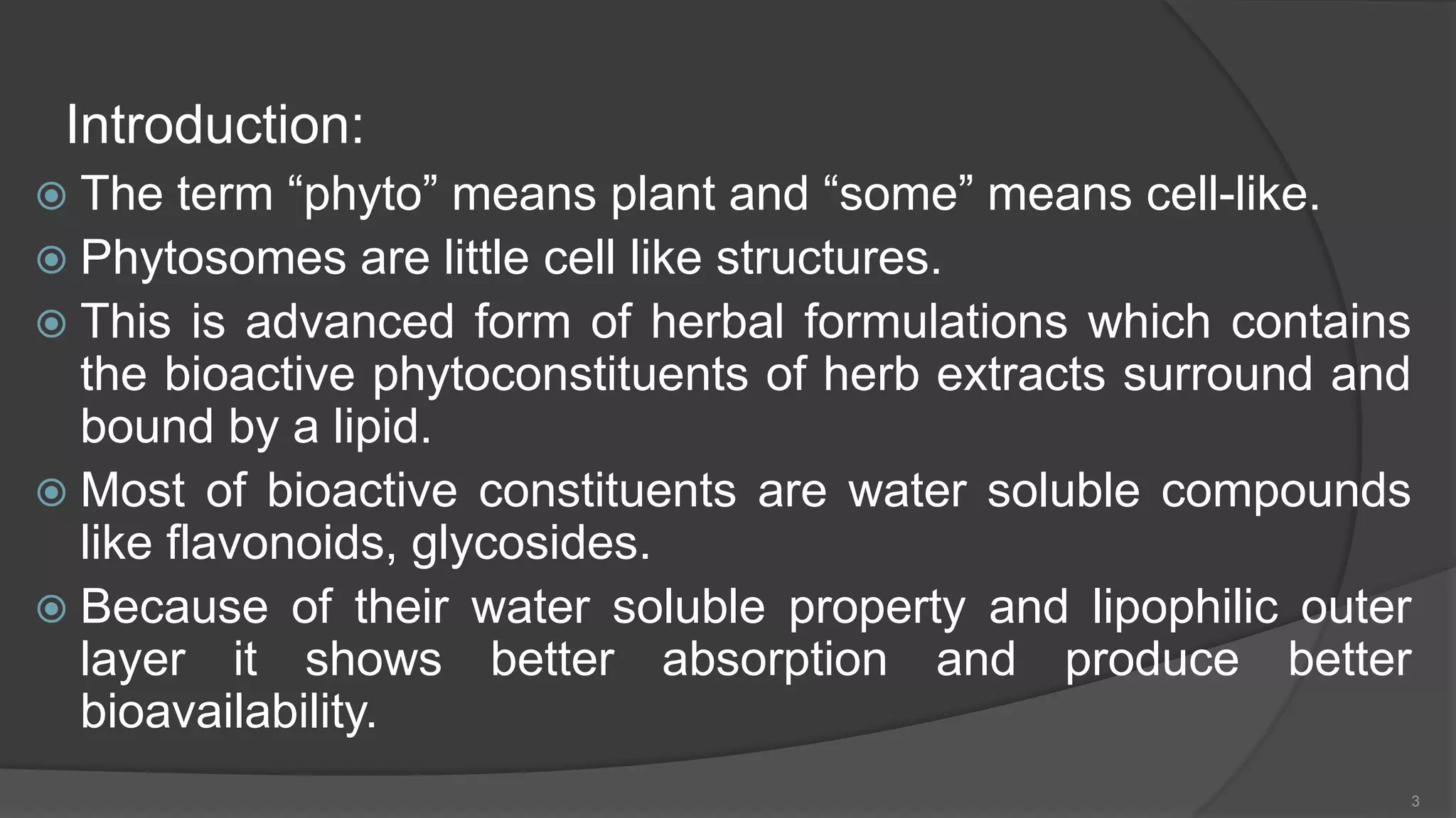
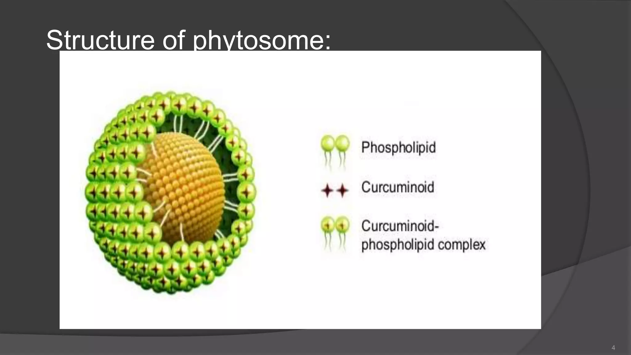
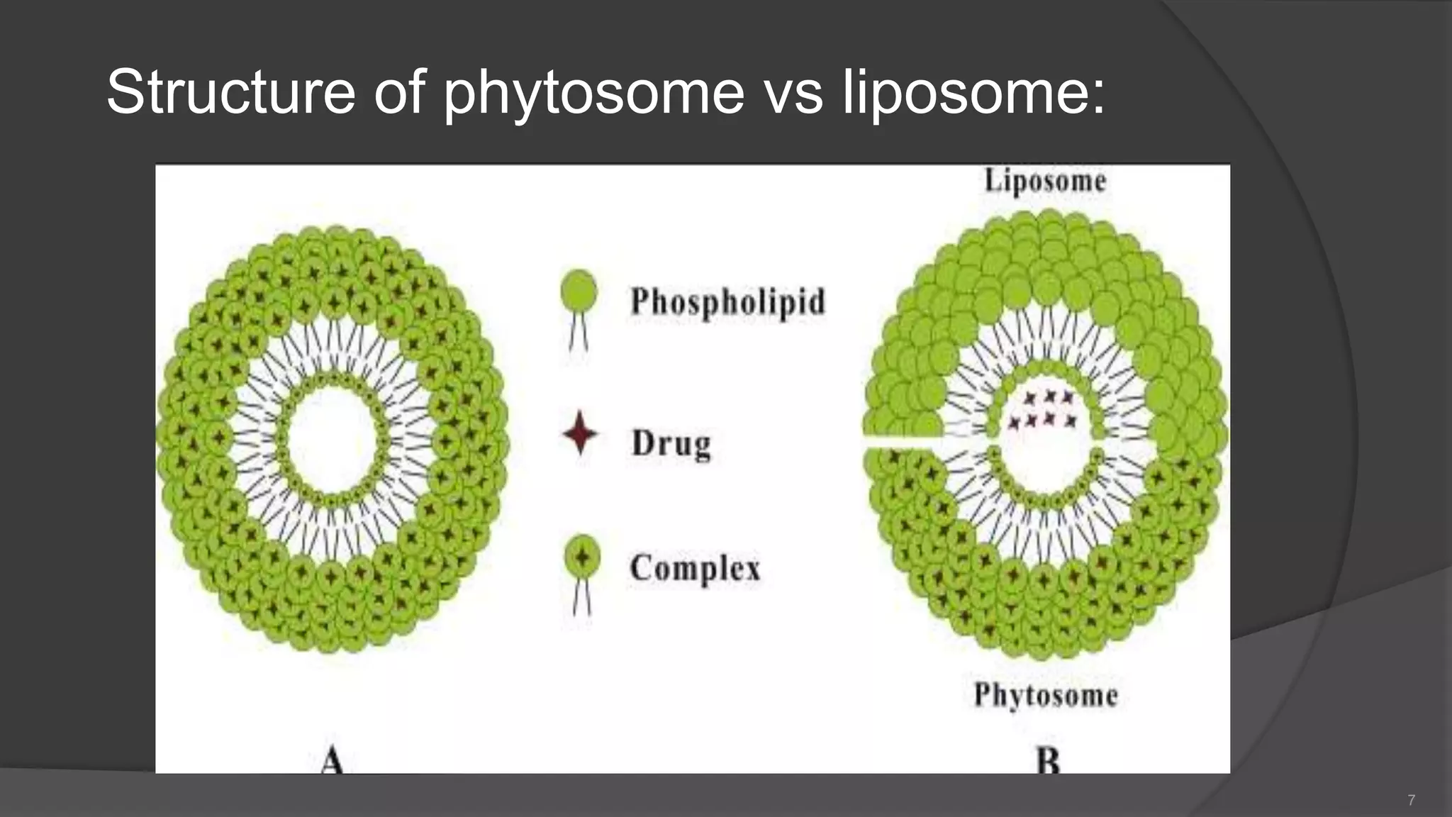
Biopharmaceutics: Mechanisms of Drug AbsorptionSURYAKANTVERMA2
Biopharmaceutics is defined as the study of factors influencing the rate and amount of drug that reaches the systemic circulation and the use of this information to optimise the therapeutic efficacy of the drug products.
It is defined as “the predictive mathematical model that describes the relationship between in vitro property (such as rate & extent of dissolution) of a dosage form and in vivo response (such as plasma drug concentration or amount of drug absorbed)”.
“It is define has an substance or Pharmaceutical material is encapsulated over the surface of solid, droplet of liquid and dispersion of medium is known has Microencapsulation”
1. Measurement of Bioavailability:
Direct and indirect methods may be used to assess drug bioavailability. The in-vivo bioavailability of a drug product is demonstrated by the rate and extent of drug absorption, as determined by comparison of measured parameters, e.g., concentration of the active drug ingredient in the blood, cumulative urinary excretion rates, or pharmacological effects.
For drug products that are not intended to be absorbed into the bloodstream, bioavailability may be assessed by measurements intended to reflect the rate and extent to which the active ingredient or active moiety becomes available at the site of action.
The design of the bioavailability study depends on the objectives of the study, the ability to analyze the drug (and metabolites) in biological fluids, the pharmacodynamics of the drug substance, the route of drug administration, and the nature of the drug product.
Pharmacokinetic and/or pharmacodynamic parameters as well as clinical observations and in-vitro studies may be used to determine drug bioavailability from a drug product.
1.1. Pharmacokinetic methods:
These are very widely used and based upon the assumption that the pharmacokinetic profile reflects the therapeutic effectiveness of a drug. Thus these are indirect methods. The two major pharmacokinetic methods are:
The major pharmacokinetic methods are:
Plasma / blood level time profile.
o Time for peak plasma (blood) concentration (t max)
o Peak plasma drug concentration (Cmax)
o Area under the plasma drug concentration–time curve (AUC)
Urinary excretion studies.
o Cumulative amount of drug excreted in the urine (Du)
o Rate of drug excretion in the urine (dDu/dt)
o Time for maximum urinary excretion (t)
C. Other biological fluids
1.2. Pharmacodynamic methods:
IT involves direct measurement of drug effect on a (patho) physiological process as a function of time. Disadvantages of it may be high variability, difficult to measure, limited choices, less reliable, more subjective, drug response influenced by several physiological & environmental factors.
They involve determination of bioavailability from:
Acute pharmacological response.
Therapeutic response.
1.3. In-vitro dissolution studies
Closed compartment apparatus
Open compartment apparatus
Dialysis systems.
1.4. Clinical observations
Well-controlled clinical trials
Mucoadhesive drug delivery system interact with the mucus layer covering the mucosal epithelial surface, & mucin molecules & increase the residence time of the dosage form at the site of the absorption.
Mucoadhesive drug delivery system is a part of controlled delivery system.
Since the early 1980,the concept of Mucoadhesion has gained considerable interest in pharmaceutical technology.
combine mucoadhesive with enzyme inhibitory & penetration enhancer properties & improve the patient complaince.
MDDS have been devloped for buccal ,nasal,rectal &vaginal routes for both systemic & local effects.
Hydrophilic high mol. wt. such as peptides that cannot be administered & poor absorption ,then MDDS is best choice.
Mucoadhesiveinner layers called mucosa inner epithelial cell lining is covered with viscoelasticfluid
Composed of water and mucin.
Thickness varies from 40 μm to 300 μm
General composition of mucus
Water…………………………………..95%
Glycoproteinsand lipids……………..0.5-5%
Mineral salts……………………………1%
Free proteins…………………………..0.5-1%
The mechanism responsible in the formation of mucoadhesive bond
Step 1 : Wetting and swelling of the polymer(contact stage)
Step 2 : Interpenetration between the polymer chains and the mucosal membrane
Step 3 : Formation of bonds between the entangled chains (both known as consolidation stage)
Electronic theory
Wetting theory
Adsorption theory
Diffusion theory
Fracture theory
Advantages over other controlled oral controlled release systems by virtue of prolongation of residence of drug in GIT.
Targeting & localization of the dosage form at a specific site
-Painless administration.
-Low enzymatic activity & avoid of first pass metabolism
If MDDS are adhere too tightlgy because it is undesirable to exert too much force to remove the formulation after use,otherwise the mucosa could be injured.
-Some patient suffers unpleasent feeling.
-Unfortunately ,the lack of standardized techniques often leads to unclear results.
-costly drug delivery system
DISSOLUTION
Dissolution is defined as a process in which a solid substance solubilises in a given solvent.
(i.e. mass transfer from the solid surface to the liquid phase.)
Three Theories:
Diffusion layer model / Film theory
Danckwert’s model / Penetration or Surface renewal theory
Interfacial barrier model / Double barrier or Limited solvation theory
It is a novel drug dosage form in herbal drugs.I have made it, because it is a part of my project work. I hope you will find helpful and knowledgeable. Thank you.
Phytosomes are one of the novel drug delivery system containing hydrophilic bioactive phyto-constituents of herbs surrounded and bounded by phospolipids.
A phytosome is a complex of a natural active ingredient and a phospholipid - mostly lecithin. It is claimed that phytosome increases absorption of "conventional herbal extracts" or isolated active principles both topically as well as orally.
The term "phyto" means plant and "some" means cell-like.Phytosomes are little cell like structures.
Structure of phytosome
Properties of phytosome - chemical and biological
Methods of preparation
PHYTOSOMES AND ELECTROSOMES of novel drug delivery system .pptxnthanuja0331
Introduction and methods of preparation, evaluation parameters of phytosomes, advantages , disadvantages and applications of phytosome and electrosomes. some of marketed products of phytosomes.
Introduction of Veterinary pharmacologyQaline Giigii
this course of Introduction of veterinary pharacology was presented by Dr. Osman Abdulahi Farah
Osman Shiine
at Gollis University faculty of Veterinary Medicine
2014
TEST BANK for Operations Management, 14th Edition by William J. Stevenson, Ve...kevinkariuki227
TEST BANK for Operations Management, 14th Edition by William J. Stevenson, Verified Chapters 1 - 19, Complete Newest Version.pdf
- Video recording of this lecture in English language: https://youtu.be/lK81BzxMqdo
- Video recording of this lecture in Arabic language: https://youtu.be/Ve4P0COk9OI
- Link to download the book free: https://nephrotube.blogspot.com/p/nephrotube-nephrology-books.html
- Link to NephroTube website: www.NephroTube.com
- Link to NephroTube social media accounts: https://nephrotube.blogspot.com/p/join-nephrotube-on-social-media.html
MANAGEMENT OF ATRIOVENTRICULAR CONDUCTION BLOCK.pdfJim Jacob Roy
Cardiac conduction defects can occur due to various causes.
Atrioventricular conduction blocks ( AV blocks ) are classified into 3 types.
This document describes the acute management of AV block.
Couples presenting to the infertility clinic- Do they really have infertility...Sujoy Dasgupta
Dr Sujoy Dasgupta presented the study on "Couples presenting to the infertility clinic- Do they really have infertility? – The unexplored stories of non-consummation" in the 13th Congress of the Asia Pacific Initiative on Reproduction (ASPIRE 2024) at Manila on 24 May, 2024.
These lecture slides, by Dr Sidra Arshad, offer a quick overview of physiological basis of a normal electrocardiogram.
Learning objectives:
1. Define an electrocardiogram (ECG) and electrocardiography
2. Describe how dipoles generated by the heart produce the waveforms of the ECG
3. Describe the components of a normal electrocardiogram of a typical bipolar leads (limb II)
4. Differentiate between intervals and segments
5. Enlist some common indications for obtaining an ECG
Study Resources:
1. Chapter 11, Guyton and Hall Textbook of Medical Physiology, 14th edition
2. Chapter 9, Human Physiology - From Cells to Systems, Lauralee Sherwood, 9th edition
3. Chapter 29, Ganong’s Review of Medical Physiology, 26th edition
4. Electrocardiogram, StatPearls - https://www.ncbi.nlm.nih.gov/books/NBK549803/
5. ECG in Medical Practice by ABM Abdullah, 4th edition
6. ECG Basics, http://www.nataliescasebook.com/tag/e-c-g-basics
New Directions in Targeted Therapeutic Approaches for Older Adults With Mantl...i3 Health
i3 Health is pleased to make the speaker slides from this activity available for use as a non-accredited self-study or teaching resource.
This slide deck presented by Dr. Kami Maddocks, Professor-Clinical in the Division of Hematology and
Associate Division Director for Ambulatory Operations
The Ohio State University Comprehensive Cancer Center, will provide insight into new directions in targeted therapeutic approaches for older adults with mantle cell lymphoma.
STATEMENT OF NEED
Mantle cell lymphoma (MCL) is a rare, aggressive B-cell non-Hodgkin lymphoma (NHL) accounting for 5% to 7% of all lymphomas. Its prognosis ranges from indolent disease that does not require treatment for years to very aggressive disease, which is associated with poor survival (Silkenstedt et al, 2021). Typically, MCL is diagnosed at advanced stage and in older patients who cannot tolerate intensive therapy (NCCN, 2022). Although recent advances have slightly increased remission rates, recurrence and relapse remain very common, leading to a median overall survival between 3 and 6 years (LLS, 2021). Though there are several effective options, progress is still needed towards establishing an accepted frontline approach for MCL (Castellino et al, 2022). Treatment selection and management of MCL are complicated by the heterogeneity of prognosis, advanced age and comorbidities of patients, and lack of an established standard approach for treatment, making it vital that clinicians be familiar with the latest research and advances in this area. In this activity chaired by Michael Wang, MD, Professor in the Department of Lymphoma & Myeloma at MD Anderson Cancer Center, expert faculty will discuss prognostic factors informing treatment, the promising results of recent trials in new therapeutic approaches, and the implications of treatment resistance in therapeutic selection for MCL.
Target Audience
Hematology/oncology fellows, attending faculty, and other health care professionals involved in the treatment of patients with mantle cell lymphoma (MCL).
Learning Objectives
1.) Identify clinical and biological prognostic factors that can guide treatment decision making for older adults with MCL
2.) Evaluate emerging data on targeted therapeutic approaches for treatment-naive and relapsed/refractory MCL and their applicability to older adults
3.) Assess mechanisms of resistance to targeted therapies for MCL and their implications for treatment selection
Title: Sense of Taste
Presenter: Dr. Faiza, Assistant Professor of Physiology
Qualifications:
MBBS (Best Graduate, AIMC Lahore)
FCPS Physiology
ICMT, CHPE, DHPE (STMU)
MPH (GC University, Faisalabad)
MBA (Virtual University of Pakistan)
Learning Objectives:
Describe the structure and function of taste buds.
Describe the relationship between the taste threshold and taste index of common substances.
Explain the chemical basis and signal transduction of taste perception for each type of primary taste sensation.
Recognize different abnormalities of taste perception and their causes.
Key Topics:
Significance of Taste Sensation:
Differentiation between pleasant and harmful food
Influence on behavior
Selection of food based on metabolic needs
Receptors of Taste:
Taste buds on the tongue
Influence of sense of smell, texture of food, and pain stimulation (e.g., by pepper)
Primary and Secondary Taste Sensations:
Primary taste sensations: Sweet, Sour, Salty, Bitter, Umami
Chemical basis and signal transduction mechanisms for each taste
Taste Threshold and Index:
Taste threshold values for Sweet (sucrose), Salty (NaCl), Sour (HCl), and Bitter (Quinine)
Taste index relationship: Inversely proportional to taste threshold
Taste Blindness:
Inability to taste certain substances, particularly thiourea compounds
Example: Phenylthiocarbamide
Structure and Function of Taste Buds:
Composition: Epithelial cells, Sustentacular/Supporting cells, Taste cells, Basal cells
Features: Taste pores, Taste hairs/microvilli, and Taste nerve fibers
Location of Taste Buds:
Found in papillae of the tongue (Fungiform, Circumvallate, Foliate)
Also present on the palate, tonsillar pillars, epiglottis, and proximal esophagus
Mechanism of Taste Stimulation:
Interaction of taste substances with receptors on microvilli
Signal transduction pathways for Umami, Sweet, Bitter, Sour, and Salty tastes
Taste Sensitivity and Adaptation:
Decrease in sensitivity with age
Rapid adaptation of taste sensation
Role of Saliva in Taste:
Dissolution of tastants to reach receptors
Washing away the stimulus
Taste Preferences and Aversions:
Mechanisms behind taste preference and aversion
Influence of receptors and neural pathways
Impact of Sensory Nerve Damage:
Degeneration of taste buds if the sensory nerve fiber is cut
Abnormalities of Taste Detection:
Conditions: Ageusia, Hypogeusia, Dysgeusia (parageusia)
Causes: Nerve damage, neurological disorders, infections, poor oral hygiene, adverse drug effects, deficiencies, aging, tobacco use, altered neurotransmitter levels
Neurotransmitters and Taste Threshold:
Effects of serotonin (5-HT) and norepinephrine (NE) on taste sensitivity
Supertasters:
25% of the population with heightened sensitivity to taste, especially bitterness
Increased number of fungiform papillae
Flu Vaccine Alert in Bangalore Karnatakaaddon Scans
As flu season approaches, health officials in Bangalore, Karnataka, are urging residents to get their flu vaccinations. The seasonal flu, while common, can lead to severe health complications, particularly for vulnerable populations such as young children, the elderly, and those with underlying health conditions.
Dr. Vidisha Kumari, a leading epidemiologist in Bangalore, emphasizes the importance of getting vaccinated. "The flu vaccine is our best defense against the influenza virus. It not only protects individuals but also helps prevent the spread of the virus in our communities," he says.
This year, the flu season is expected to coincide with a potential increase in other respiratory illnesses. The Karnataka Health Department has launched an awareness campaign highlighting the significance of flu vaccinations. They have set up multiple vaccination centers across Bangalore, making it convenient for residents to receive their shots.
To encourage widespread vaccination, the government is also collaborating with local schools, workplaces, and community centers to facilitate vaccination drives. Special attention is being given to ensuring that the vaccine is accessible to all, including marginalized communities who may have limited access to healthcare.
Residents are reminded that the flu vaccine is safe and effective. Common side effects are mild and may include soreness at the injection site, mild fever, or muscle aches. These side effects are generally short-lived and far less severe than the flu itself.
Healthcare providers are also stressing the importance of continuing COVID-19 precautions. Wearing masks, practicing good hand hygiene, and maintaining social distancing are still crucial, especially in crowded places.
Protect yourself and your loved ones by getting vaccinated. Together, we can help keep Bangalore healthy and safe this flu season. For more information on vaccination centers and schedules, residents can visit the Karnataka Health Department’s official website or follow their social media pages.
Stay informed, stay safe, and get your flu shot today!
Ozempic: Preoperative Management of Patients on GLP-1 Receptor Agonists Saeid Safari
Preoperative Management of Patients on GLP-1 Receptor Agonists like Ozempic and Semiglutide
ASA GUIDELINE
NYSORA Guideline
2 Case Reports of Gastric Ultrasound
Title: Sense of Smell
Presenter: Dr. Faiza, Assistant Professor of Physiology
Qualifications:
MBBS (Best Graduate, AIMC Lahore)
FCPS Physiology
ICMT, CHPE, DHPE (STMU)
MPH (GC University, Faisalabad)
MBA (Virtual University of Pakistan)
Learning Objectives:
Describe the primary categories of smells and the concept of odor blindness.
Explain the structure and location of the olfactory membrane and mucosa, including the types and roles of cells involved in olfaction.
Describe the pathway and mechanisms of olfactory signal transmission from the olfactory receptors to the brain.
Illustrate the biochemical cascade triggered by odorant binding to olfactory receptors, including the role of G-proteins and second messengers in generating an action potential.
Identify different types of olfactory disorders such as anosmia, hyposmia, hyperosmia, and dysosmia, including their potential causes.
Key Topics:
Olfactory Genes:
3% of the human genome accounts for olfactory genes.
400 genes for odorant receptors.
Olfactory Membrane:
Located in the superior part of the nasal cavity.
Medially: Folds downward along the superior septum.
Laterally: Folds over the superior turbinate and upper surface of the middle turbinate.
Total surface area: 5-10 square centimeters.
Olfactory Mucosa:
Olfactory Cells: Bipolar nerve cells derived from the CNS (100 million), with 4-25 olfactory cilia per cell.
Sustentacular Cells: Produce mucus and maintain ionic and molecular environment.
Basal Cells: Replace worn-out olfactory cells with an average lifespan of 1-2 months.
Bowman’s Gland: Secretes mucus.
Stimulation of Olfactory Cells:
Odorant dissolves in mucus and attaches to receptors on olfactory cilia.
Involves a cascade effect through G-proteins and second messengers, leading to depolarization and action potential generation in the olfactory nerve.
Quality of a Good Odorant:
Small (3-20 Carbon atoms), volatile, water-soluble, and lipid-soluble.
Facilitated by odorant-binding proteins in mucus.
Membrane Potential and Action Potential:
Resting membrane potential: -55mV.
Action potential frequency in the olfactory nerve increases with odorant strength.
Adaptation Towards the Sense of Smell:
Rapid adaptation within the first second, with further slow adaptation.
Psychological adaptation greater than receptor adaptation, involving feedback inhibition from the central nervous system.
Primary Sensations of Smell:
Camphoraceous, Musky, Floral, Pepperminty, Ethereal, Pungent, Putrid.
Odor Detection Threshold:
Examples: Hydrogen sulfide (0.0005 ppm), Methyl-mercaptan (0.002 ppm).
Some toxic substances are odorless at lethal concentrations.
Characteristics of Smell:
Odor blindness for single substances due to lack of appropriate receptor protein.
Behavioral and emotional influences of smell.
Transmission of Olfactory Signals:
From olfactory cells to glomeruli in the olfactory bulb, involving lateral inhibition.
Primitive, less old, and new olfactory systems with different path
2. Contents:
Introduction
Structure of phytosome
Physical properties
Structure of phytosome vs liposome
Advantages
Disadvantages
Preparation of phytosome
Evaluation
Application
Marketed products
2
3. Introduction:
The term “phyto” means plant and “some” means cell-like.
Phytosomes are little cell like structures.
This is advanced form of herbal formulations which contains
the bioactive phytoconstituents of herb extracts surround and
bound by a lipid.
Most of bioactive constituents are water soluble compounds
like flavonoids, glycosides.
Because of their water soluble property and lipophilic outer
layer it shows better absorption and produce better
bioavailability.
3
5. Chemical properties:
Phyosome is a complex between natural product and natural
phospholipid.
The phytosome complex is obtained by reaction of suitable
amount of phospholipid and the substrate in appropriate
solvent such as glycerol.
The main phospholipid-substrate interaction is due to
formation of hydrogen bonds between polar head of
phospholipid and polar functionalities of substrate.
When treated with water, they assume a micelle shape,
forming structures which resembles liposomes.
5
6. Biological properties:
Phytosomes are advanced forms of herbal products
that are better absorbed, utilized and as a result
produce better results than conventional herbal
extracts.
Freely soluble in non-polar and aprotic solvent ;
Solvents in which the hydrophilic moiety is not present.
Moderately soluble in fats.
Insoluble in water.
6
8. Phytosome vs Liposome
PHYTOSOME LIPOSOME
In this the active chemical constituent molecules
are anchored through chemical bonds to the polar
head of phospholipids.
In this active principles is dissolved in the medium
of cavity or in the layer of membrane.
Chemical bonds are formed. No chemical bonds are formed.
In phytosome, phosphatidylcholine and plant
compound form 1:1 or 2:1 complex depending on
substance.
In this hundred or thousands of
phosphatidylcholine molecules surround the
water soluble molecule.
Phytosomes are much better absorbed than
liposomes showing better bioavailability.
Bioavailability of liposomes is less than
phytosomes.
Contents of phospholipids is less higher. Contents of phospholipid is much higher.
8
9. Advantages:
Enhanced absorption of herbal constituent.
As the absorption of active constituents is improved, its dose
requirement is also reduced.
Phosphotidylcholine acts as hepatoprotective, giving synergistic effect.
It shows better stability profile.
It assures proper delivery of drugs to the respective tissues.
Entrapment efficiency is high.
Phyotosomes are also superior to liposomes in skin care products.
Better bioavailability.
Nutritional benefit.
Enhanced permeation of drug through skin.
9
10. Disadvantages:
When administered orally or topically they limit their
bioavailability.
Phytoconstituents is quickly eliminated from phytosome.
Stability problem.
10
11. Different additives used in the formulation of
phytosomes:
1) phospholipids: soya phosphatidyl choline, egg phosphatidyl
choline, dipalmityl phosphatidyl choline, phosphatidylcholine,
phosphatidylethanolamine.
2) Aprotic solvent: Dioxane, acetone, methylene chloride.
3) Non-solvent: n-hexane.
4) Alcohol: ethanol, methanol.
11
12. Preparation of Phytosome:
Phospholipid
↓
Dissolved in organic solvent containing drug extract
↓
Solution of phospholipid in organic solvent with drug extract
↓
Drying/ solvent evaporation
↓
Formation of thin film
↓
Hydration
↓
Formation of phytosomal suspension
12
13. 1) Antisolvent precipitation technique:-
Drug + soya lecithin
↓
Refluxed with20ml dichloromethane at 60 degree for 2hrs.
↓
Concentrate mixture to 5-10ml
↓
Add hexane 20ml
↓
Filter the ppt formed. Dry, crush and pass through #100
13
14. 2) Rotary evaporation technique:
Drug and soya lecithin
↓
Dissolved in 30ml of tetrahydrofuran
↓
stirring for 3hours at a temperature not exceeding 40degree
↓
Thin film is formed
↓
Add n-hexane with stirring
↓
Precipitate obtained
↓
Dry and pass through mesh
↓
Phytosomes formed
14
15. 3) Solvent evaporation method:
Drug and soya lecithin
↓
Refluxed with 20ml of acetone at a temperature 50-60 degree for
2hours
↓
Concentrate mixture to 5-10ml
↓
Obtain the precipitate
↓
Filter and collect
↓
phytosomes obtained
15
16. Evaluation:
1) Determination of entrapment efficiency:- the entrapment
efficiency of a phytosomal formulation can be determined by
subjecting the formulation to ultracentrifugation technique.
2) Determination of vesicle size and zeta potential:- It can be
determined by dynamic light scattering which uses a
computerized inspection system and photon correlation
spectroscopy.
3) Surface tension activity measurement:- It can be measured by
ring method using Du Nouy ring tensiometer. The surface
tension of drug is measured.
4) Spectroscopic evaluation:- it is employed in order to confirm the
formation of complex as well as to study interactions between 16
17. 5) Determination of drug content:- drug content of phytosome
complex was determined by dissolving 100mg of complex in
10ml of methanol. After dilution, absorbance was determined by
UV spectrophotometer at 269nm.
6) In-vitro and In-vivo evaluation:- this can be done according to
therapeutic activity measurement parameters of biologically
active phytoconstituents present in phytosomes.
17
18. Applications:
1) Silymarin phytosome:
Most of the phytosomes are focused to silybum marianum which
contains liver protective flavonoids.
The fruit of the milk thistle plant ( S. marianum family-
steraceae) contains flavonoids for hepato protective effect.
Silymarin has been shown to have positive effects in treating
liver diseases of various kinds including hepatitis, cirrhosis, fatty
infiltration of the liver and inflammation of the bile duct.
18
19. 2) Phytosomes of grape seed:
Grape seed phytosome is composed of oligomeric polyphenols of
varying molecular size complexed with phospholipids.
The main properties of procyanidin flavonoids of grape seed are
an increase in total antioxidant capacity and stimulation of
physiological defense of plasma.
3) Phytosome of green tea:
Green tea leaves is characterized by presence of polyphenolic
compound epigallocatechin-3-0-gallate as the key component.
These are potent modulator of several biochemical process
linked to the breakdown of homeostasis in major chronic
degenerative diseases like cancer and atherosclerosis.
It has also beneficial activities like antioxidant, anticarcinogenic,19
20. 4) Phytosomes of curcumin:
Maiti et al developed the phytosomes of curcumin ( flavonoid
from turmeric, curcuma longa linn) and naringenin ( flavonoid
from grape, vitis vinifera).
Phytosome of naringenin produced better antioxidant activity
than the free compound with a prolonged deviation of action.
20
23. Reference:
Semalty A, Semalty M, Singh R, Rawat MSM. Phytosomes in herbal drug
delivery. Indian drugs 2006; page no: 937-946.
Zhang J, Tang Q, Xu X, Li N. Development and evaluation of a novel
phytosome -loaded chitosan microsphere system for curcumin delivery.
International Journal of Pharmaceutics 2013; 448:168– 174. 14.
Habbu P, Madagundi S, Kulkarni R, Jadav S, Vanakudri R, Kulkarni V, et al.
Preparation and evaluation of Bacopa phospholipids complex for antiamnesic
activity in rodents. Drug invention today 2013; 5(1): 13 - 21.
Parris K, Kathleen H, A review of the bioavailability and clinical efficacy of milk
thistle phytosome: a silybinphosphatidylcholine complex, Altern Med Rev,
10(3), 2005, 193-203.
Gupta A, Ashawat MS, Saraf S, Phytosome: A novel approach towards
functional cosmetics, J Plant sci, 2(6), 2007, 644-649.
23