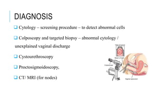
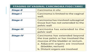
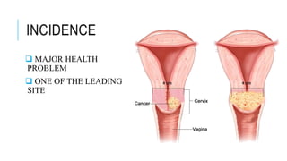
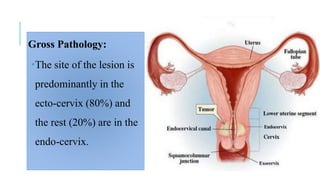
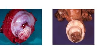
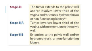
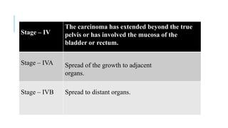
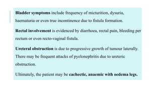
VAGINAL CARINOMA
INCIDENCE
1 percent of genital malignancies.
ETIOLOGY
 HPV
 Progression from vaginal intraepithelial neoplasia
 Women with history of cervical cancer
 Diethyl stilboesterol (DES)
PATHOLOGY
Site:
The commonest site is in the upper-third of the posterior wall.
Naked eye:
The growth may be ulcerative or fungative.
TYPE
 Squamous cell
 Adeno carcinoma
 Fibrosarcoma
 Melanoma
SPREAD
 Direct spread- ascending / descending
 Lymphatics - inguinofemoral lymph nodes and pelvic
lymph nodes
Blood borne (rare) – lungs, liver and bones
CLINICAL FEATURES
 Age: 55 years
 SYMPTOMS
 Asymptomatic
 Abnormal vaginal bleeding including post coital bleeding
 Foul smelling discharge per vaginum.
SIGNS
Speculum examination reveals an ulcerative, nodular or
exophytic growth.
The cervix looks apparently normal.
DIAGNOSIS
 Cytology – screening procedure – to detect abnormal cells
 Colposcopy and targeted biopsy – abnormal cytology /
unexplained vaginal discharge
 Cystourethroscopy
 Proctosigmoidoscopy,
 CT/ MRI (for nodes)
CARCINOMA
CERVIX
INCIDENCE
 MAJOR HEALTH
PROBLEM
 ONE OF THE LEADING
SITE
Gross Pathology:
The site of the lesion is
predominantly in the
ecto-cervix (80%) and
the rest (20%) are in the
endo-cervix.
Exophytic: These arise from
the ecto-cervix and form friable
masses almost filling up the
upper vagina in late cases.
Ulcerative: The lesion excavates the
cervix and often involves the vaginal
fornices
Infiltrative: These are found in
endo-cervical growth. They cause
expansion of the cervix, so that it
becomes barrel-shaped.
Squamous cell carcinoma most commonest – (80%) - ectocervix
Adenocarcinoma (10–15%) develops from the endo-cervical canal,
either from the lining epithelium or from the glands.
MODE OF SPREAD
Direct extension: to vagina, and to the body of the uterus
Lymphatic: parametrial nodes, internal iliac nodes,
obturator, external iliac nodes and sacral nodes
Hematogenous: Blood borne metastasis is late. Lungs, liver or
bone are usually involved.
STAGE INVASIVE CARCINOMA
Stage – I The carcinoma is strictly confined to the cervix.
Stage – IA CA which can be only diagnosed by microscopy with
deepest invasion < 5mm and largest extension < 7mm
Stage – IB Clinically visible lesions limited to the cervix uteri or
preclinical cancers greater than IA
Stage – IV
The carcinoma has extended beyond the true
pelvis or has involved the mucosa of the
bladder or rectum.
Stage – IVA Spread of the growth to adjacent
organs.
Stage – IVB Spread to distant organs.
SIGNS
 A red granular area extending from the external os
 A nodular growth or an ulcer.
The lesion bleeds on friction
 Friable
Bladder symptoms include frequency of micturition, dysuria,
haematuria or even true incontinence due to fistula formation.
Rectal involvement is evidenced by diarrhoea, rectal pain, bleeding per
rectum or even recto-vaginal fistula.
Ureteral obstruction is due to progressive growth of tumour laterally.
There may be frequent attacks of pyelonephritis due to ureteric
obstruction.
Ultimately, the patient may be cachectic, anaemic with oedema legs.

Vaginal & cervical carcinoma

  • 1.
  • 2.
    INCIDENCE 1 percent ofgenital malignancies.
  • 4.
    ETIOLOGY  HPV  Progressionfrom vaginal intraepithelial neoplasia  Women with history of cervical cancer  Diethyl stilboesterol (DES)
  • 5.
    PATHOLOGY Site: The commonest siteis in the upper-third of the posterior wall. Naked eye: The growth may be ulcerative or fungative.
  • 6.
    TYPE  Squamous cell Adeno carcinoma  Fibrosarcoma  Melanoma
  • 7.
    SPREAD  Direct spread-ascending / descending  Lymphatics - inguinofemoral lymph nodes and pelvic lymph nodes Blood borne (rare) – lungs, liver and bones
  • 8.
    CLINICAL FEATURES  Age:55 years  SYMPTOMS  Asymptomatic  Abnormal vaginal bleeding including post coital bleeding  Foul smelling discharge per vaginum.
  • 9.
    SIGNS Speculum examination revealsan ulcerative, nodular or exophytic growth. The cervix looks apparently normal.
  • 10.
    DIAGNOSIS  Cytology –screening procedure – to detect abnormal cells  Colposcopy and targeted biopsy – abnormal cytology / unexplained vaginal discharge  Cystourethroscopy  Proctosigmoidoscopy,  CT/ MRI (for nodes)
  • 12.
  • 13.
  • 14.
    Gross Pathology: The siteof the lesion is predominantly in the ecto-cervix (80%) and the rest (20%) are in the endo-cervix.
  • 15.
    Exophytic: These arisefrom the ecto-cervix and form friable masses almost filling up the upper vagina in late cases.
  • 16.
    Ulcerative: The lesionexcavates the cervix and often involves the vaginal fornices Infiltrative: These are found in endo-cervical growth. They cause expansion of the cervix, so that it becomes barrel-shaped.
  • 18.
    Squamous cell carcinomamost commonest – (80%) - ectocervix Adenocarcinoma (10–15%) develops from the endo-cervical canal, either from the lining epithelium or from the glands.
  • 19.
    MODE OF SPREAD Directextension: to vagina, and to the body of the uterus Lymphatic: parametrial nodes, internal iliac nodes, obturator, external iliac nodes and sacral nodes
  • 20.
    Hematogenous: Blood bornemetastasis is late. Lungs, liver or bone are usually involved.
  • 21.
    STAGE INVASIVE CARCINOMA Stage– I The carcinoma is strictly confined to the cervix. Stage – IA CA which can be only diagnosed by microscopy with deepest invasion < 5mm and largest extension < 7mm Stage – IB Clinically visible lesions limited to the cervix uteri or preclinical cancers greater than IA
  • 24.
    Stage – IV Thecarcinoma has extended beyond the true pelvis or has involved the mucosa of the bladder or rectum. Stage – IVA Spread of the growth to adjacent organs. Stage – IVB Spread to distant organs.
  • 25.
    SIGNS  A redgranular area extending from the external os  A nodular growth or an ulcer. The lesion bleeds on friction  Friable
  • 27.
    Bladder symptoms includefrequency of micturition, dysuria, haematuria or even true incontinence due to fistula formation. Rectal involvement is evidenced by diarrhoea, rectal pain, bleeding per rectum or even recto-vaginal fistula. Ureteral obstruction is due to progressive growth of tumour laterally. There may be frequent attacks of pyelonephritis due to ureteric obstruction. Ultimately, the patient may be cachectic, anaemic with oedema legs.