A Text Book of
P
PH
HA
AR
RM
MA
AC
CO
OL
LO
OG
GY
Y -
- I
II
I
As Per PCI Regulations
Third Year B. Pharm.
Semester - V
Dr. Manjunatha P. Mudagal
Professor & H.O.D.
Department of Pharmacology,
Acharya and B.M. Reddy College of Pharmacy
Bengaluru
Dr. Uday Raj Sharma
Associate Professor
Department of Pharmacology,
Acharya Dr. Sarvepalli Radhakrishna Road
Acharya PO, Soladevanahalli,
Bengaluru-560 107, Karnataka, India
N3998
Pharmacology - II ISBN 978-93-89533-11-8
First Edition : September 2019
© : Authors
The text of this publication, or any part thereof, should not be reproduced or transmitted in any form or stored in any computer
storage system or device for distribution including photocopy, recording, taping or information retrieval system or reproduced on any disc,
tape, perforated media or other information storage device etc., without the written permission of Author with whom the rights are
reserved. Breach of this condition is liable for legal action.
Every effort has been made to avoid errors or omissions in this publication. In spite of this, errors may have crept in. Any mistake, error
or discrepancy so noted and shall be brought to our notice shall be taken care of in the next edition. It is notified that neither the publisher
nor the authors or seller shall be responsible for any damage or loss of action to any one, of any kind, in any manner, therefrom.
Published By : Polyplate
NIRALI PRAKASHAN
Abhyudaya Pragati, 1312, Shivaji Nagar,
Off J.M. Road, PUNE – 411005
Tel - (020) 25512336/37/39, Fax - (020) 25511379
Email : niralipune@pragationline.com
DISTRIBUTION CENTRES
PUNE
Nirali Prakashan : 119, Budhwar Peth, Jogeshwari Mandir Lane, Pune 411002, Maharashtra
(For orders within Pune) Tel : (020) 2445 2044; Mobile : 9657703145
Email : niralilocal@pragationline.com
Nirali Prakashan : S. No. 28/27, Dhayari, Near Asian College Pune 411041
(For orders outside Pune) Tel : (020) 24690204; Mobile : 9657703143
Email : bookorder@pragationline.com
MUMBAI
Nirali Prakashan : 385, S.V.P. Road, Rasdhara Co-op. Hsg. Society Ltd.,
Girgaum, Mumbai 400004, Maharashtra; Mobile : 9320129587
Tel : (022) 2385 6339 / 2386 9976, Fax : (022) 2386 9976
Email : niralimumbai@pragationline.com
DISTRIBUTION BRANCHES
JALGAON
Nirali Prakashan : 34, V. V. Golani Market, Navi Peth, Jalgaon 425001, Maharashtra,
Tel : (0257) 222 0395, Mob : 94234 91860; Email : niralijalgaon@pragationline.com
KOLHAPUR
Nirali Prakashan : New Mahadvar Road, Kedar Plaza, 1st
Floor Opp. IDBI Bank, Kolhapur 416 012
Maharashtra. Mob : 9850046155; Email : niralikolhapur@pragationline.com
NAGPUR
Nirali Prakashan : Above Maratha Mandir, Shop No. 3, First Floor,
Rani Jhanshi Square, Sitabuldi, Nagpur 440012, Maharashtra
Tel : (0712) 254 7129; Email : niralinagpur@pragationline.com
DELHI
Nirali Prakashan : 4593/15, Basement, Agarwal Lane, Ansari Road, Daryaganj
Near Times of India Building, New Delhi 110002 Mob : 08505972553
Email : niralidelhi@pragationline.com
BENGALURU
Nirali Prakashan : Maitri Ground Floor, Jaya Apartments, No. 99, 6th
Cross, 6th
Main,
Malleswaram, Bengaluru 560003, Karnataka; Mob : 9449043034
Email: niralibangalore@pragationline.com
Other Branches : Hyderabad, Chennai
Note: Every possible effort has been made to avoid errors or omissions in this book. In spite this, errors may have crept in. Any type of error
or mistake so noted, and shall be brought to our notice, shall be taken care of in the next edition. It is notified that neither the publisher, nor
the author or book seller shall be responsible for any damage or loss of action to any one of any kind, in any manner, therefrom. The reader
must cross check all the facts and contents with original Government notification or publications.
niralipune@pragationline.com | www.pragationline.com
Also find us on www.facebook.com/niralibooks
Preface
It is our immense pleasure to bring out the “First Edition” of this book which is
dedicated to the students and faculty of B. Pharma. institutes of this country. This book is
designed and edited in accordance to the syllabus requirement of “Pharmacology-II” of
third year (5th
semester) B. Pharm course in pharmacy prescribed in “Bachelor of Pharmacy
(B. Pharm) course regulations 2014” by Pharmacy council of India.
Sincere efforts have been made to present theoretical aspects in details along with
flowcharts/ pictorial for easy understanding the mechanism of actions of drugs and its
pharmacological aspects. Most aspects are described stating examples with an intention to
scaffold theoretical concepts and easy attempted during Pharmacology Practical sessions to
various global organizations.
The major objective of this book is to provide students, collective information about
subject in simple and lucid language. We have kept in mind the difficulties which the
students generally face.
The salient features of the book are:
1. It covers all the topics prescribed in “Bachelor of Pharmacy (B. Pharm) course
regulations 2014” by Pharmacy council of India.
2. The language used is simple and lucid.
3. Questions: The book contain MCQs, short, long questions on each chapter.
We hope that this book shall be found useful by the students and quick lessons for
teaching faculty.
We are thankful to the management of Acharya & BM Reddy College of Pharmacy,
Bengaluru for their keen interest and timely encouragement that made it possible to bring
out this first volume.
We are highly indebted to Dr. Divakar Goli, Campus director, Acharya Institutes,
Bengaluru for his constant motivation and guidance.
Suggestions and comments are always welcome and they shall be gratefully
acknowledged.
Manjunatha P Mudagal
Uday Raj Sharma
https://youtube.
com/@Pharma
masti
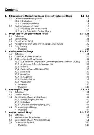
Syllabus
Unit I [10 Hrs.]
1. Pharmacology of Drugs Acting on Cardio Vascular System
(a) Introduction to Hemodynamic and Electrophysiology of Heart.
(b) Drugs used in Congestive Heart Failure.
(c) Anti-hypertensive Drugs.
(d) Anti-anginal Drugs.
(e) Anti-arrhythmic Drugs.
(f) Anti-hyperlipidemic Drugs.
Unit II [10 Hrs.]
1. Pharmacology of Drugs Acting on Cardio Vascular System
(a) Drug used in the Therapy of Shock.
(b) Hematinics, Coagulants and Anticoagulants.
(c) Fibrinolytics and Anti-platelet Drugs.
(d) Plasma Volume Expanders.
2. Pharmacology of Drugs Acting on Urinary System
(a) Diuretics.
(b) Anti-diuretics.
Unit III [10 Hrs.]
3. Autocoids and Related Drugs
(a) Introduction to Autacoids and Classification.
(b) Histamine, 5-HT and their antagonists.
(c) Prostaglandins, Thromboxanes and Leukotrienes.
(d) Angiotensin, Bradykinin and Substance P.
(e) Non-steroidal Anti-inflammatory Agents.
(f) Anti-gout Drugs.
(g) Antirheumatic Drugs.
Unit IV [08 Hrs.]
5. Pharmacology of Drugs Acting on Endocrine System
(a) Basic Concepts in Endocrine Pharmacology.
(b) Anterior Pituitary Hormones-analogues and their Inhibitors.
(c) Thyroid Hormones-analogues and their Inhibitors.
(d) Hormones regulating Plasma Calcium LEVEL-Parathormone, Calcitonin and
Vitamin-D.
(d) Insulin, Oral Hypoglycemic Agents and Glucagon.
(e) ACTH and Corticosteroids.
Unit V [07 Hrs.]
5. Pharmacology of Drugs Acting on Endocrine System
(a) Androgens and Anabolic Steroids.
(b) Estrogens, Progesterone and Oral Contraceptives.
(c) Drugs acting on the Uterus.
6. Bioassay
(a) Principles and Applications of Bioassay.
(b) Types of Bioassay
(c) Bioassay of Insulin, Oxytocin, Vasopressin, ACTH, d-Tubocurarine, Digitalis, Histamine
and 5-HT.
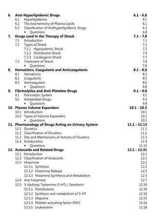
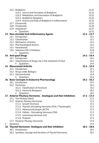
Contents
1. Introduction to Hemodynamic and Electrophysiology of Heart 1.1 - 1.7
1.1 Cardiovascular Hemodynamics 1.1
1.1.1 Introduction 1.1
1.1.2 Coronary Blood Flow 1.1
1.2 Electrophysiology of Heart 1.4
1.2.1 Physiology of Cardiac Muscle 1.4
1.2.2 Action Potential in Cardiac Muscle 1.5
2. Drugs used in Congestive Heart Failure 2.1 - 2.11
2.1 Definition 2.1
2.2 Epidemiology 2.1
2.3 Classification of CHF 2.1
2.4 Pathophysiology of Congestive Cardiac Failure (C.C.F) 2.2
2.5 Drug Therapy 2.4
• Questions 2.11
3. Antihypertensive Drugs 3.1 - 3.10
3.1 Definition 3.1
3.2 Classification of Hypertension 3.2
3.3 Antihypertensive Drug Classes 3.2
3.3.1 ACE Inhibitors (Angiotensin Converting Enzyme Inhibitors (ACEIs)) 3.3
3.3.2 Angiotensin II Receptor Antagonists 3.4
3.3.3 Diuretics 3.5
3.3.4 Calcium Channel Blockers (CCB) 3.6
3.3.5 β-blockers 3.7
3.3.6. α-blockers 3.8
3.3.7 α-2 Agonists 3.8
3.3.8 Renin Inhibitors 3.8
3.3.9 Vasodilator 3.9
• Summary 3.10
• Questions 3.10
4. Anti-Anginal Drugs 4.1 - 4.7
4.1 Definition 4.1
4.2 Types of Angina 4.1
4.3 Classification of Anti-anginal Drugs 4.2
4.3.1 Nitrates/Organic Nitrates 4.2
4.3.2 β-Blockers 4.3
4.3.3 Calcium Channel Blockers (CCBs) 4.4
4.4 Other Antianginal Drugs 4.7
• Questions 4.7
5. Anti-Arrhythmic Drugs 5.1 - 5.10
5.1 Definition 5.1
5.2 Mechanisms of Arrhythmias 5.1
5.3 Classification of Anti-Arrhythmic Drugs 5.1
5.4 Other Anti-arrhythmic 5.10
• Questions 5.10
6. Anti-Hyperlipidemic Drugs 6.1 - 6.8
6.1 Hyperlipidemia 6.1
6.2 The biochemistry of Plasma Lipids 6.1
6.3 Classification of Antihyperlipidemic Drugs 6.3
• Questions 6.8
7. Drugs used in the Therapy of Shock 7.1 - 7.8
7.1 Introduction 7.1
7.2 Types of Shock 7.1
7.2.1 Hypovolemic Shock 7.2
7.2.2 Distributive Shock 7.4
7.2.3 Cardiogenic Shock 7.7
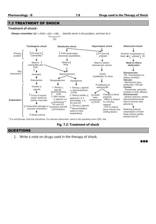
7.3 Treatment of Shock 7.8
• Questions 7.8
8. Hematinics, Coagulants and Anticoagulants 8.1 - 8.8
8.1 Hematinics 8.1
8.2 Coagulants 8.5
8.3 Anticoagulant 8.6
• Questions 8.8
9. Fibrinolytics and Anti-Platelets Drugs 9.1 - 9.8
9.1 Fibrinolytic System 9.1
9.2 Antiplatelet Drugs 9.3
• Questions 9.8
10. Plasma Volume Expanders 10.1 - 10.5
10.1 Introduction 10.1
10.2 Types of Volume Expanders 10.1
• Questions 10.5
11. Pharmacology of Drugs Acting on Urinary System 11.1 - 11.13
11.1 Diuretics 11.1
11.2 Classification of Diuretics 11.2
11.3 Site and Mechanisms of Actions of Diuretics 11.3
11.4 Antidiuretics 11.9
• Questions 11.13
12. Autocoids and Related Drugs 12.1 - 12.31
12.1 Introduction 12.1
12.2 Classification of Autacoids 12.2
12.3 Histamine 12.2
12.3.1 Synthesis 12.2
12.3.2 Histamine Release 12.2
12.3.3 Histamine-Synthesis and Metabolism 12.3
12.4 Anti histamine 12.7
12.5 5-Hydroxy Trptamine (5-HT) / Serotonin 12.10
12.5.1 Distributions 12.10
12.5.2 Synthesis and metabolism of 5-HT 12.10
12.5.3 Migraine 12.15
12.5.4 Platelet-activating factor (PAF) 12.16
12.5.5 Leukotriene 12.18
12.6 Bradykinin 12.21
12.6.1 Source and Formation of Bradykinin 12.21
12.6.2 Metabolism and Inactivation of Bradykinin 12.21
12.6.3 Bradykinin Receptors 12.22
12.6.4 Actions and Role of Bradykinin in Inflammation 12.22
12.7 Eicosanoids 12.23
12.8 Prostanoids 12.24
12.9 Angiotensin 12.27
• Questions 12.31
13. Non-steroidal Anti-Inflammatory Agents 13.1 - 13.7
13.1 Introduction 13.1
13.2 Classification 13.1
13.3 Mechanism of Action 13.2
13.4 Pharmacological Actions 13.2
13.5 Paracetamol 13.5
13.6 Selective COX-2 Inhibitors 13.6
• Questions 13.7
14. Anti-gout Drugs 14.1 - 14.4
14.1 Introduction 14.1
14.2 Classifications of Drugs use in the treatment of Gout 14.2
• Questions 14.4
15. Rheumatoid Arthritis 15.1 - 15.4
15.1 Introduction 15.1
15.2 Drugs under Biologics 15.3
15.3 Glucocorticoids 15.4
• Questions 15.4
16. Basic Concepts in Endocrine Pharmacology 16.1 - 16.2
16.1 Introduction 16.1
16.2 Hormones 16.1
16.2.1 Classification of Hormone 16.2
16.2.2 Hormone Receptors 16.2
• Questions 16.2
17. Anterior Pituitary Hormones - Analogues and their Inhibitiors 17.1 - 17.4
17.1 The Pituitary Gland 17.1
17.2 Anterior Pituitary Hormones 17.2
17.2.1 Growth Hormone 17.2
17.2.2 Thyroid-stimulating Hormone (TSH) / Thyrotrophin 17.2
17.2.3 Adrenocorticotropin (ACTH) 17.3
17.2.4 Follicle – Stimulating Hormone (FSH) 17.3
17.2.5 Leutinizing Hormone (LH) 17.3
17.2.6 Prolactin 17.3
17.3 Posterior Pituitary Hormones 17.4
• Questions 17.4
18. Thyroid Hormones- Analogues and their Inhibitors 18.1 - 18.8
18.1 Introduction 18.1
18.2 Synthesis, Storage and Secretion of Thyroid Hormones 18.1
18.3 Regulation of Thyroid Function 18.2
18.4 Actions of the Thyroid Hormones 18.3
18.5 Anti-thyroid Drugs 18.6
• Questions 18.8
19. Hormone Regulating Plasma Calcium Level 19.1 - 19.5
19.1 Calcium 19.1
19.2 Calcitonin (Thyrocalcitonin) 19.2
19.2.1 Synthesis 19.2
19.2.2 Pharmacological Action 19.2
19.2.3 Mechanism 19.2
19.3 Parathormone (PTH) 19.3
19.3.1 Mechanism 19.3
19.3.2 Pharmacological Action 19.3
19.4 Vitamine-D 19.4
19.4.1 Pharmacological Action 19.4
• Questions 19.5
20. Insulin, Oral Hypoglycemic Agents and Glucagon 20.1 - 20.14
20.1 Diabetes Mellitus 20.1
20.2 Treatment of Diabetes Mellitus 20.3
20.3 Classification 20.4
20.3.1 Oral Hypoglycemic Drugs 20.4
20.3.2 Anti Hyperglycemic Drugs 20.5
20.4 Insulin 20.8
20.4.1 Synthesis and Secretion 20.8
20.4.2 Pharmacological Action 20.9
20.5 Glucagon 20.12
20.5.1 Synthesis and Secretion 20.12
20.5.2 Control of Blood Glucose 20.13
20.5.2.1 Somatostatin 20.13
20.5.2.2 Amylin (Islet Amyloid Polypeptide) 20.14
• Questions 20.14
21. ACTH and Corticosteroids 21.1 - 21.4
21.1 ACTH (Adenocorticotropic Hormone/Corticotropin) 21.1
21.2 Corticosteroids 21.1
21.2.1 Glucocorticoid 21.1
21.2.2 Mineralocorticoid 21.3
• Questions 21.4
22. Oral Contraceptives 22.1 - 22.16
22.1 Introduction 22.1
22.2 General Physiology of Human Reproduction 22.1
22.3 Oral Contraceptive 22.2
22.3.1 Types of Oral Contraceptive 22.3
22.4 Oestrogens 22.3
22.4.1 Introduction 22.5
22.4.2 Physiological Roles 22.6
22.4.3 Classifications 22.7
22.4.4 Preparations 22.7
22.5 Progesterone 22.9
22.5.1 Introduction 22.9
22.5.2 Classifications 22.9
22.5.3 Physiological Actions 22.10
22.5.4 Progestogens and Antiprogestogens 22.12
22.5.5 Antiprogestogens 22.12
22.6 Androgens and Anabolic Steroids 22.12
22.6.1 Introduction 22.12
22.6.2 Physiological Actions 22.13
22.6.3 Mechanism of Action 22.13
22.7 Anabolic Steroids 22.14
22.7.1 Classification 22.15
22.7.2 Pharmacological Actions 22.15
22.7.3 Therapeutic Uses 22.15
• Questions 22.16
23. Drugs Acting on the Uterus 23.1 - 23.6
23.1 Introduction 23.1
23.2 Uterine Stimulants 23.1
23.2.1 Oxytocin 23.1
23.2.2 Ergometrine 23.4
23.3 Uterine Relaxants 23.5
• Questions 23.6
24. Bio-Assays General Aspects 24.1 - 24.10
24.1 Introduction 24.1
24.2 Reservations of Bioassays 24.1
24.3 Use of Standards 24.2
24.4 Principles 24.2
24.5 Biological Variation 24.3
24.6 Classification 24.3
• Questions 24.10
25. Bioassay 25.1 - 25.12
25.1 Biological Assay of Insulin 25.1
25.1.1 Rabbit Method 25.1
25.1.2 Mouse Method 25.2
25.1.3 Rat Diaphragm Method 25.3
25.2 Biological Assay of Oxytocin 25.3
25.3 Bioassay of Digitalis 25.6
25.4 Bioassay of D-Tubocurarine 25.7
25.4.1 Rabbit Head-drop Method 25.7
25.4.2 Frog’s Rectus Abdominis Muscle Preparation 25.8
25.5 Bioassay of Histamine 25.8
25.6 Bioassay of Vasopressin 25.9
25.7 Bioassay of ACTH 25.10
25.8 Bioassay of 5HT 25.12
• Questions 25.12
Multiple Choice Questions Q.1 - Q.11
(1.1)
Unit I
Chapter
Chapter
Chapter
Chapter ...
...
...
... 1
1
1
1
Introduction to Hemodynamic and
Electrophysiology of Heart
♦ LEARNING OBJECTIVES ♦
After completing this chapter, reader should be able to understand:
• Pathophysiology of hypertension, atherosclerosis, arrhythmia.
1.1 CARDIOVASCULAR HEMODYNAMICS
Haemodynamics is the term used to describe the interactions of the physiological
parameters that govern the behaviour of the CVS.
1.1.1 Introduction
• The cardiovascular system is concerned with the circulation of the blood. Essentially it
consists of heart, which works as pump, and the blood vessels, which carry the blood.
The blood carries oxygen and nutrients, and circulates through various tissue of the
body.
• The word hemodynamics – means circulation of blood in the human body.
• Cardiovascular hemodynamics comprises of blood circulation to the heart and in turn
the blood circulation regulated by the heart.
1.1.2 Coronary Blood Flow
• Resting coronary blood flow in human average is approximately 225ml/minute, which is
0.7 to 0.8 ml per gram of the heart muscle.
• During the diastole, cardiac muscle relaxes completely and no longer obstructs the
blood flow through left ventricular capillaries.
• This is phasic changes in coronary blood flow during cardiac muscle compression.
• During cardiac contraction – Intra myocardial pressure in the inner layer of the heart
muscle is so much greater than the outer layer.
• It compresses the sub endocardial blood vessels far more than it compresses the outer
vessel.
Control of Coronary Blood Flow:
• Oxygen demand is a major factor in local blood flow regulation.
• Determinants of oxygen consumption.
• Importance of increase in coronary blood flow in response to myocardial oxygen usage.
• Reactive hyperemia in coronary system.
Pharmacology - II 1.2 Introduction to Hemodynamic & …
…
…
…
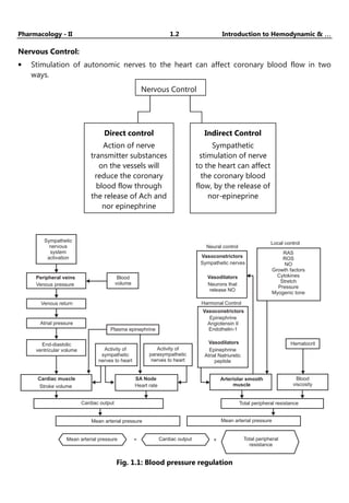
Nervous Control:
• Stimulation of autonomic nerves to the heart can affect coronary blood flow in two
ways.
Sympathetic
nervous
system
activation
Blood
volume
Peripheral veins
Venous pressure
Venous return
Atrial pressure
End-diastolic
ventricular volume
Plasma epinephrine
Cardiac muscle
Stroke volume
SA Node
Heart rate
Activity of
sympathetic
nerves to heart
Activity of
parasympathetic
nerves to heart
Cardiac output
Mean arterial pressure
Mean arterial pressure Cardiac output
= × Total peripheral
resistance
Mean arterial pressure
Total peripheral resistance
Blood
viscosity
Hematocrit
Arteriolar smooth
muscle
Vasoconstrictors
Epinephrine
Angiotensin II
Endothelin-1
Vasodilators
Epinephrine
Atrial Natriuretic
peptide
Harmonal Control
Vasoconstrictors
Sympathetic nerves
Vasodilators
Neurons that
release NO
Neural control
Local control
RAS
ROS
NO
Growth factors
Cytokines
Stretch
Pressure
Myogenic tone
Fig. 1.1: Blood pressure regulation
Nervous Control
Direct control
Action of nerve
transmitter substances
on the vessels will
reduce the coronary
blood flow through
the release of Ach and
nor epinephrine
Indirect Control
Sympathetic
stimulation of nerve
to the heart can affect
the coronary blood
flow, by the release of
nor-epineprine
Pharmacology - II 1.3 Introduction to Hemodynamic & …
…
…
…
Stroke Volume
• The amount of blood pumped by the left ventricle of the heart in one contraction.
Normally only about 2/3rd of the blood in the ventricle is expelled with each beat.
Cardiac Output
• Flow of blood is usually measured in l/min.
• Total amount of blood flowing through the circulation = Cardiac output (CO)
Cardiac Output = Stroke Volume × Heart Rate = 5 L/min.
• Influenced by blood pressure (force of blood against side wall) & resistance (Blood
viscosity, Vessel length, Vessel Elasticity, Vasoconstriction/Vasodilation).
ADH production
in hypothalamus
(osmoreceptors)
Concentrated
urine excreted
In
h
i
b
i
t
s
S
tim
ulates
Stim
u
l
a
t
e
s
Blood volume
Serum osmolality
( Thirst and water intake)
Arterial B.P.
(stimulates baroreceptors)
Sympathetic discharge
Renal perfusion
Renin release ( GFR)
Diuresis results H O and Na
filtered by kidney
2
+
Blood volume
Serum osmolality
Na+
2
and H O excretion; blood pressure
Angiotensin and
Aldosterone by
adrenal cortex
Urine excretion
Circulating volume of H O and Na (loss of K )
2
+ +
ADH release into
bloodstream from storage
in posterior pituitary
Reabsorption of H O
by kidneys
2
Fig. 1.2
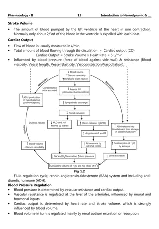
Fluid regulation cycle, rennin angiotensin aldosterone (RAA) system and including anti-
diuretic hormone (ADH).
Blood Pressure Regulation
• Blood pressure is determined by vascular resistance and cardiac output.
• Vascular resistance is regulated at the level of the arterioles, influenced by neural and
hormonal inputs.
• Cardiac output is determined by heart rate and stroke volume, which is strongly
influenced by blood volume.
• Blood volume in turn is regulated mainly by renal sodium excretion or resorption.
Pharmacology - II 1.4 Introduction to Hemodynamic & …
…
…
…
• Renin, a major regulator of blood pressure, is secreted by the kidneys in response to
decreased blood pressure in afferent arterioles. In turn, renin cleaves angiotensinogen to
angiotensin I; subsequent peripheral catabolism produces angiotensin II, which regulates
blood pressure by increasing vascular smooth muscle cell tone and by increasing adrenal
aldosterone secretion and, consequently, renal sodium resorption.
1.2 ELECTROPHYSIOLOGY OF HEART
1.2.1 Physiology of Cardiac Muscle
• Three Major types of cardiac muscle fiber:
o Atrial muscle.
o Ventricular muscle.
o Specialized excitatory and conductive muscle fibers.
o Atrial and ventricular muscle contract in a same way as skeletal muscle but the
duration of contraction is longer.
o The specialized excitatory and conductive fibers contract feebly because they
contain few contractile fibrils. Exhibit automatic rhythmical electrical discharge in
the form of action potentials.
Dyslipidemia Hypertension Tobacco
smoking
Diabetes
mellitus
Obesity Age Male gender Hereditary
LDL
modifica--
tions
Vascular
permeability
Intimal LDL
retention
LDL Binding
to SMC
LDL
Endothelial
dysfunction
Insulin
resistance
hypertension
Dyslipidemia
Estrogen
mediated
atheroprotection
HDL
LDL
Modifiable risk factors Non-modifiable risk factors
Atherosclerosis development and
progression
Complications
Calcification Rupture Hemorrhage
Embolization
Distal sites
Aneurysm
AAA
Embolic stroke
Thrombotic
stroke
Myocardial
infarction
Peripheral
vascular
disease
Angina
Renal artery
stenosis
LDL
glycation
Endothelial
dysfunction
Fig. 1.3: Risk factors and complications of atherosclerosis
Atherosclerosis:
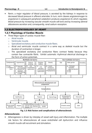
• Atherogenesis is driven by interplay of vessel wall injury and inflammation. The multiple
risk factors for atherosclerosis all cause endothelial cell dysfunction and influence
smooth muscle cell recruitment and stimulation.
Pharmacology - II 1.5 Introduction to Hemodynamic & …
…
…
…
• Atherosclerotic plaques develop and grow slowly over decades. Stable plaques can
produce symptoms related to chronic ischemia by narrowing vessels, whereas unstable
plaques can cause dramatic and potentially fatal ischemic complications related to acute
plaque rupture, thrombosis, or embolization.
• Stable plaques tend to have a dense fibrous cap, minimal lipid accumulation, and little
inflammation, whereas “vulnerable” unstable plaques have thin caps, large lipid cores,
and relatively dense inflammatory infiltrates.
1.2.2 Action Potential in Cardiac Muscle
+25
1
2
3
4
Transmembrane
potential,
mV
0
-25
-50
-75
-100
Na
influx
+
Ca
influx
+
K
efflux
+
Na
efflux
+
K
influx
+
Fig. 1.4
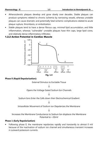
Phase 0 (Rapid Depolarization):
External Stimulus to Excitable Tissue
↓
Opens the Voltage Gated Sodium Ion Channels
↓
Sodium Ions Enter the Cells down their Electrochemical Gradient
↓
Intracellular Movement of Sodium ion Depolarizes the Membrane
↓
Increases the Membrane Conductance to Sodium Ion displaces the Membrane
Potential to +30mV
Phase 1 (Early Repolarization)
• Following phase 0, the membrane repolarizes rapidly and transiently to almost 0 mV
because of the inactivation of sodium ion channel and simultaneous transient increases
in outward potassium currents.
Pharmacology - II 1.6 Introduction to Hemodynamic & …
…
…
…
Phase 2 (Plateau)
• Membrane potential remains approximately 0 mV for a relatively prolonged duration.
• A balance between slow inward Ca2+
and outward K+
currents mediates the plateau
phase of the action potential.
Phase 3 (Repolarization)
• Inactivation of Ca2+
channels and a simultaneous increase in outward K+ current
through K+
channels produces a net outward movement of positive charge and
repolarization of the membrane.
Phase 4 (Resting Membrane Potential)
• The membrane potential of ventricular myocytes remains at the resting membrane
potential until the cell is stimulated again.
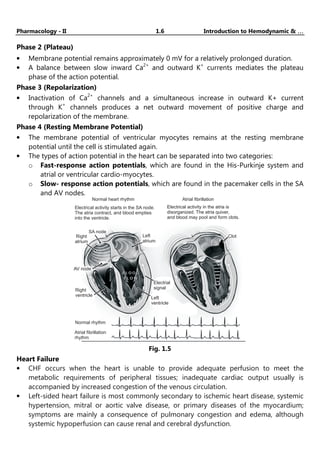
• The types of action potential in the heart can be separated into two categories:
o Fast-response action potentials, which are found in the His-Purkinje system and
atrial or ventricular cardio-myocytes.
o Slow- response action potentials, which are found in the pacemaker cells in the SA
and AV nodes.
Normal heart rhythm
Electrical activity starts in the SA node.
The atria contract, and blood empties
into the ventricle.
Electrical activity in the atria is
disorganized. The atria quiver,
and blood may pool and form clots.
Atrial fibrillation
Right
atrium
SA node
Left
atrium
AV node
Electrial
signal
Left
ventricle
Right
ventricle
Normal rhythm
Atrial fibrillation
rhythm
Clot
Fig. 1.5
Heart Failure
• CHF occurs when the heart is unable to provide adequate perfusion to meet the
metabolic requirements of peripheral tissues; inadequate cardiac output usually is
accompanied by increased congestion of the venous circulation.
• Left-sided heart failure is most commonly secondary to ischemic heart disease, systemic
hypertension, mitral or aortic valve disease, or primary diseases of the myocardium;
symptoms are mainly a consequence of pulmonary congestion and edema, although
systemic hypoperfusion can cause renal and cerebral dysfunction.
Pharma Masti
YouTube channel :-
Pharmacology - II 1.7 Introduction to Hemodynamic & …
…
…
…
• Right-sided heart failure is most often due to left heart failure and, less commonly, to
primary pulmonary disorders; signs and symptoms are related chiefly to peripheral
edema and visceral congestion.
Arrhythmias
• Arrhythmias can be caused by ischemic or structural changes in the conduction system
or by myocyte electrical instability. In structurally normal hearts, arrhythmias more often
are due to mutations in ion channels that cause aberrant repolarization or
depolarization.
• Sudden cardiac death (SCD) most frequently is due to coronary artery disease leading to
ischemia. Myocardial irritability typically results from non-lethal ischemia or from
pre-existing fibrosis from previous myocardial injury. SCD less often is due to acute
plaque rupture with thrombosis that induces a rapidly fatal arrhythmia.
YouTube channel :-
Pharma Masti
(2.1)
Chapter
Chapter
Chapter
Chapter ...
...
...
... 2
2
2
2
Drugs used in Congestive Heart Failure
♦ LEARNING OBJECTIVES ♦
After completing this chapter, reader should be able to:
• Classify drugs.
• Illustrate mechanism of action and ADR.
2.1 DEFINITION
Congestive heart failure (CHF) is a clinical syndrome during which the center is unable to
pump ample blood to fulfill the metabolic necessities of the body, or will do thus solely at an
elevated filling pressure.
2.2 EPIDEMIOLOGY
• Heart failure may be a burgeoning downside worldwide, with over twenty million
folks affected.
• The overall prevalence of HF within the adult population in developed countries is
2%.
• HF prevalence follow an exponential pattern, rising with age, and affects 6-10% of
people over age 65.
• The incidence of HF is lower in women than in men.
• Although HF once was through to arise primarily within the setting of a depressed
left chamber (LV) ejection fraction (EF), medical specialty studies have shown that
around simple fraction of patients WHO develop HF have a standard or preserved EF
(EF 40-50%).
• Accordingly, HF patients are now broadly categorized into one of two groups: (1) HF
with a depressed EF (systolic failure) or (2) HF with a preserved EF (diastolic failure).
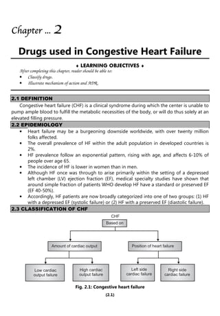
2.3 CLASSIFICATION OF CHF
CHF
Based on
Amount of cardiac output Position of heart failure
Low cardiac
output failure
High cardiac
output failure
Left side
cardiac failure
Right side
cardiac failure
Fig. 2.1: Congestive heart failure
Pharmacology - II 2.2 Drugs used in Congestive Heart Failure
Based on amount of Cardiac Output:
Low cardiac Output Failure High cardiac Output Failure
Most frequent. Very rarely.
Metabolic demands of the body organs for
oxygen are normal and within limits.
Metabolic demands of the body for
oxygen is very high.
Myocardial fraction is prominent factor
leading to the failure of systolic and diastolic
function of the ventricles, ultimately results
in low cardiac output failure.
Hyperthyroidism, anaemia, arteriovenous
shunt causes high cardiac output failure.
Based on the position of Heart Failure:
Left side Cardiac Failure Right side Cardiac Failure
Is the result of right side cardiac failure. Is the result of left side cardiac failure
Inefficient pumping action of left ventricle is
responsible for the accumulation of blood in
the ventricles.
Inefficient pumping action of right
ventricle is responsible for the accumu-
lation of blood in right ventricle.
Left ventricle fails to accept/collect the blood
from lungs due to back pressure.
Right ventricle fails to accept/collect the
blood from peripheral organs.
Pulmonary congestion/oedema is the final
result.
Peripheral generalized oedema is the final
result.
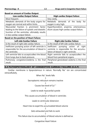
2.4 PATHOPHYSIOLOGY OF CONGESTIVE CARDIAC FAILURE (C.C.F)
Cardiac membrane is lipoproteinous in nature. Normally Na+
ion are concentrated
extracellularly.
When Na+
levels falls
↓
Sacroplasmic reticulum remains inactive
↓
Causes low level of Ca++
↓
Leads to weak myocardial systole
↓
This causes accumulation of blood in ventricles
↓
Leads to ventricular distension
↓
Heart tries to expel this accumulated blood volume
↓
Gets exhausted and fails to work
↓
This failure due to accumulation of blood volume is defined as
↓
Congestive Heart Failure
Pharmacology - II 2.3 Drugs used in Congestive Heart Failure
Compensatory Mechanisms of CHF
To enhances the cardiac output, body compensates for the intrinsic cardiac effects in the
following manner.
1. Increased sympathetic discharge.
2. To complete the remittent B.P., baroreceptors set within the arch of artery arterial
blood vessel sinuses and walls of the center get excited and causes activation of
beta-adrenergic receptors resulting in an increase in rate and force of contraction of
heart.
3. An increase in blood vessel comes back (preload) is additionally seen because of the
activation of alpha adrenergic receptors.
4. Increased rate associated force of contraction at the side of the enhanced preload
ends up in an initial increase within the flow.
5. Vasoconstriction of the arteries due to alpha stimulation also causes an increase in
after load, leading to fall in ejection fraction.
Activation of Renin Angiotensin Aldosterone (RAA)
• Fall within the flow decreases the urinary organ perfusion rate; as a result the RAA
system gets activated.
• Angiotensin II may cause atrophic response in vascular smooth muscle (with
vasoconstriction) and myocardial hypertrophy, attempting to restore wall stress to
normal.
Cardiac Remodeling:
It is most vital mechanism by that body stipendiary for the intrinsic internal organ
effects.
• It involves changes within the form of the center (from traditional to spherical)
because of cardiac muscle hypertrophy.
• During cardiac remodeling, the connective tissue cells as well as the abnormal
myocardial cells undergo proliferation and dilation instead of stretching under the
influences of angiotensin-2.
• In the early stages, the remodeled heart maintains the cardiac performances.
• But later on, hypertrophy may exert certain adverse effects like ischaemic changes,
decrease in the rate and force of contraction of heart. After certain period of time
the antagonistic mechanisms get exhausted and worsen the cardiac performances.
The stress on heart increases and a stage is reached where these mechanisms fails to
maintain the adequate cardiac output.
Clinical manifestations/signs and symptoms
• Fluid retention
• Pulmonary congestion
• Dyspnoea and orthopnoea
Pharmacology - II 2.4 Drugs used in Congestive Heart Failure
CVS Manifestations
• Resting tachycardia
• Ventricular arrhythmias
• Enlargement of heart
Renal Manifestations
• Nocturia
• Oliguria
Other Manifestations
• Reduced cardiac output lead to poor perfusion of skeletal muscle resulting in
fatigue.
• Reduced flow result in poor perfusion of muscle leading to fatigue.
• Reduced perfusion to brain results in altered mental states and confusion.
• Reduced perfusion might also cause the patient to seem pale with cold and
perspiring hands.
Treatment
Non drug treatment/ non pharmacological approach:
• Physical exercise
• Salt intake
• Fluid intake
• Alcohol consumption
• Liquorice
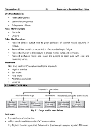
2.5 DRUG THERAPY
Drug used in heart failure
Cardiac
glycosides
(digoxin)
Positive inotropic drugs Vasodillators Miscellaneous drugs for chronic failure
Beta
agonists
(dobutamine)
PDE inhibitors
(milrinone)
Nitroprusside
Nitrates
Hydralazine
Loop diuretics
ACE inhibitors
Nesiritide
Beta
blockers
Spironolactone
Fig. 2.2: Drugs used in heart failure
Inotropes:
• Increase force of contraction.
• All increase intracellular cardiac Ca++
concentration.
E.g. Digitalis (cardiac glycoside), Dobutamine (β-adrenergic receptor agonist), Milrinone.
Pharmacology - II 2.5 Drugs used in Congestive Heart Failure
Non-pharmacological Approaches:
CARDIAC GLYCOSIDES
Mechanism of action:
“Effect on cardio vascular function in CHF”.
1. Digitalis derivatives when administrated to individuals suffering from CHF:
• Acts directly on the myocardium and increases conductivity, automaticity,
rhythmicity and causes forceful contraction of heart. Because of powerful
contractions, ventricular blood is forced from right side in to artery and from left
ventricles to aorta.
This causes complete emptying of heart.
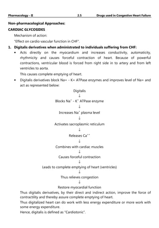
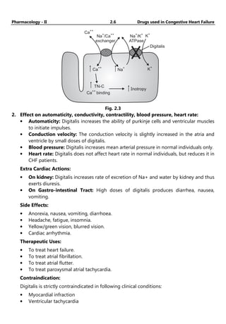
• Digitalis derivatives block Na+ - K+ ATPase enzymes and improves level of Na+ and
act as represented below:
Digitalis
↓
Blocks Na+
- K+
ATPase enzyme
↓
Increases Na+
plasma level
↓
Activates sacroplasmic reticulum
↓
Releases Ca++
↓
Combines with cardiac muscles
↓
Causes forceful contraction
↓
Leads to complete emptying of heart (ventricles)
↓
Thus relieves congestion
↓
Restore myocardial function
Thus digitalis derivatives, by their direct and indirect action, improve the force of
contractility and thereby assure complete emptying of heart.
Thus digitalized heart can do work with less energy expenditure or more work with
some energy expenditure.
Hence, digitalis is defined as “Cardiotonic”.
Pharmacology - II 2.6 Drugs used in Congestive Heart Failure
Ca
++
Na /Ca
exchanger
+ ++
Na /K
ATPase
+ +
K
+
K
+
Na
+
Ca
++
Ca binding
++
TN-C
Inotropy
Digitalis
Fig. 2.3
2. Effect on automaticity, conductivity, contractility, blood pressure, heart rate:
• Automaticity: Digitalis increases the ability of purkinje cells and ventricular muscles
to initiate impulses.
• Conduction velocity: The conduction velocity is slightly increased in the atria and
ventricle by small doses of digitalis.
• Blood pressure: Digitalis increases mean arterial pressure in normal individuals only.
• Heart rate: Digitalis does not affect heart rate in normal individuals, but reduces it in
CHF patients.
Extra Cardiac Actions:
• On kidney: Digitalis increases rate of excretion of Na+ and water by kidney and thus
exerts diuresis.
• On Gastro-intestinal Tract: High doses of digitalis produces diarrhea, nausea,
vomiting.
Side Effects:
• Anorexia, nausea, vomiting, diarrhoea.
• Headache, fatigue, insomnia.
• Yellow/green vision, blurred vision.
• Cardiac arrhythmia.
Therapeutic Uses:
• To treat heart failure.
• To treat atrial fibrillation.
• To treat atrial flutter.
• To treat paroxysmal atrial tachycardia.
Contraindication:
Digitalis is strictly contraindicated in following clinical conditions:
• Myocardial infraction
• Ventricular tachycardia
Pharmacology - II 2.7 Drugs used in Congestive Heart Failure
• Partial heart block
• Previous digitalis therapy
• Calcium administration.
Digitalis interaction:
• Digitalis, Calcium: Calcium ions increase the force of contraction of heart. High
plasma calcium levels stimulate the myocardium so much, that it leads to cardiac
arrest during systole. Digitalis is also known to increase the force of contraction of
heart. Thus digitalis and calcium act synergetically and may prove to be toxic. Hence
during digitalis therapy Ca++
ion administration must be avoided.
• Digitalis, Quinidine: Quinidine, when administrated in individuals taking digitalis,
increases serum digoxin levels. This may cause adverse effects of digitalis and other
clinical complexities. Hence must be avoided.
Treatment of over digitalisation:
• Immediately stop the administration of digitalis.
• Stop if any diuretic administration is in continuation.
• Mild tachycardia can be treated with atropine.
• Mild toxicity can be treated by administration of potassium salts – 5 to 7.5 g of
potassium chloride orally daily.
• Ventricular tachycardia can be treated with phenytoin (250 mg well diluted).
Rapidly Acting Inotropic Agents
In critically sick infants with CHF, in those with renal dysfunction (e.g., infants or in
postoperative cardiac patients with heart failure) quickly acting catecholamines with a short
duration of action is preferable to digoxin.
• dopamine,
• dobutamine
• epinephrine
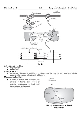
β1 Blockers:
β1-Blockers-bisoprolol, carvedilol, metoprolol.
Mechanism of Action:
• Heart failure is accompanied by an increase activation of sympathetic nervous
system.
• This brings about structural and functional modification in the myocardium.
• β Blockers inhibit the sympathetic outflow of nor epinephrine and counteract the
changes produced.
• The ventricular remodeling in heart failure is also reversed by β Blockers.
• Increases beta receptor sensitivity.
Pharmacology - II 2.8 Drugs used in Congestive Heart Failure
Cell
membrane
-
+
B1
GS
PDE
5' AMP
cAMP
ATP
b1-Blockers
PKA active
+
Ca
2+
+
RyR
L-VDDC
AC
Ca
2+
RyR
Ca
2+
Sequestered Ca
2+
Sarcoplasmic
reticulum
Actin/tropomyosin-troponin
Myosin
Sarcomere
Fig. 2.4
Adverse drug reaction:
• Hypotension
• Bradycardia.
VASODILATORS
• Isosorbide dinitrate, isosorbide mononitrate, and hydralazine also used specially in
patients who cannot tolerate ACE inhibitors.
Mechanism of Action:
• It directly relaxes the arterioles and
arteries reducing the peripheral
vascular resistances, preload and
help to reduce after load.
Sodium
Nitroprusside
NO
NO
GC
GTP cGMP
Relaxation
Smooth
Muscle
Fig. 2.5: Mechanism of Action of
Vasodilators
Pharmacology - II 2.9 Drugs used in Congestive Heart Failure
Adverse drug reaction:
• Nausea
• Palpitation
• Tachycardia
• Salt and water retention on prolong therapy.
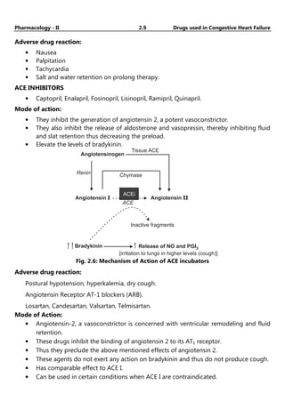
ACE INHIBITORS
• Captopril, Enalapril, Fosinopril, Lisinopril, Ramipril, Quinapril.
Mode of action:
• They inhibit the generation of angiotensin 2, a potent vasoconstrictor.
• They also inhibit the release of aldosterone and vasopressin, thereby inhibiting fluid
and slat retention thus decreasing the preload.
• Elevate the levels of bradykinin.
Angiotensinogen
Angiotensin I Angiotensin II
Renin Chymase
Tissue ACE
Bradykinin Release of NO and PGI2
[irritation to lungs in higher levels (cough)]
Inactive fragments
ACE
ACEi
Fig. 2.6: Mechanism of Action of ACE incubators
Adverse drug reaction:
Postural hypotension, hyperkalemia, dry cough.
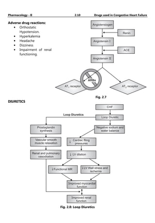
Angiotensin Receptor AT-1 blockers (ARB).
Losartan, Candesartan, Valsartan, Telmisartan.
Mode of Action:
• Angiotensin-2, a vasoconstrictor is concerned with ventricular remodeling and fluid
retention.
• These drugs inhibit the binding of angiotensin 2 to its AT₁ receptor.
• Thus they preclude the above mentioned effects of angiotensin 2.
• These agents do not exert any action on bradykinin and thus do not produce cough.
• Has comparable effect to ACE I.
• Can be used in certain conditions when ACE I are contraindicated.
Pharmacology - II 2.10 Drugs used in Congestive Heart Failure
Adverse drug reactions:
• Orthostatic
Hypotension.
• Hyperkalemia
• Headache
• Dizziness
• Impairment of renal
functioning.
Angiotensiogen
Renin
Angiotensin I
Angiotensin II
ACE
AT receptor
2
AT receptor
1
Fig. 2.7
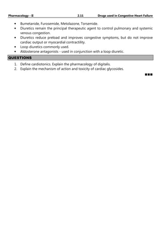
DIURETICS
Loop Diuretics
CHF
Loop Diuretic
Negative sodium and
water balance
Prostaglandin
synthesis
Vascular smooth
muscle relaxation
Cardiac flling
pressures
LV dilation
Renal and pulmonary
vasodilation
LV Wall stress and
ischemia
Functional MR
Improved myocardial
function
Improved renal
function
Fig. 2.8: Loop Diuretics
Pharmacology - II 2.11 Drugs used in Congestive Heart Failure
• Bumetanide, Furosemide, Metolazone, Torsemide.
• Diuretics remain the principal therapeutic agent to control pulmonary and systemic
venous congestion.
• Diuretics reduce preload and improves congestive symptoms, but do not improve
cardiac output or myocardial contractility.
• Loop diuretics commonly used.
• Aldosterone antagonists - used in conjunction with a loop diuretic.
QUESTIONS
1. Define cardiotonics. Explain the pharmacology of digitalis.
2. Explain the mechanism of action and toxicity of cardiac glycosides.
YouTube channel:- Pharma Masti
(3.1)
Chapter
Chapter
Chapter
Chapter ...
...
...
... 3
3
3
3
Antihypertensive Drugs
♦ LEARNING OBJECTIVES ♦
After completing this chapter, reader should be able to:
• Describe the classes of antihypertensive and their indications for use.
• Explain mechanism of action and ADR.
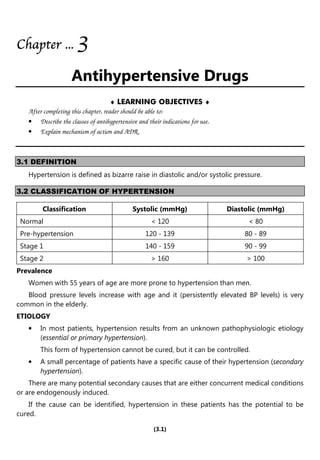
3.1 DEFINITION
Hypertension is defined as bizarre raise in diastolic and/or systolic pressure.
3.2 CLASSIFICATION OF HYPERTENSION
Classification Systolic (mmHg) Diastolic (mmHg)
Normal < 120 < 80
Pre-hypertension 120 - 139 80 - 89
Stage 1 140 - 159 90 - 99
Stage 2 > 160 > 100
Prevalence
Women with 55 years of age are more prone to hypertension than men.
Blood pressure levels increase with age and it (persistently elevated BP levels) is very
common in the elderly.
ETIOLOGY
• In most patients, hypertension results from an unknown pathophysiologic etiology
(essential or primary hypertension).
This form of hypertension cannot be cured, but it can be controlled.
• A small percentage of patients have a specific cause of their hypertension (secondary
hypertension).
There are many potential secondary causes that are either concurrent medical conditions
or are endogenously induced.
If the cause can be identified, hypertension in these patients has the potential to be
cured.
Pharmacology - II 3.2 Antihypertensive Drugs
Essential Hypertension:
• More than 90% of individuals with hypertension have essential hypertension.
• Numerous mechanisms have been identified that may contribute to the
pathogenesis of this form of hypertension, so identifying the exact underlying
abnormality is not possible.
• Genetic factors may play an important role in the development of essential
hypertension.
Secondary Hypertension:
• Fewer than 10% of patients have secondary hypertension where either a comorbid
disease or drug is responsible for elevating BP.
• In most of these cases, renal dysfunction resulting from severe chronic kidney
disease or renovascular disease is the most common secondary cause.
• Certain drugs, either directly or indirectly, can cause hypertension or exacerbate
hypertension by increasing BP.
Diseases: Chronic kidney disease, Obstructive sleep apnea, Parathyroid disease,
Pheochromocytoma, Thyroid disease.
Prescription drugs: Adrenal steroids, Amphetamines/anorexiants, Decongestants,
Nonsteroidal anti-inflammatory drugs.
Food substances: Sodium, Ethanol, Licorice.
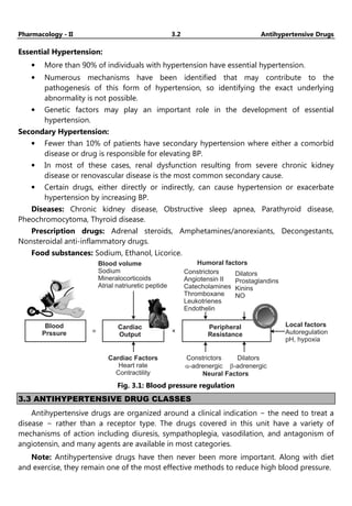
Blood
Prssure
Cardiac
Output
= ×
Cardiac Factors
Heart rate
Contractility
Constrictors
-adrenergic
a
Dilators
-adrenergic
b
Neural Factors
Peripheral
Resistance
Local factors
Autoregulation
pH, hypoxia
Blood volume
Sodium
Mineralocorticoids
Atrial natriuretic peptide
Humoral factors
Constrictors
Angiotensin II
Catecholamines
Thromboxane
Leukotrienes
Endothelin
Dilators
Prostaglandins
Kinins
NO
Fig. 3.1: Blood pressure regulation
3.3 ANTIHYPERTENSIVE DRUG CLASSES
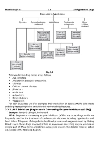
Antihypertensive drugs are organized around a clinical indication − the need to treat a
disease − rather than a receptor type. The drugs covered in this unit have a variety of
mechanisms of action including diuresis, sympathoplegia, vasodilation, and antagonism of
angiotensin, and many agents are available in most categories.
Note: Antihypertensive drugs have then never been more important. Along with diet
and exercise, they remain one of the most effective methods to reduce high blood pressure.
Pharmacology - II 3.3 Antihypertensive Drugs
Drugs used in hypertension
Diuretics Sympathoplegics-
blockers of
Angiotensin
antagonists
Renin
inhibitor
(aliskiren)
Vasodilators
Alpha or beta
receptors
(prazosin,
propranolol)
ACE
inhibitors
(captopril)
Receptor
blockers
(losartan)
Nerve
terminals
(guanethidine)
Ganglia
(hexamethonium)
Older oral
vasodilators
(hydralazine)
Calcium
blockers
(nifedipine)
Parenteral
vasodilators
(nitroprusside)
CNS
sympathetic
outflow
(clonidine)
Fig. 3.2
Antihypertensive drug classes are as follows:
• ACE inhibitors
• Angiotensin II receptor antagonists
• Diuretics
• Calcium channel blockers
• β-blockers
• α-blockers
• α2-blockers
• Renin inhibitors
• Vasodilators
For each drug class, we offer examples, their mechanism of actions (MOA), side effects
and drug interaction profiles and any other relevant clinical features.
3.3.1. ACE Inhibitors (Angiotensin Converting Enzyme Inhibitors (ACEIs))
Example: Ramipril, Lisinopril, Perindopril.
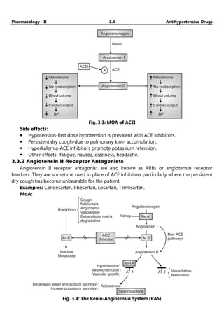
MOA: Angiotensin converting enzyme inhibitors (ACEIs) are those drugs which are
frequently used for the treatment of cardiovascular disorders including hypertension and
heart failure. This group of drugs diminishes blood pressure and oxygen demand by dilating
blood vessels. These drugs principally inhibit an angiotensin converting enzyme which is an
integral part of RAAS (Renin-angiotensin-aldosterone system). The detailed mode of action
is described in the following diagram.
Pharmacology - II 3.4 Antihypertensive Drugs
Angiotensinogen
Renin
ACE
X
ACEI
Angiotensin I
Angiotensin II
Aldosterone
Na reabsorption
Blood volume
Cardiac output
BP
Aldosterone
Na reabsorption
Blood volume
Cardiac output
BP
Fig. 3.3: MOA of ACEI
Side effects:
• Hypotension-first dose hypotension is prevalent with ACE inhibitors.
• Persistent dry cough-due to pulmonary kinin accumulation.
• Hyperkalemia-ACE inhibitors promote potassium retension.
• Other effects- fatigue, nausea, dizziness, headache.
3.3.2 Angiotensin II Receptor Antagonists
Angiotensin II receptor antagonist are also known as ARBs or angiotensin receptor
blockers. They are sometime used in place of ACE inhibitors particularly where the persistent
dry cough has become unbearable for the patient.
Examples: Candesartan, lrbesartan, Losartan, Telmisartan.
MoA:
Bradykinin
Cough
Natriuresis
Angiodema
Vasodilaton
Extracellular matrix
degradation
ACE
ACE
Inhibitor
Inactive
Metabolite
Hypertension
Vasoconstriction
Vascular growth
AIIRA
AT 1 AT 2
ACE
Renin
Angiotensinogen
Non-ACE
pathways
Angiotensin II
Angiotensin I
Kidney
Vasodilation
Natriuresis
Aldosterone
Decerased water and sodium secretion
Increse potassium secretion
Spironolactone
Fig. 3.4: The Renin-Angiotensin System (RAS)
Pharmacology - II 3.5 Antihypertensive Drugs
Side effects: Hypotension, Hyperkalemia
Renal failure (as with ACE inhibitors):
• Cough-though less likely than with an ACE inhibitors.
• Due to risk of hyperkalemia, other potassium-elevating drugs should not be
prescribed.
• This includes potassium supplements, potassium-sparing diuretics.
• As with ACE inhibitors, taking ARBs with NSAIDs increase risk of renal failure.
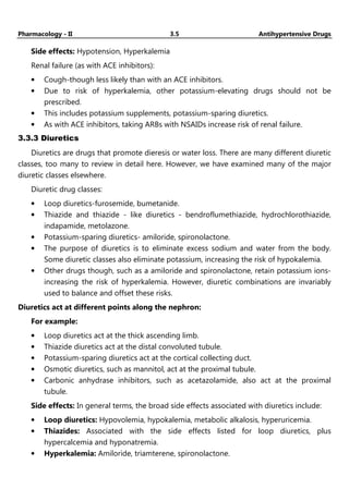
3.3.3 Diuretics
Diuretics are drugs that promote dieresis or water loss. There are many different diuretic
classes, too many to review in detail here. However, we have examined many of the major
diuretic classes elsewhere.
Diuretic drug classes:
• Loop diuretics-furosemide, bumetanide.
• Thiazide and thiazide - like diuretics - bendroflumethiazide, hydrochlorothiazide,
indapamide, metolazone.
• Potassium-sparing diuretics- amiloride, spironolactone.
• The purpose of diuretics is to eliminate excess sodium and water from the body.
Some diuretic classes also eliminate potassium, increasing the risk of hypokalemia.
• Other drugs though, such as a amiloride and spironolactone, retain potassium ions-
increasing the risk of hyperkalemia. However, diuretic combinations are invariably
used to balance and offset these risks.
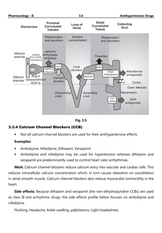
Diuretics act at different points along the nephron:
For example:
• Loop diuretics act at the thick ascending limb.
• Thiazide diuretics act at the distal convoluted tubule.
• Potassium-sparing diuretics act at the cortical collecting duct.
• Osmotic diuretics, such as mannitol, act at the proximal tubule.
• Carbonic anhydrase inhibitors, such as acetazolamide, also act at the proximal
tubule.
Side effects: In general terms, the broad side effects associated with diuretics include:
• Loop diuretics: Hypovolemia, hypokalemia, metabolic alkalosis, hyperuricemia.
• Thiazides: Associated with the side effects listed for loop diuretics, plus
hypercalcemia and hyponatremia.
• Hyperkalemia: Amiloride, triamterene, spironolactone.
Pharmacology - II 3.6 Antihypertensive Drugs
Glomerulus
Proximal
Convoluted
Tubules
Loop of
Henle
Distal
Convoluted
Tubule
Collecting
Duct
Reabsorption
and Secretion
Afferent
arteriole
NSAIDs
PGI & PGE
Vasodilators
2 2
Reabsorption
and secretion
Solution
concentration
carbonic
Anhydrase
Inhibitors
Efferent
arteriole
Ang-II
vasoconstrictor
ACE-Is
Osmotic
diuretics
Thiazides
Loop
diuretics Aldosterone
antagonists
Cortex
Outer Medulla
Vasopressin
ADH
antagonists
Descending
Limb
Ascending
Limb
K
H O
(ADH)
2
NaHCO
Glucose
3
H O
2
Na/K/2Cl
ENaC
(Aldosterone)
NaCl
Na
Fig. 3.5
3.3.4 Calcium Channel Blockers (CCB)
• Not all calcium channel blockers are used for their antihypertensive effects.
Examples:
• Amlodipine, Nifedipine, Diltiazem, Verapamil.
• Amlodipine and nifedipine may be used for hypertension whereas diltiazem and
verapamil are predominantly used to control heart rate/ arrhythmias.
MoA: Calcium channel blockers reduce calcium entry into vascular and cardiac cells. This
reduces intracellular calcium concentration which, in turn causes relaxation on vasodilation
in atrial smooth muscle. Calcium channel blockers also reduce myocardial contractility in the
heart.
Side effects: Because diltiazem and verapamil (the non-dihydropyridine CCBs) are used
as class III anti-arrhythmic drugs, the side effects profile below focuses on amlodipine and
nifedipine.
Flushing, Headache, Ankle swelling, palpitations, Light headedness.
Pharmacology - II 3.7 Antihypertensive Drugs
CCB MOA
CCB
Ca
++
L-type Ca Channel
++
Arterial vasodilation
BP
Fig. 3.6: MoA of CCB
3.3.5 β-blockers
β-blockers are not just used in the treatment of hypertension (though not generally
given as initial therapy), they also used in the treatment of ischemic heart disease, chronic
heart failure, atrial fibrillation and supraventricular tachycardia.
Examples: Metoprolol, Bisoprolol, Labetalol, Nebivolol.
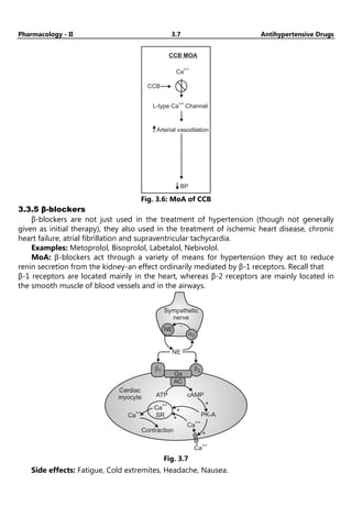
MoA: β-blockers act through a variety of means for hypertension they act to reduce
renin secretion from the kidney-an effect ordinarily mediated by β-1 receptors. Recall that
β-1 receptors are located mainly in the heart, whereas β-2 receptors are mainly located in
the smooth muscle of blood vessels and in the airways.
Sympathetic
nerve
NE
ATP cAMP
PK-A
Cardiac
myocyte
Ca
++
Ca
++
Ca
++
Ca
++
Contraction
+
+
+
+
SR
-
NE
a2
b2
b1
Gs
AC
Fig. 3.7
Side effects: Fatigue, Cold extremites, Headache, Nausea.
Pharmacology - II 3.8 Antihypertensive Drugs
3.3.6. α-blockers
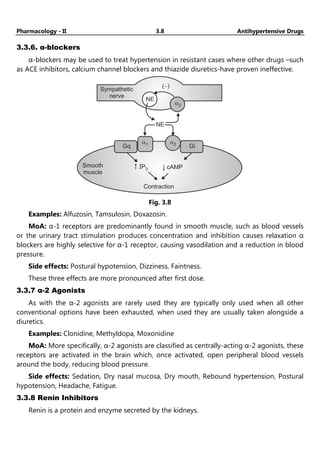
α-blockers may be used to treat hypertension in resistant cases where other drugs –such
as ACE inhibitors, calcium channel blockers and thiazide diuretics-have proven ineffective.
Sympathetic
nerve
Smooth
muscle
Contraction
cAMP
IP3
NE
( )
-
NE
a2
Gq
a1 a2
Gi
Fig. 3.8
Examples: Alfuzosin, Tamsulosin, Doxazosin.
MoA: α-1 receptors are predominantly found in smooth muscle, such as blood vessels
or the urinary tract stimulation produces concentration and inhibition causes relaxation α
blockers are highly selective for α-1 receptor, causing vasodilation and a reduction in blood
pressure.
Side effects: Postural hypotension, Dizziness, Faintness.
These three effects are more pronounced after first dose.
3.3.7 α-2 Agonists
As with the α-2 agonists are rarely used they are typically only used when all other
conventional options have been exhausted, when used they are usually taken alongside a
diuretics.
Examples: Clonidine, Methyldopa, Moxonidine
MoA: More specifically, α-2 agonists are classified as centrally-acting α-2 agonists, these
receptors are activated in the brain which, once activated, open peripheral blood vessels
around the body, reducing blood pressure.
Side effects: Sedation, Dry nasal mucosa, Dry mouth, Rebound hypertension, Postural
hypotension, Headache, Fatigue.
3.3.8 Renin Inhibitors
Renin is a protein and enzyme secreted by the kidneys.
Pharmacology - II 3.9 Antihypertensive Drugs
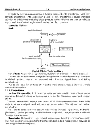
It works by cleaving angiotensinogen (hepatic-produced) into angiotensin I. ACE then
converts angiotensin-I into angiotensin-II and, in turn angiotensin-II causes increased
secretion of aldosterone-increasing blood pressure. Renin inhibitors are then, an effective
way to block the effects of angiotensin-II and reduce blood pressure.
Examples: Aliskiren
MoA:
Angiotensinogen
Aliskiren
-
Renin
Bradykinin
(active vasodilator)
Angiotensin
(inactive decapeptide)
I
- ACE inhibitors
Angiotensin
(active vasoconstrictor)
II Inactive metabolites
AT receptor blockers
1
-
AT receptor
1
Angiotensin-
converting
nzyme
Fig. 3.9: MOA of Renin inhibitors
Side effects: Angioedema, Hyperkalemia, Hypotension, Diarrhea, Headache, Dizziness.
Aliskiren should not be taken alongside an angiotensin-receptor blocker or ACE inhibitor
in diabetic patients due to an increased risk of stroke, hyperkalemia and kidney
complications.
Due to the above risk and side effect profile, many clinicians regard aliskiren as more
harmful than beneficial.
3.3.9 Vasodilator
Sodium Nitroprusside: Sodium nitroprusside has been used in cases of hypertensive
emergency. It is administered via intravenous route and for this reason, has a rapid onset of
effects.
Sodium nitroprusside deploys nitric oxide for its antihypertensive effect. Nitric oxide
works to reduce total peripheral resistance and venous return. This reduces both preload
and afterload.
Side effects associated with sodium nitroprusside include: Hypotension, Methemo-
globinemia, Cyanide poisoning, Bradyarrhythmia, Palpitation, Tachyarrhythmia, Confusion,
Dizziness, Renal azotemia.
Hydralazine: Hydralazine is used to treat hypertension, though it is more often used to
treat high blood pressure, gestational hypertension. Like sodium nitropruside, it may also be
used in hypertensive emergency.
Pharmacology - II 3.10 Antihypertensive Drugs
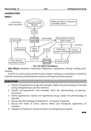
VASODILATORS:
DIRECT
a1-AR blockers
Direct vasodilators
VASODILATING
DRUGS
PVR
Arterial
pressure
Natriuresis
Aldosterone
Renin
release
AT-III
Na+ retension
and plasma volume
PVR
Baroreceptor Sympathetic nervous
system outflow
Reflex stimulation in response to
vasodilating agents
b-blockers
Venous
capacitance
Heart rate,
cardiac contractility
Arterial
pressure
Cardiac output
Fig. 3.10: MOA of Vasodilators
Side effects: Headache, Tachycardia, Palpitations, Hypotension, Aching/ swelling joints,
Flushing.
It works as a direct acting smooth muscle relaxant, working as a vasodilator in resistance
arterioles-decreasing total peripheral resistance and lowering blood pressure.
QUESTIONS
1. Classify antihypertensive drugs with examples. Explain the pharmacology of centrally
acting antihypertensive and ACE inhibitors.
2. Classify anti-hypertensive with examples. Write the pharmacology of phenoxy-
benzamine.
3. Define hypertension. Classify anti hypertensive drugs. Explain the pharmacology of
Atenolol.
4. Discuss the pharmacology of Angiotensin – II receptor antagonist.
5. Discuss the mode of action, adverse effects and therapeutic applications of
clonidine.
6. Highlight on Potassium channel activators as antihypertensive agents.
YouTube channel :-
https://youtube.com/@Pharmamasti
(4.1)
Chapter
Chapter
Chapter
Chapter ...
...
...
... 4
4
4
4
Anti-Anginal Drugs
♦ LEARNING OBJECTIVES ♦
After completing this chapter, reader should be able to:
• Recall classification of Anti-Anginal drugs.
• State the mechanism of action and ADR.
4.1 DEFINITION
Angina Pectoris is a symptom of myocardial ischemia and occurs due to an imbalance
between oxygen demand and oxygen supply of myocardium.
• Characteristic abrupt, severe, pressing-like substernal chest pain radiating towards
neck, jaw, back, and arms. Patients may also experience dyspnea or atypical
symptoms such as indigestion, nausea, vomiting, or diaphoresis.
• Usually precipitated by exercise, excitement or a heavy meal.
• Transient episodes (15 seconds to 15 minutes) of myocardial ischemia (stable
angina) do not results cellular death; as occurs in myocardial infarction (MI).
4.2 TYPES OF ANGINA
Typical Angina (Classical Angina)
• Pain is commonly induced by exercise, excitement or a heavy meal.
• Secondary to advanced atherosclerosis of coronary vessels.
• Associated with ST-segment depression on ECG.
• Usually lasts 1-15 minutes.
Variant Angina (Prinzmetal Angina)
• Pain is induced while at rest.
• Symptoms are caused by decreased blood flow to the heart muscle from the spasm
of the coronary artery.
• Although individuals with this form of angina may have significant coronary
atherosclerosis, the angina attacks are unrelated to physical activity, heart rate, or
blood pressure.
• Associated with ST-segment elevation on ECG.
• Generally responds promptly to coronary vasodilators, such as nitroglycerin and
calcium-channel blockers.
• But β-blockers are contraindicated.
Pharmacology - II 4.2 Anti-Anginal Drugs
Unstable angina (Acute Coronary Syndrome)
• May involve coronary spasm and may also have the component of atherosclerosis.
• The duration of manifestation is longer than the first two and has the manifestation
of myocardial infarction (MI).
• Lies between stable angina and MI.
• The pathology is similar to that involved in MI: a platelet-fibrin thrombus associated
with a ruptured atherosclerotic plaque but without complete occlusion of the blood
vessel.
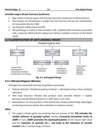
4.3 CLASSIFICATION OF ANTI-ANGINAL DRUGS
Antiaginal drugs by class
Calcium channel
blockers
Nondihydropyridine Dihydropyridine
Organic nitrates
·
·
·
·
Amyl nitrile
Nitroglycerine
sosorbide dinitrate
I
Isosorbide mononitrate
·
·
Verapamil
Diltiazem
·
·
·
Nifedipine
Nicardipine
Amlodipine
Propranolol
Atenolol
Metoprolol
Nadolol
Pindolol
b-
blocker
Fig. 4.1: Antianginal Drugs
4.3.1 Nitrates/Organic Nitrates
Nitroglycerine, Isosorbide dinitrate, Isosorbide mononitrate.
• Preload reduction: Peripheral pooling of blood → decreased venous return (preload
reduction).
• After load reduction: Nitrates also produce some arteriolar dilation → slightly
decrease total peripheral resistance or after load on the heart.
• Redistribution of coronary flow: In the arterial tree, nitrates preferentially relax bigger
conducting coronary arteries than arterioles or resistance vessels.
MOA:
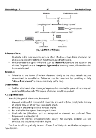
• The organic nitrate agents are prodrugs that are sources of NO. NO activates the
soluble isoforms of guanylyl cyclase, thereby increasing intracellular levels of
cGMP. In turn, cGMP promotes the dephosphorylation of the myosin light chain
and the reduction of cytosolic Ca++ and leads to the relaxation of smooth
muscles cells in a broad range of tissues.
Pharmacology - II 4.3 Anti-Anginal Drugs
Nitrates Endothelial cells
Guanylyl cyclase* Guanylyl cyclase*
NO
+
-
GTP cGMP GMP
PDE
?
MLCK*
Myosin-LC Myosin-LC-PO4 Myosin-LC
Actin
Contraction Relaxation
Sildenafil
Fig. 4.2: MOA of Nitrates
Adverse effects:
• Headache is the most common adverse effect of nitrates. High doses of nitrates can
also cause postural hypotension, facial flushing and tachycardia.
• Phosphodiesterase type 5 inhibitors such as sildenafil potentiate the action of the
nitrates. To preclude the dangerous hypotension that may occur, this combination
is contraindicated.
Tolerance:
• Tolerance to the action of nitrates develops rapidly as the blood vessels become
desensitized to vasodilation. Tolerance can be overcome by providing a daily
“nitrate free interval” to restore sensitivity to the drug.
Dependence:
• Sudden withdrawal after prolonged exposure has resulted in spasm of coronary and
peripheral blood vessels. Withdrawal of nitrates should be gradual.
4.3.2 β-Blockers
Atenolol, Bisoprolol, Metoprolol, Proranolol
• Atenolol, metoprolol, propranolol, bisoprolol are used only for prophylactic therapy
of angina; they are of no value in an acute attack.
• Effective in preventing exercise-induced angina.
• But are ineffective against the vasospastic form.
• Cardioselective β-blockers, such as metoprolol or atenolol, are preferred. Thus,
Propranolol is not preferred.
• Agents with intrinsic sympathomimetic activity (for example, pindolol) are less
effective and should be avoided in angina.
The dose should be gradually tapered off over 5 to 10 days to avoid rebound angina or
hypertension.
Pharmacology - II 4.4 Anti-Anginal Drugs
MOA: Suppress the activation of the heart by blocking B1 receptors.
I. Decrease the heart rate, resulting in:
1. decreased myocardial oxygen demand.
2. Increased oxygen delivery to the heart.
II. Decrease myocardial contractility, helping to conserve energy/ decrease demand.
III. Reduce the work of the heart by decreasing COP and causing a slight decrease in BP
• ↓ HR,
• ↓ contractility,
• ↓ systolic wall tension,
• ↑ perfusion time
Reasons for Using Nitrates and β-Blockers in Combination in Angina
• β-Blockers prevent reflex tachycardia and contractility produced by nitrate-induced
hypotension.
• Nitrates prevent any coronary vasospasm produced by β- Blockers.
• Nitrates prevent increases in left ventricular filling pressure or preload resulting from
the negative inotropic effects produced by β-Blockers.
4.3.3 Calcium Channel Blockers (CCBs)
Amlodipine, Diltiazem, Felodipine, Nicardipine, Nifedipine, Verapamil.
MOA
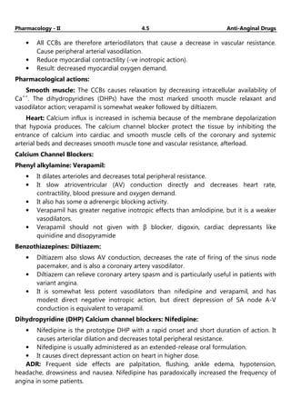
• Calcium is essential for muscular contraction.
• The CCBs protect the tissue by inhibiting the entrance of Ca+2
into cardiac and
smooth muscle cells of the coronary and systemic arterial beds.
Ca channels
2+
Ca
2+ -
channel
blockers
Ca (intracellular)
2+
Calmodulin
Ca -calmodulin complex cAMP
2+
ATP
+
+
b2
agonists
MLCK
*
MLCK-(PO )
4 2
Myosin-LC kinase
(MLCK)
Myosin
light chain
(myosin-LC)
Myosin-LC PO4
Actin
Myosin-LC
+
Contraction Relaxation
Fig. 4.3: MOA of Ca
+
Channel Blockers
Pharmacology - II 4.5 Anti-Anginal Drugs
• All CCBs are therefore arteriodilators that cause a decrease in vascular resistance.
Cause peripheral arterial vasodilation.
• Reduce myocardial contractility (-ve inotropic action).
• Result: decreased myocardial oxygen demand.
Pharmacological actions:
Smooth muscle: The CCBs causes relaxation by decreasing intracellular availability of
Ca++
. The dihydropyridines (DHPs) have the most marked smooth muscle relaxant and
vasodilator action; verapamil is somewhat weaker followed by diltiazem.
Heart: Calcium influx is increased in ischemia because of the membrane depolarization
that hypoxia produces. The calcium channel blocker protect the tissue by inhibiting the
entrance of calcium into cardiac and smooth muscle cells of the coronary and systemic
arterial beds and decreases smooth muscle tone and vascular resistance, afterload.
Calcium Channel Blockers:
Phenyl alkylamine: Verapamil:
• It dilates arterioles and decreases total peripheral resistance.
• It slow atrioventricular (AV) conduction directly and decreases heart rate,
contractility, blood pressure and oxygen demand.
• It also has some α adrenergic blocking activity.
• Verapamil has greater negative inotropic effects than amlodipine, but it is a weaker
vasodilators.
• Verapamil should not given with β blocker, digoxin, cardiac depressants like
quinidine and disopyramide
Benzothiazepines: Diltiazem:
• Diltiazem also slows AV conduction, decreases the rate of firing of the sinus node
pacemaker, and is also a coronary artery vasodilator.
• Diltiazem can relieve coronary artery spasm and is particularly useful in patients with
variant angina.
• It is somewhat less potent vasodilators than nifedipine and verapamil, and has
modest direct negative inotropic action, but direct depression of SA node A-V
conduction is equivalent to verapamil.
Dihydropyridine (DHP) Calcium channel blockers: Nifedipine:
• Nifedipine is the prototype DHP with a rapid onset and short duration of action. It
causes arteriolar dilation and decreases total peripheral resistance.
• Nifedipine is usually administered as an extended-release oral formulation.
• It causes direct depressant action on heart in higher dose.
ADR: Frequent side effects are palpitation, flushing, ankle edema, hypotension,
headache, drowsiness and nausea. Nifedipine has paradoxically increased the frequency of
angina in some patients.
Pharmacology - II 4.6 Anti-Anginal Drugs
Other dihydropyridine (DHP) calcium channel blockers:
• Amlodipine, an oral dihydropyridine, functions mainly as an arteriolar vasodilators.
• Nitrendipine is a calcium channel blocker with additional action of vasodilation
action. Vasodilation action is due to release NO from the endothelium and inhibits
cAmp phosphodiesterase.
• Lacidipine, is a highly vasoselective newer DHP.
• Nimodipine is short-acting DHP which penetrates blood-brain barrier very efficiently
due to high lipid solubility.
• DHP with long duration of action: Lercanidipine, Benidipine.
Uses:
• Calcium channel blockers can be safely given to patients with obstructive lung
disease and peripheral vascular disease in whom β blocker are contraindicated.
• CCB are used for the treatment of:
o Angina pectoris
o Hypertension
o Cardiac arrhythmias
o Hypertrophic cardiomyopathy
ADR: Lightheadedness. Low blood pressure. Slower heart rate.
Potassium Channel Openers:
Nicorandil:
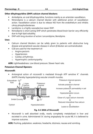
• Antianginal action of nicorandil is mediated through ATP sensitive K+
channels
(KATP) thereby hyperpolarizing vascular smooth muscles.
Extracellular
fluid
Ca
2+
L-type
calcium
channel
Potassium
channel
K
+
Cytosol
Inhibit Ca entry
Nicorandil
Hyperpolarization
- +
Potassium channel openers
Activate potassium channel
Increase potassium permeability in cell
Hyperpolarisation occurs
Closer of L-type calcium channels
Reduced intracellular free calcium
Leads to vasodilatation
Fig. 4.4: MOA of Nicorandil
• Nicorandil is well absorbed orally, nearly completely metabolized in liver and
excreted in urine. Administered I.V. during angioplasty for acute MI, it is believed to
improve outcome.
ADR: Flushing, palpitation, weakness, headache, dizziness, nausea and vomiting.
Pharmacology - II 4.7 Anti-Anginal Drugs
4.4 OTHER ANTIANGINAL DRUGS
Drug Pharmacological Action
Dipyridamol • Dipyridamol inhibit platelet aggregation.
• It is a powerful coronary dilator.
Trimetazidine • This antianginal drugs act by nonhaemodynamic mechanisms.
• The MOA of trimetazidine is uncertain, but it may improve
cellular tolerance to ischemia by inhibiting mitochondrial long
chain 3-ketoacyl-CoAhiolase.
Ranolazine • This novel antianginal drug primarily act by inhibiting late Na+
current in the myocardium.
Ivabradine • This ‘pura’ heart rate lowering antianginal drugs has been
introduced recently as an alternative to β blocker.
• It blocks cardiac pacemaker (sino-atrial) cell ‘f’ channels.
Oxyphedrine • Improve myocardial metabolism.
Questions
1. Classify anti-anginal agents and write pharmacology of nitroglycerine.
2. What is angina pectoris? Classify anti-anginal agents. Describe the pharmacology of
nitrates.
3. Explain the pharmacology of organic nitrates.
https://youtube.com/@Pharmamasti
(5.1)
Chapter
Chapter
Chapter
Chapter ...
...
...
... 5
5
5
5
Anti-Arrhythmic Drugs
♦ LEARNING OBJECTIVES ♦
After completing this chapter, reader should be able to:
• Enlist the anti-arrhythmic drugs.
• Describe mechanism of action and ADR.
5.1 DEFINITION
Arrhythmia is defined as the variation of heart from normal rhythm.
Disturbances in the rhythm, heart rate, impulse generation or conduction of electrical
impulses is responsible for membrane depolarization. These changes can escort to
alterations in overall cardiac function that can be life-threatening.
5.2 MECHANISMS OF ARRHYTHMIAS
• Disturbances in generation of impulse may be due to:
o Abnormal automaticity.
o Delayed after depolarization.
• Disturbances in conduction of impulse:
o By causing repeated activation (re-entry), impulse may re-circulate in heart.
o Blocks conduction.
5.3 CLASSIFICATION OF ANTI-ARRHYTHMIC DRUGS
(Vaughan-Williams-Singh 1969)
Class 1
Na channel blocker
1a (moderate):
Quinidine, procainamide
1b (weak):
lidocaine, phenytoin
1c (strong):
Flecainide, propafenone
+
Class 4
Ca
channel blocker
verapamil
diltiazem
2+
Class 3
K channel blocker
amiodarone
sotalol
+
Class 2
-blocker
propranolol
metoprolol
b
K /Cl (out)
-
+
Na (in)
+ K (out)
+
K (rectifier)
+
4
0
1 2
3
4
Ca (in)
2+
Fig. 5.1
Pharmacology - II 5.2 Anti-Arrhythmic Drugs
Class I: Block Na+ channels Ia (quinidine, Disopyramide, Lidocaine, Mexiletine,
procainamide, Propafenone)
Ib (lignocaine, phenytoin, mexiletine)
Ic (flecainide)
Class II: ß-adrenoceptor antagonists Atenolol, Esmolol, Metoprolol, Propranalol.
Class III: K+
channel blocker-prolong
action potential and prolong
refractory period.
Amiodarone, Dofetilide, Sotalol, Dronedarone,
Ibutilide.
Class IV: Ca2+
channel antagonists Verapamil, Diltiazem.
Other anti-arrhythmic drugs Adenosine.
Classification based on Clinical use:
• Drugs used for supraventricular arrhythmia`s.
o Adenosine, verapamil, diltiazem.
• Drugs used for ventricular arrhythmias.
o Lignocaine, mexelitine, bretylium.
• Drugs used for both supraventricular and ventricular arrhythmias.
o Amiodarone, β-blockers, disopyramide, procainamide.
Na+ Channel Blocker:
• Bind to and block Na+ channels (and K+ also).
• Act on initial rapid depolarisation (slowing effect).
• Local Anaesthetic (higher concentration): block nerve conduction.
• Do not alter resting membrane potential (Membrane Stabilisers).
• At times, post repolarization refractoriness.
• Bind preferentially to the open channel state.
• Use Dependence: The more the channel is in use, the more drug is bound.
CLASS IA:
No drug
0mV
Phase 0
( )
INa
-85 mV Effective
refractory
period
Phase 3 ( )
IK
Class 1A drugs slow
phase 0 depolarization,
prolong action potential,
and slow conduction.
Fig. 5.2
Quinidine:
• D-isomer of quinine obtained from cinchona bark.
• Historically first antiarrhythmic drug used.
Pharmacology - II 5.3 Anti-Arrhythmic Drugs
MOA:
• Blocks sodium channels.
• Decreases automaticity, conduction velocity and prolong S repolarization.
• Decreases phase 0 depolarization, increases action potential duration (APD) and
effective refractory period (ERP).
Other actions:
• Decreases BP (α-block), skeletal muscle relaxation.
Clinical Pharmacokinetics
• Well absorbed.
• 80% bound to plasma proteins (albumin).
• Extensive hepatic oxidative metabolism.
• 3-hydroxyquinidine, is nearly as potent as quinidine in blocking cardiac Na+
Channels and prolonging cardiac action potentials.
Uses:
• To maintain sinus rhythm in patients with atrial flutter or atrial fibrillation.
• To prevent recurrence of ventricular tachycardia or VF.
Adverse Effect:
Non-cardiac
• Diarrhea, thrombocytopenia.
• Cinchonism and skin rashes.
Cardiac:
• Marked QT-interval prolongation and torsades de pointes (2-8%).
• Hypotension.
• Tachycardia.
Drug Interactions:
• Metabolized by CYP450.
• Increases digoxin levels.
• Cardiac depression with beta blockers.
• Inhibits CYP2D6.
Disopyramide:
MOA:
Disopyramide produces a negative ionotropic effects that is greater than weak effect
exerted by quinidine and procainamide, and unlike the latter drugs, disopyramide causes
peripheral vasoconstriction.
Adverse Effects:
• Precipitation of glaucoma.
• Constipation, dry mouth.
• Urinary retention.
• Myocardial depression.
Pharmacology - II 5.4 Anti-Arrhythmic Drugs
Drug Interactions:
Both metabolism of disopyramide and the accumulation of its metabolite are increased, in
the presence of phenytoin, thus escalating the probability of anti-cholinergic properties.
Use:
• Ventricular tachycardia.
• Atrial flutter (AF) and Atrial fibrillation (AFI).
Procainamide:
• Procaine derivative, quinidine like action.
MOA:
• Procainamide binds to open and inactivated Na+ channels and prevents sodium
influx, slowing the rapid upstroke during phase 0.
Adverse Effect:
• Hypotension.
• Hypersensitivity reaction.
Drug interactions:
• Cimitidine inhibits the metabolism of procainamide.
Uses:
• Premature atrial contractions.
• Paroxysmal atrial tachycardia.
CLASS IB
• They shorten Phase 3 repolarization.
• ↓ the duration of the cardiac action potential.
• Prolong phase 4.
IBL:
5.3
No
drugs
0 mV
-85 mV
Phase 3 ( )
IK
Effective
refractory period
Class 1B drugs shorten
phase 3 repolarization
and decrease the duration
of the action potential
Phase 0
( )
INa
Fig. 5.3
Lidocaine
MOA
• It shorten phase 3 repolarization and decreases the duration of action potential.
Pharmacology - II 5.5 Anti-Arrhythmic Drugs
Pharmacokinetics:
• High first pass metabolism.
• Metabolism dependent on hepatic blood flow.
• T½ = 8 min – distributive, 2 hrs – elimination.
• Propranolol decreases half life of lignocaine.
• Dose = 50-100 mg bolus followed by 20-40 mg every 10-20 min i.v.
Adverse Effects:
• Drowsiness.
• Slurred speech.
• Confusion and convulsions.
Drug Interaction:
• Propranolol increases its toxicity.
• The myocardial depressant effect of lidocaine is enhanced by phenytoin
administration.
Uses:
• VA.
• Digitalis toxicity.
Phenytoin:
Phenytoin was originally introduced for the control of convulsive disorders but now also
been shown to be effective in the treatment of cardiac arrythmias.
Adverse Effects:
• Respiratory arrest.
• Hypotension.
Drug Interaction:
• In the presence of chloramphenicol, disulfiram, and isoniazid, the plasma
concentration of phenytoin is increased, since the later drugs inhibit the hepatic
metabolism of phenytoin.
Uses:
• Anaesthesia.
• Open heart surgery.
• Digitalized induced and ventricular arrythmia in children.
Mexiletine
MOA
• By the oral route, it acts as local anesthetic as well as an active antiarrythmic;
chemically and pharmacologically similar to lidocaine.
• It reduces automaticity in Purkinje Fibres (PF), both by decreasing phase IV slow and
by increasing threshold voltage.
• It may convert one-way block to two-way block by reducing the rate of 0 phase
depolarization in ischemic PF.
Pharmacology - II 5.6 Anti-Arrhythmic Drugs
Adverse Effects:
• Tremor
• Hypotension
• Bradycardia
Drug Interactions:
• When mexiletine is administered with phenytoin or rifampin, since these drugs
stimulate the hepatic metabolism of mexiletine, reducing its plasma concentration.
Uses:
• VA
• Congenital long QT syndrome.
CLASS IC
• Markedly slow Phase 0 depolarization.
• Slow conduction in the myocardial tissue.
• Minor effects on the duration of action potential and ERP.
• Reduce automaticity by increasing threshold potential rather than decreasing slope
of Phase 4 depolarization.
No
drugs
0 mV
Phase 0
( )
INa
-85 mV
Phase 3 ( )
IK
5.3
Effective
refractory period
Class C drugs markedly
slow phase 0
depolarization
I
Fig. 5.4
FLECAINIDE
MOA
• Flecainide suppresses phase 0 upstroke in purkinje and myocardial fibers.
• This causes marked slowing of conduction in all cardiac tissues, with a minor effect
on the duration of the action potential and refractoriness.
• Automaticity is reduced by an increase in the threshold potential rather than a
decrease in the slope of phase 4 depolarization.
• Potent blocker of Na & K channels with slow unblocking kinetics.
• Maintain sinus rhythm in supraventricular arrhythmias.
Pharmacology - II 5.7 Anti-Arrhythmic Drugs
Adverse Effect:
• Torsades de point, visual disturbances & headache.
Uses
• Ventricular arrhythmia.
Class II Drugs: β- Blockers
PROPRANOLOL, METOPROLOL, ESMOLOL, ACEBUTOLOL
β-receptor stimulation:
• Increase automaticity.
• Decrease AV conduction velocity.
• Increase refractory period.
• β-adrenergic blockers competitively block catecholamine induced stimulation of
cardiac β-receptors.
β-blockers:
• Depress phase 4 depolarization of pacemaker cells,
• Slow sinus as well as AV nodal conduction: Decrease Heart Rate (HR), Increase Pulse
Rate (PR).
• Increase ERP, prolong Action Potential (AP) Duration by Decrease AV conduction
• Reduce myocardial oxygen demand.
Propranolol
MOA
• Propanolol decreases the slope of phase 4 depolarization.
• Prolong the ERP of A-V node.
Adverse Effect:
• Hypoglycemia (infants)
• Asthma
• Bronchospasm
Uses:
• Atrial Fibrillation.
• Digitalis induced arrhythmias
Esomolol
MOA
• Esomolol is a short-acting β1-selective adrenoreceptor blocker.
• It doesn’t possess membrane-stabilizing activity.
• Route of administration is through i.v.
Adverse Effects:
• Nausea
• Hypotension
• Headache
Pharmacology - II 5.8 Anti-Arrhythmic Drugs
Uses:
• Supra ventricular tachyarrythmias.
Class III Drugs:
5.3
Effective
refractory period
Class drugs prolong
phase 3 repolarization,
without altering phase 0.
III
Phase 3 ( )
IK
Phase 0
( )
INa
-85 mV
0 mV
No
drug
Fig. 5.5
Amiodarone:
• Iodine containing long acting drug.
MOA: (Multiple actions)
• Prolongs APD by blocking K+
channels.
• Blocks inactivated sodium channels.
• β blocking action , Blocks Ca2+
channels.
• Decrease conduction, decrease ectopic automaticity.
Pharmacokinetics:
• Variable absorption 35-65%.
• Slow onset 2 days to several weeks.
• Duration of action: weeks to months.
• Many drug interactions.
Uses:
• Can be used for both supraventricular and ventricular tachycardia.
Adverse effects:
• Cardiac: hypotension, bradycardia, QT prolongation, heart block , cardiac failure.
• Pulmonary: pneumonitis leading to pulmonary fibrosis.
• Bluish discoloration of skin.
• GIT disturbances, hepatotoxicity.
• Causes hypothyroidism or hyperthyroidism by blocking peripheral conversion of T4
to T3.
Bretylium:
• Adrenergic neuron blocker used in ventricular arrhythmias.
Pharmacology - II 5.9 Anti-Arrhythmic Drugs
Sotalol:
• Beta blocker.
Dofetilide:
• Selective K+
channel blocker, less adverse events.
• Oral use in AF to convert or maintain sinus rhythm.
Ibutilide:
• K+
channel blocker used as IV infusion in AF.
Class IV: Ca2+
channel Blockers
• Inhibit the inward movement of calcium.
• Decrease contractility, automicity and AV conduction.
Effective
refractory period
-75 mV
Note
No
drugs
Phase 0
( )
ICa
0 mV
Phase 2 ( and )
I
ICa K
Group
action
IV
Class V drugs slow phase 4 spontaneous
depolarization and slow conduction in
tissues dependent on calcium
currents, such as the AV node
I
Fig. 5.6
Verapamil:
Uses:
• Terminate PSVT.
• Control ventricular rate in atrial flutter or fibrillation.
Drug interactions:
• Displaces digoxin from binding sites.
• Decrease renal clearance of digoxin.
Anti Arrhythmic Drugs:
Class Mechanism Action Notes
I. Na+
channel blocker Change the slope of
phase 0.
Can abolish
tachyarrhythmia caused
by reentry circuit.
II. β-blocker Increase heart rate and
conduction velocity.
Can indirectly alter K+
and
Ca2+
conductance.
contd. …
…
…
…
Pharmacology - II 5.10 Anti-Arrhythmic Drugs
III. K+
channel blocker 1. Increase action
potential duration
(APD) or effective
refractory period
(ERP).
2. Delay repolarization.
Inhibit re-entry
tachycardia
IV. Ca++
channel blocker Slowing the rate of rise in
phase 4 of SA node.
Decrease conduction
velocity in SA and AV
node.
5.4 OTHER ANTI-ARRHYTHMIC
Adenosine:
• Purine nucleotide having short and rapid action.
MOA:
• It activates ACh sensitive K+
channels and causes membrane hyperpolarization
through interaction with A1 type of adenosine GPCRs on SA node coronaries.
Adverse events:
• Nausea, dyspnoea, flushing, headache.
QUESTIONS
1. Enlist anti-arrhythmic drugs with examples and mechanism of action of quinidine.
2. Discuss the action and uses of Propranolol.
3. Discuss about Lidocaine as an anti-arrhythmic agents.
4. Mention types of arrhythmias and classify anti-arrhythmic drugs.
5. Discuss the pharmacology of calcium channel blockers.
https://youtube.com/@Pharmamasti
(6.1)
Chapter
Chapter
Chapter
Chapter ...
...
...
... 6
6
6
6
Anti-Hyperlipidemic Drugs
♦ LEARNING OBJECTIVES ♦
After completing this chapter, reader should be able to:
• Enlist the drugs.
• Summarize mechanism of action and ADR.
6.1 HYPERLIPIDEMIA
Hyperlipidemia is a broad term, it is also called as hyperlipoproteinemia, it is a common
disorder in developed countries and major cause of coronary heart disease.
It is a result of abnormalities in lipid metabolism or plasma lipid transport or a disorder
in the synthesis and degradation of plasma lipoprotein.
Causes of Hyperlipidemia:
• Most of the time hyperlipidemia is caused due to lifestyle habits or treatable medical
conditions.
• Obesity, not exercising, and smoking, diabetes, obstructive jaundice, and an under
active thyroid gland inherit hyperlipidemia.
6.2 THE BIOCHEMISTRY OF PLASMA LIPIDS
Lipids: Lipids are the heterogenous mixtures of fatty acids and alcohol that are present
in the body. The major lipids in the bloodstream are cholesterol and it’s esters,
triglycerides and phospholipids.
Cholesterol: It is C27− steroid that serves as an important component of all cell
membranes and important precursor molecule for the biosynthesis of bile acids, steroid
hormones, and several fat-soluble vitamins.
Normal functions of cholesterol in the body:
• It is compulsory for new cells to form and for older cells to repair themselves after
injury.
• It is also used by the adrenal glands to form hormones such as cortisol, by the
testicles to form testosterone, and by the ovaries to produce estrogen and
progesterone.
Normal functions of triglycerides and phospholipids in the body.
• Triglycerides supply energy for the body. Triglycerides are required to meet
immediate energy needs in muscles or to store fat for future energy requirements.
• Phospholipids are compounds that are used to make cell membranes, generate
second messengers, and store fatty acids for the use in generation of prostaglandins.
Pharmacology - II 6.2 Anti-Hyperlipidemic Drugs
Lipoproteins:
• Blood and other body fluids are watery, so fats require a special transport system to
travel around the body.
• Fats are travelling from one place to another mixing with protein particles, called
lipoproteins.
• Four (or five) types of lipoproteins are there, each having particular distinct job.
• A lipoprotein contains both proteins and lipids, bound to another protein which is
called apolipoproteins, which allow fats to move through the water inside and
outside cells. Provide structural support and stability, binds to receptors.
Classification of Lipoproteins:
Classification Composition Primary function
Chylomicrons Triglyceride TGs 99%, 1% protein. Transport dietary TGs to adipose
tissue and muscle.
VLDL Newly synthesized TGs.
Lipid 90%, 10% protein.
Transport endogenous TGs to
adipose tissue and muscle.
IDL Intermediate between VLDL and
LDL.
They are not usually detectable in
the blood.
LDL Lipid 80%, 20% protein. Transport endogenous cholesterol
from liver to tissues.
HDL Lipid 60%, 40% protein. Collect cholesterol from the
body's tissues, and take it back to
the liver.
Lipoproteins
Liver produces
lipoproteins
LDL
Molecule
VLDL
Molecule
IDL
Molecule
Fat cells store
Triglyceride (fat)
Protein Shell
Cholesterol
Triglycerise (fat)
Liprotein Molecule
Protein Shell
Cholesterol
Triglyceride (fat)
Lipoprotein Molecule
Muscle cells burn
Triglyceride (fat)
Fig. 6.1: Life cycle of Cholesterol-Carrying Lipoproteins
Pharmacology - II 6.3 Anti-Hyperlipidemic Drugs
Synthesis of
lipoproteins
(VLDL)
Liver
Small Intestine
Synthesis of
chylomicrons from
dietary lipids
VLDL with TG
transported
Chylomicrons
with
TG transported
Stored
triacylglycerol
Adipose
tissue
Fatty
acids
Triacylglycerols
and fatty acids utilized
Muscle, liver, heart etc.
ENERGY
Fig. 6.2: Overview of Fat Metabolism
6.3 CLASSIFICATION OF ANTIHYPERLIPIDEMIC DRUGS
Sr. No. Class Example
1. HMG CoA Reductase Inhibitors Lovastatin, Simvastatin, Metastatin,
Pravastatin, Fluvastatin, Atorvastatin,
Pitavastatin, Rosuvastatin
2. Fibrates Clofibrate, Fenofibrate, Gemfibrozil,
Ciprofibrate, benzafibrate, Fluvestatin.
3. Bile acid sequestrants Cholestyramine, Colestipol
4. LDL oxidation inhibitor Probucol
5. Pyridine derivatives Nicotinic acid, Nicotinamide
6. Cholesterol absorption inhibitors Ezetimibe
7. Miscellaneous agents β-Sitosterol, Dextrothyroxine
1. HMG- CoA Reductase inhibitors (STATINS):
• 3-hydroxy-3-methylglutaryl-coenzyme A (HMG-CoA).
• The enzyme that catalyzes the conversion of HMG-CoA to mevanolate.
• This reaction is the rate determining step in the synthetic pathway of cholesterol.
Statins:
• Lovastatin was isolated from Aspergillus terreus.
Pharmacology - II 6.4 Anti-Hyperlipidemic Drugs
• There are two classes of statins:
o Natural Statins: Lovastatin(mevacor), Pravastatin (pravachol), Simvastatin
(Zocor).
o Synthetic Statins: Atorvastatin (Lipitor), Fluvastatin (Lescol).
• Statins are competitive inhibitors of HMG-CoA reductase. They are bulky and “stuck”
in the active site.
• Statins prevents the binding of enzyme with its substrate, HMG CoA.
Acetyl CoA and Acetoacetyl CoA
Statins Stop
Squalene
HMG-CoA
HMG-CoA reductase
Mevalonate
Farnesylpyrophosphate
Cholesterol
(in humans)
Ergosterol
(in fungi)
Geranylgeranyl pyrophosphate
Fig. 6.3: MoA of Statins
Drug ADR Uses
Lovastatin Increased creatinine phosphor-
kinase, Flatulence, Nausea.
Antihyperlipoproteinemic agent.
Simvastatin Headache, nausea, flatulence,
heartburn, abdominal pain.
Antihyperlipdemic agent.
Pravastatin GI disturbances, headache, insomnia,
chest pain, rash.
Antihyperlipoproteinemic agent.
Atorvastatin Headache, flatulence, diarrhea. Primmary hyperlipidemia and
secondary hyper cholesterolemia.
Rosuvastatin Headache, dizziness, constipation,
nausea, vomiting.
High LDL, total cholesterol, TGs.
2. Fibrates:
• Fibrates are antihyperlipidemic agents, these are used in the treatment of various
forms of hyperlipidemia and hypercholesterolemia.
• These are 2-phenoxy-2-methyl propanoic acid derivatives.
Pharmacology - II 6.5 Anti-Hyperlipidemic Drugs
• These drugs stimulate β-oxidation of fatty acids in mitochondria.
• These are specially using for decreasing plasma levels of fatty acid and
triacylglycerol.
MOA:
• Fibrates are decreasing plasma TGs levels.
• This class of drugs acting through lowering of the blood triglyceride levels by
decreasing the production of liver’s VLDL (the triglyceride-carrying particle that
circulates in the blood) by activation of lipoprotein lipase and enhances the removal
of TGs from the blood. It is due to activation of PPAR-α.
• These drugs are acts by increasing blood HDL cholesterol and not effective in
lowering LDL cholesterol.
a
a
HDL synthesis Decreased
Triglycerides
Decreased in
inflammation
b
Fig. 6.4: MoA of Fibrates
Drug ADR Uses
Clofibrate Cholecystitis, gall stone,
eosinophilia, pneumonia.
Type III Hyperlipoproteinemias.
Gemfibrozil Myositis syndrome, Cholelithiasis,
GI disturbances, rash and
headache.
Hyperlipidemia.
Fenofibrate Headache, dizziness, asthaenia,
fatigue, arrhythmia.
More potent hypercholesterolemic
and triglycerides lowering agent.
Pharmacology - II 6.6 Anti-Hyperlipidemic Drugs
3. Bile Acid Sequestrants:
• Cholestyramine (Questran)
• Colestipol hydrochloride (Colestid)
• Colesevelam (tablet form)
Also called bile acid–binding resins and ion-exchange resins.
Bile Acid Sequestrants
Increased Bile
Synthesis
Statins Ezetimibe
Inhibition of HMG-CoA
Reductase
Decreased Intestinal
Cholesterol Absorption
Decrease in Hepatic Cholesterol
Increase in Hepatic LDL Receptors
Decrease in Plasma LDL
Fig. 6.5: MoA of Anti-hyper lipidemic drug
4. Cholesterol Absorption Inhibitor:
Ezetimibe (Zetia):
It is a drug that lowers plasma cholesterol levels. It acts by decreasing cholesterol
absorption in the intestine.
MOA:
• Ezetimibe lowers the plasma cholesterol levels by inhibiting the absorption from
intestine.
• This drug decreases the cholesterol delivery to the liver which in turn clears more
cholesterol from the blood.
• It has selective action (( not interfere with TGs, lipidsoluble vitamins absorption)
• The levels of LDL-C in the serum are reduced by bile acid sequestrants.
Therapeutic Uses:
• As mono therapy or in combination with HMGRI for reduction of elevated total
cholesterol.
5. Pyridine Derivatives:
• Nicotinic Acid (Niacin): Nicotinic acid is a water soluble vitamin of the B family;
after converted into the amide, it is incorporated as NAD. For the effective action the
dose should be 1.5 to 3.5 gm daily. It is available as sustained release dosage form.
Vitamin B3:
• For Lipid-lowering action it requires higher doses than when used as a vitamin.
• Effective, inexpensive, also used in combination with other lipid-lowering drugs.
Pharmacology - II 6.7 Anti-Hyperlipidemic Drugs
MOA:
• It Increases the activity of lipase, which breaks down the lipids.
• It decreases the metabolism of cholesterol and triglycerides.
Indications:
• It is effective in the lowering of triglycerides, total serum cholesterol, and LDL levels.
• It also increases the HDL levels.
• It is also effective in the treatment of types IIa, IIb, III, IV, and V hyperlipidemias
Adverse Effect:
• Flushing (due to histamine release)
• Pruritus
• GI distress
• Liver dysfunction and jaundice. Serious liver damage is the most important risk.
6. LDL Oxidation Inhibitor:
Probucol:
HO
(H C) C
3 3
CH3
CH3
S C S
(H C) C
3 3
HO
C(CH )
3 3
C(CH )
3 3
• Probucol has two tertiary butyl phenol groups linked by the help of dithiopropy-
lidene bridge and gives a high lipophilic character along with high antioxidant
properties.
• In humans, it reduces both liver and serum cholesterol levels and it does not alter
plasma triglycerides.
• It reduces LDL levels.
• It decreases the extent of HDL levels by a unique mechanism that is still not clearly
delineated.
• The decreased HDL level may be caused by the ability of probucol to inhibit the
synthesis of apoprotein A-1, a major protein component of HDL.
• It is effective in reducing the levels of LDL and it is also used in hyper-
lipoproteinemias characterized by increased LDL levels.
ADR:
• GI disorders and prolongation of GI intervals.
Use:
• It is used as antihyperlipoproteinemic agent.
7. Miscellaneous Agent:
β-Sitosterol:
• Sitosterol is a plant sterol, whose structure is identical with that of cholesterol, except
for the substituted ethyl group on C-24 of its side chain.
Pharmacology - II 6.8 Anti-Hyperlipidemic Drugs
• Its hypolipidemic effect is not clearly understood, it is claimed that the drug inhibits
absorption of dietary cholesterol from the gastrointestinal tract.
• It is absorbed poorly from the mucosal lining and it is competing with cholesterol for
absorption site in the intestine.
ADR:
• Diarrhoea, constipation, GI disturbances.
Use:
• Anti cholesteremic agent and treatment of prostatic edema.
Questions
1. Classify hypolipidemic agents with examples, write the pharmacology of
Atorvastatin.
2. Write the classification of hypolipidemics. Explain the complications of dyslipidemia.
3. Write the classification of hypolipidemics with examples. Describe the mechanism of
Niacin.
Pharma Masti
(7.1)
Unit II
Chapter
Chapter
Chapter
Chapter ...
...
...
... 7
7
7
7
Drugs used in the Therapy of Shock
♦ LEARNING OBJECTIVES ♦
After completing this chapter, reader should be able to:
• Discuss the general concepts associated with shock states, including physiologic response to shock, and
shock progression.
• Classify drugs and illustrate mechanism of action and ADR
7.1 INTRODUCTION
It is abnormal physiological state resulting from widespread and serious reduction of
tissue perfusion that if prolonged will lead to generalized impairment of cellular function.
• A life-threatening clinical syndrome of cardiovascular collapse characterized by:
o An acute reduction of effective circulating blood volume (hypotension).
o An inadequate perfusion of cells and tissues (hypoperfusion).
• If uncompensated, these mechanisms may lead to impaired cellular metabolism and
death.
• The clinical manifestations of shock are the result of stimulation of the sympathetic
and neuroendocrine stress responses, inadequate oxygen delivery, end-organ
dysfunction.
7.2 TYPES OF SHOCK
• Hypovolemic shock
• Cardiogenic shock
• Distributive shock
o Septic shock
o Neurogenic shock
o Anaphylatic shock
Management of Shock:
• Ionotrope: An agent that changes myocardial contractility.
• Vasopressor: An agent that increases blood pressure.
• Chronotrope: An agent that changes heart rate.
• Dromotrope: An agent that increases cardiac conduction velocity.
Pharmacology - II 7.2 Drugs used in the Therapy of Shock
Norepinephrine:
• Most widely used vasopressor.
• Potent α1 agonist causing vasoconstriction in tissue beds.
• Resultant increase in SVR causes rise in blood pressure.
• Standard dose: 4 mg in 50 ml (0.08 mg/ml).
Epinephrine:
• Nature’s vasopressor.
• Most commonly used during resuscitation cardiac arrest and anaphylaxis.
α1: Increases SVR.
β1: Increases HR and myocardial contractility.
β2: Bronchial smooth muscle relaxation.
Standard dose: 10 mg in 50 ml (0.2mg/ml).
Dopamine:
• Vasopressor agent.
• Use in cardiogenic and septic shock.
• Receptor stimulation depend on dose given.
Dobutamine:
• A synthetic cathecholamine.
• An inodilator.
• β1 stimulation: Increase HR and increase cardiac contractility.
• β2 mediated vasodilatation.
• Reduction in MAP is common with dobutamine.
• NE usually needed to offset vasodilatation.
Vassopressin:
• Peptide hormone released from posterior pituitary.
• Causes increase permeability of DCT and CT, increases water retention.(V2 receptor).
• V1 receptor present in the smooth muscle of a arteriolar wall and stimulation causes
smooth muscle contraction and vasoconstriction.
7.2.1 Hypovolemic Shock
Improper tissue perfusion as a result of severe loss of blood or other fluid from the body
or inadequate fluid intake, any of which decrease intravascular volume.
Causes of Hypovolemic Shock;
• Haemorragic (acute blood loss)
• Burns
• Excessive vomiting and diarrhea
Pharmacology - II 7.3 Drugs used in the Therapy of Shock
Pathophysiology of Hypovolemic shock
Hemorrhage from small venules and veins (50%)
↓
Decreased filling of right heart
↓
Decreased filling of pulmonary vasculature
↓
Decreased filling of left atrium and ventricle
↓
Left ventricular stroke volume decreases (Frank Starling )
↓
Drop in arterial blood pressure and tachycardia
↓
Poor perfusion to pulmonary arteries
↓
Cardiac depression and pump failure
Classsification of Hypovolemic Shock:
• Hemorrhagic: Trauma, gastrointestinal bleeding.
• Non-Hemorrhagic: external fluid loss, diarrhoea, vomiting, polyurea, fluid
redistribution, burns, anaphylaxis.
Signs and Symptoms:
• Anxiety, restlessness, altered mental state.
• Hypotension.
• A rapid, weak, thready pulse.
• Cool, clammy skin.
• Rapid and shallow respirations.
• Hypothermia.
• Thirst and dry mouth.
• Distracted look in the eyes.
Compensatory Mechanisms:
1. Adrenergic discharge.
2. Hyperventilation.
3. Vasoactive hormones Angiotensin, Vasopressin, Epinephrine.
4. Collapse.
5. Re-absorption of fluid from interstitial tissue.
6. Resorption of fluid from intracellular to extracellular space.
7. Renal conservation of body water and electrolyte.
Clinical Monitoring:
• Blood pressure
• Respiration
• Urine output
• Central venous pressure
Pharmacology - II 7.4 Drugs used in the Therapy of Shock
• ECG
• Swan-Ganz catheter
o cardiac output.
o mixed venous oxygen level.
o vascular pressure.
• Pulmonary artery wedge pressure.
Diagnosis:
• In management of trauma patients, understanding the patterns of injury of the
patient in shock will help direct the evaluation and management.
• Blood loss sufficient to cause shock is generally of a large volume (e.g. external,
intrathoracic, intra-abdominal, retroperitoneal, and long bone fractures).
• Diagnostic and therapeutic tube thoracotomy may be indicated in unstable patients
based on clinical findings and clinical suspicion.
• Chest radiographs, pelvic radiography, diagnostic ultrasound or diagnostic
peritoneal lavage.
MANAGEMENT:
Objectives:
(a) Increase Cardiac Output
(b) Increase Tissue Perfusion
The plan of action should be based on:
(a) Primary problem
(b) Adequate fluid replacement
(c) Improving myocardial contractility
(d) Correcting acid-base disturbances
• Resuscitation
• Immediate control of bleeding: Rest, Pressure Packing.
Operative Methods:
• Extracellular fluid replacement:
o Infusion of fluid is the fundamental treatment.
o Crystalloids, for initial resuscitation for most forms of hypovolemic shock.
o After the initial resuscitation, with up to several liters of crystalloid fluid, use of
colloids.
Drugs:
1. Sedatives
2. Chronotropic agents
3. Inotropic
7.2.2 Distributive Shock
• As in hypovolemic shock, there is an insufficient intravascular volume of blood.
• This form of "relative" hypovolemia is the result of dilation of blood vessels which
diminishes systemic vascular resistance.
• Examples of this form of shock:
1. Septic shock
2. Anaphylactic shock
3. Neurogenic shock
Pharmacology - II 7.5 Drugs used in the Therapy of Shock
1. Septic Shock: A type of distributive shock resulting from sepsis.
• Sepsis: An abnormal body wide inflammatory response to an infection that can
result in death.
Pathophysiology of Septic Shock:
Nidus of infection Organism
Pathogenesis of septic shock
Exotoxin
TSST-1
Toxin-A
Gut release of
endotoxin
Structural component
Abscess
Pneumonia
Peritonitis
Pyelonephritis
Cellulitis
Plasma Monocyte-macrophage Endothelial cells Neutrophils
Teichoic acid antigen
Peptidoglycan
Endotoxin (LPS)
Complement Kinins Cytokines
Coagulation
- Extrinsic/intrinsic
pathways
- Protein C; S
- TFPI
- TNF
- Interleukins
- Interferons
Platelet activating factor
Nitric oxide
Selection, Icams
Renin-angiotensin system
Prostaglandins
Leukotrienes
Prostacyclin
Thromboxane
Endothelin
Lysosomes
Oxygen free radicals
(superoxides)
Granulocyte colony
stimulating factor
(G-CSF)
Cellular dysfunction
Cell death Apoptosis
?
Membrane
receptor
Membrane
channel
Mitochondria lysosome
Actin/myosin
Nucleus
- Vasodilation
- Vasoconstriction
- Leukocyte aggregation
- Endothelial cell dysfunction
Vasculature Myocardium
- Dysfunction
- Metabolic abnormalities
Organs
Shock
- Depression
- Dilation
Refractory hypotension Multiple organ dysfunction Recovery
Death
Fig. 7.1: Pathogenesis of Septic Shock
Pharmacology - II 7.6 Drugs used in the Therapy of Shock
Clinical Signs:
• Hyperthermia
• Tachycardia
• Wide pulse pressure
• Low blood pressure (SBP < 90)
• Mental status changes
Treatment of Septic Shock:
• Fluid replacement.
• Supplemental oxygen.
• Antibiotics: Survival correlates with how quickly the correct drug given cover gram
positive and gram negative bacteria:
o Ceftriaxone 1 gram IV BD or
o Imipenem 1 gram IV TDS.
Add additional coverage for:
• Pseudomonas: Gentamicin or Cefepime.
2. Anaphylactic Shock:
It develops following exposure to:
• Allergen and cross links IgE on mast cells causing mediator release (release of
Histamine, Eicosanoids-LTs, PGs).
Clinical Presentation:
• Urticaria and angioedema.
• Bronchospasm.
• Hypertension and CV collapse.
Treatment:
Epinephrine is 1st
line drug:
• Standard Dose: Inj. 0.5 ml (1:1000) IM.
• Repeat every 5-10 min if not improve.
• Inj. 0.5 ml (1: 10000),(1:100000) IV.
Antihistaminic:
• Diphenhydramine (H1) administered IV.
• Ranitidine (H2) administered IV.
• β2 agonist: Salbutamol.
• Corticosteroid: Hydrocortisone 200 mg IV followed by oral prednisolone for 3 days.
3. Neurogenic Shock:
Develops secondary to a sudden loss of ANS functions following spinal cord injury
resulting in vasomotor tone and impaired cellular metabolism.
Pharmacology - II 7.7 Drugs used in the Therapy of Shock
Features:
• Hypotension
• Bradycardia
• Poikilothermia
Management:
• Airway support.
• Fluid replacement.
• Dopamine (>10 mcg/kg/min).
• Ephedrine (12.5 - 25 mg IV every 3-4 hr).
• Atropine for bradycardia. (0.5 mg IV every 3 to 5 mins − 3 mg).
• Treatment of the underlying cause.
7.2.3 Cardiogenic Shock
A state of inadequate cardiac output despite of adequate intravascular volume ,
resulting in hypoxia.
• Cool, mottled skin
• Tachypnea
• Hypotension
• Altered mental status
• Narrowed pulse pressure
Causes of Cardiogenic Shock:
• Acute myocardial infarction
• Myocarditis
• Myocardial contusion
• Aortic or mitral stenosis
• Acute aortic insufficiency
Pathophysiology of Cardiogenic Shock:
• Often after ischemia, loss of LV function
• CO reduction = lactic acidosis, hypoxia
• Stroke volume is reduced
• Tachycardia develops as compensation
• Ischemia and infarction worsens
Treatment of Cardiogenic Shock:
• Aspirin, beta blocker, morphine, heparin
• If no pulmonary edema, IV fluid
• If pulmonary edema
• Dopamine – will ↑ HR and thus cardiac work
• Dobutamine – May drop blood pressure
• Combination therapy may be more effective
• Thrombolytics(streptokinase, rt-PA)
Pharmacology - II 7.8 Drugs used in the Therapy of Shock
7.3 TREATMENT OF SHOCK
Primary
problem
*Cardiogenic shock
Key
intervention
SV (due to
contractility
Need to
contractility and
PVR
Inotropes
Dobutamine
Cardiac
agonist
b1
Force of myosin
power stroke ion
cardiac myocytes
Explanation
Contractility (strength of
myocyte contraction)
Stroke volume
* For arrhythmias, treat the arrhythmia. For valvular dysfunction, send to the operating room (OR), stat
Treatment of shock:
Always remember: BP = PVR × (SV × HR) - dentify which is the problem, and then fix it
I
CO
Distributive shock Hypovolemic shock Obstructive shock
PVR (profoundly)
abnormal vasodilation)
Need to
PVR
Vasoconstrictors:
Norepinephrine Epinephrine
1. Strong
agonist
a1
vasoconstriction,
PVR
2. Mild cardiac
agonist
contractivity
SV and CO
(inotropic) and HR
(chronotropic)
b1
1. Strong agonist
vasoconstriction,
PVR
2. Strong cardiac
agobnist
contractility,
SV and CO
3. Strong agonist
Bronchodilation
useful to treat
anaphylaxis)
a
b
1
b1
2
SV (due to
preload)
Need to restore
intravascular volume
Fluids:
crystalloids: 2L bolus
If ineffective at
restoring BP:
Consider
vaso-
condstrictors
to PVR
Change to blood
products:
- RBCs to restore
O carrying.
capacity
- FFP to restore
blood volume and
clotting factors
2
Relieving ecternal
compression of heart
helps restore cardiac
preload and SV
Need to relieve
obstruction
Intravascular:
Valvular:
Cardiac:
Extravascular:
-PE: Thrombolytics to
destroy emoblus
- Mechanical valve
obstruction: OR stat
- Temponade: pericadio-
centesis to drain effusion
- tension pneumo: needle
decompression/chest
tube to remove intra-
pleural air
Extrinsic compression of
heart preload, SV
Fig. 7.2: Treatment of shock
QUESTIONS
1. Write a note on drugs used in the therapy of shock.
https://youtube.com/@Pharmamasti
(8.1)
Chapter
Chapter
Chapter
Chapter ...
...
...
... 8
8
8
8
Hematinics, Coagulants and
Anticoagulants
♦ LEARNING OBJECTIVES ♦
After completing this chapter, reader should be able to understand:
• The mechanism of drug action and its relevance in the treatment.
8.1 HEMATINICS
• Agent that tends to stimulate blood cell formation or to increase the hemoglobin in
the blood.
• Or used for the prevention and treatment of anemia.
Haemoglobin:
• Formed in red bone marrow.
• It is a conjugated protein, consisting of an iron containing pigment combined with
histone (protein) is known as Globin.
• The iron containing protein is a porphyrin consisting of 4 pyrrole rings.
• This porphyrin is designated as Heam.
• Folic acid and vitamin B12 are capable of increasing the rate of Heam synthesis in
the red cells.
Anemia:
• A condition in which the blood is deficient in the RBC (erythrocytes), in hemoglobin.
• Or deficiency in quality or in the quantity of blood.
• Erythrocytes are mainly responsible for the delivering oxygen to the tissues, less RBC
means less oxygen to tissues.
4 types
• Microcytic anemia: Deficiency of iron (Fe).
• Macrocytic anemia: Deficiency of folic acid and B12.
• Hemolytic anemia: Abnormal breakdown of RBCs.
• Aplastic anemia: Body stops producing new blood cells.
Hematinics:
• IRON.
• FOLIC ACID (pteroylglutamic acid).
• VITAMIN B12 (cyanocobalamin).
Pharmacology - II 8.2 Hematinics, Coagulants and Anticoagulants
IRON:
• The human body contains about 3.5 gm of iron of which about 2/3 is contained in
the blood.
• 5 - 10% of ingested iron is absorbed.
• Once ingested the acid in the stomach:
1. Aids in ionization of iron
2. Splits chelated food iron from chelator
3. Maintains iron in soluble form
4. Allows iron to remain in the absorbable form Fe3+
.
Mechanism of Iron Absorption:
• Iron absorption occurs all over the intestine.
• In the stomach, which contains HCL and reducing agent, convert the ferric to ferrous.
• Two separate iron transporters in the intestinal mucosal cells function to effect iron
absorption.
• At the luminal membrane the divalent metal transporter 1 (DMT) carries ferrous iron
into the mucosal cell.
• The ferroportin are bound with ferrous iron and pass through mucosal cell directly
into the blood steam.
Fe
3+
Haeme
Fe
2+
Acid,
reducing agents
DMT 1
FP 1
Exfoliation
Ferritin
Mucosal cell
Fe
3+
Fe
2+
Dietary iron
Ferritin
RE cell
( ron stores)
I Tf R
Tf
Fe
3+
Fe
2+
Plasma
Fig. 8.1: MoA of Iron Absorption
Transport, Utilization, Storage and Excretion:
• As such, on entering plasma it is immediately converted to the ferric form and
complexed with a glycoprotein transferring (Tf).
Pharmacology - II 8.3 Hematinics, Coagulants and Anticoagulants
• Iron is transported into erythropoietic and other cells through attachment of
transferring receptor (Tf Rs).
• The complex is engulfed by receptor mediated endocytosis.
• Iron dissociates from the complex at the acidic pH of the intracellular vesicles.
• The released iron is utilized for haemoglobin synthesis or other purposes.
• Tf and Tf R are returned to the cell surface to carry fresh loads.
Storage:
• Reticulo endothelial cells
• Spleen
• Bone marrow
• Hepatocytes and myocytes.
Therapeutic uses of Iron:
• Iron Deficient Anemia
• Pregnancy
• Premature Babies
• Blood loss
• Hookworn infestation
• Malabsorption Syndrome
• GI Bleeding due to:
o Ulcers, Aspirin, Excess consumption of coffee.
Iron Preparations:
Oral Iron:
• Ferrous Sulfate (Feosol) – 300 mg tid.
• Side Effects are extremely mild:
o Nausea, upper abdominal pain, constipation or diarrhea.
o Cheapest form of Iron and one of the most widely used.
Parenteral
• Iron Dextran (Imferon) – IM or IV
• Indicated for patients who cannot tolerate or absorb oral iron or where oral iron is
insufficient to treat the condition ie. Malabsorption syndrome, prolonged salicylate
therapy, dialysis patients.
FOLIC ACID
• Source in food – yeast, egg yolk, liver and leafy vegetables.
• Folic Acid (F.A.) is absorbed in the small intestines.
• F.A. is converted to tetrahydrofolate by dihydrofolate reductase.
• Folic Acid deficiency (F.A. Deficiency) is also called Will’s Disease.
• Deficiency may produce megaloblastic anemia; neural tube defect in fetus.
Pharmacology - II 8.4 Hematinics, Coagulants and Anticoagulants
Therapeutic Uses of Folic Acid
1. Megaloblastic Anemia due to inadequate dietary intake of folic acid:
o Can be due to chronic alcoholism, pregnancy, infancy, impaired utilization:
uremia, cancer or hepatic disease.
2. To alleviate anemia that is associated with dihydrofolate reductase inhibitors:
o i.e. Methotrexate (Cancer chemotherapy), Pyrimethamine (Antimalarial)
o Administration of citrovorum factor (methylated folic acid) alleviates the anemia.
3. Ingestion of drugs that interfere with intestinal absorption and storage of folic
acid:
o Mechanism: Inhibition of the conjugases that break off folic acid from its food
chelators.
o Example: Phenytoin, Progestin/estrogens (oral contraceptives)
4. Malabsorption: Sprue, Celiac disease, partial gastrectomy.
5. Rheumatoid arthritis: Increased folic acid demand or utilization.
Dose:
• Synthetic folic acid daily 10-30mg orally is given.
Toxicity:
• Non toxic to man.
VITAMIN B12:
Source:
• In food, especially in liver and kidneys. GI Microorganism synthesis, Vitamin Supple-
ments (Cyanocobalamin).
• Necessary for normal DNA synthesis.
Absorption of B12:
1. Intrinsic Factor (low dose): A protein made by stomach parietal cells that binds to
B12 and delivers it from the ileum via a calcium mediated event.
2. Mass Action (High dose): 1000 mg/day, absorbed via passive diffusion.
Distribution of B12:
Vitamin B12 is distributed to various cells bound to a plasma glycoprotein,
Transcobalamin II.
Storage of B12:
• Excess vitamin B12 (upto 300-500 microgram) is stored in liver.
Therapeutic Uses of B12
• Daily Requirements - 0.6 - 1.0mh/day; T1/2 ~ 1 year.
• Pernicious Anemia.
Pharmacology - II 8.5 Hematinics, Coagulants and Anticoagulants
• Impaired GI absorption of B12.
• Gastrectomy.
• Corrosive Injury of GI mucosa.
• Fish tape worm: worm siphons off B12.
• Placebo abuse with B12, especially in elderly patients.
• Malabsorption syndrome.
8.2 COAGULANTS
Haemostasis (arrest of blood loss) and blood coagulation involve complex interactions
between the injured vessels wall, platelets and coagulation factors.
Coagulants:
Vitamin K : K1 (from fat-soluble): phytonadione (phylloquinone)
: K3 (synthetic)
• Fat soluble (Menadione, Acetomenaphthone)
• Water soluble (Menadione sod. Bisulfite, Menadione sod. Diphosphate.
• Miscellaneous: Fibrinogen (human), Antihaemophilic factors, Desmopressin,
Adrenochrome monosemicarbazone, Rutin, Ethamsylate.
Vitamin K:
• Vit. K is a fat-soluble dietary principle required for the synthesis of clotting factors.
• Daily requirements: Vit. K2 produced by colonic bacteria and 3-10 µg/day external
source may be sufficient. The total requirement of Vit. K for an adult has been
estimated to be 50-100 µg/day.
Mechanism of Action:
• Vit. K acts as a cofactor at a late stage in the synthesis by liver of coagulation
proteins – prothrombin, factors VII, IX and X.
Vitamin K, produced by
the body, helps to form
blood-clotting proteins
Blood-clotting
proteins hold
blood cells
together to
form clots
Lower levels of clotting
proteins makes blood cells
less likely to clot
Warfarin reduces the body's
ability to make vitamin K which
interferes with protein creation
Blood cell
K
K
K
K
K
K K
K
K
K
K
K
K K
Fig. 8.2: Role of Vitamin K in Blood Coagulation
Pharmacology - II 8.6 Hematinics, Coagulants and Anticoagulants
Uses:
• The only use of Vitamin K is in prophylaxis and treatment of bleeding due to
deficiency of clotting factors.
Plasma Fractions:
• Deficiencies in plasma coagulation factors can cause bleeding.
• Factor VIII deficiency (classic hemophilia or hemophilia A) and factor IX deficiency
(Christmas disease, or hemophilia B) account for most of the heritable coagulation
defects. Concentrated plasma fractions and recombinant protein preparations are
available for the treatment of these deficiencies.
Desmopressin Acetate:
• Desmopressin (DDAVP) stimulates the release of von willebrand factor (Vwf) from the
Weibel-palade bodies of endothelial cells, thereby increasing the levels of Vwf (as well
as coagulant factor VIII) 3 to 5 fold.
• It is also used to promote the release of Vwf in patients with coagulation disorders
such as von willebrand disease, mild hemophilia A and thrombocytopenia.
Cryoprecipitate:
• It is a plasmaprotein fraction obtainable from whole blood. It is used to treat
deficiencies or qualitative abnormalities of fibrinogen.
• It may also be used for patients with factor VIII deficiency and Willbrand disease.
8.3 ANTICOAGULANT
Drugs that prevent blood coagulation and stop the occurrence or expansion of a
thrombus.
Classification:
1. Vitamin K antagonist - Warfarin.
2. Heparin and related drugs:
(a) Heparin.
(b) LMWH (Enoxaparin, Dalteparin, Tinzaparin).
(c) Synthetic heparin derivatives (Fondaparinux – longer acting).
3. Direct thrombin inhibitors:
(a) Parenteral → Hirudin, Lepirudin, Argatroban, Bivalirudin.
(b) Oral → Dabigatran.
4. Active factor Xa inhibitor → Rivaroxaban, Apixaban.
Warfarin:
• Competitively inhibits vitamin K epoxide reductase and inhibits the post-translational
carboxylation of glutamate residues on vitamin K dependent coagulation factors II
(prothrombin), VII, IX, and X.
Pharmacology - II 8.7 Hematinics, Coagulants and Anticoagulants
OH
OH
R
O
R
O
O
Vitamin K 2, 3-epoxide
(inactive form)
Vitamin K-reduced
(active form)
O2
CO2
Glutamate residue in
coagulation factor
Vitamin K-dependent
carboxylase
g-Carboxyglutamate residue in
coagulation factor
Epoxide
reductase
NAD
+
NADH
Oral anticoagulants
(a)
Aspirin
inhibition of platelet
aggregation
Potentiation of
anticoagulation
inhibition of
metabolism
of warfarin
Acute alcohol
intoxication
Amiodarone
Fluconazole
Metronidazole
Sulfamethoxazole /
trimethoprin
Chronic alcohol
ingestion
Barbiturates
Dicloxacillin
Rifampin
Warfarin
Stimulation of
metabolism
of warfarin
Attenuation of
anticoagulation
(b)
Fig. 8.3
Pharmacology - II 8.8 Hematinics, Coagulants and Anticoagulants
Heparin:
• Antithrombin III-Irreversibly inactivates thrombin and factor Xa.
• Heparin potentiates anti-thrombin III activity.
Advantages of LMWH:
1. Can be administered s.c.
2. Effects are consistent and dosing less frequent (Long t1/2 and elimin. By 1st order
kinetics).
3. Dose is given in mg (not in units) can be easily calculated on body weight basis.
4. Chance of haemorrhage is less.
5. Risk of osteoporosis is decreased.
Heparin Warfarin
Route of administration I.v., S.c. Oral
Onset of action Immediate Delayed
Mechanism Activ. Of AT-III Decrease activ. Of c.f.
2,7,9,10
Antagonist Protamine sulphate Vitamin K
Use To initiate therapy For maintenance
Uses of anti-coagulants:
1. Myocardial infarction
2. Unstable angina
3. Rheumatic heart disease
4. Cerebrovascular disease
5. Haemodialysis
6. Defibrination syndrome (DIC).
QUESTIONS
1. Write a note on haematinics agents.
2. Write a note on oral anticoagulants.
3. Discuss the factors in blood coagulation.
4. Explain drugs used for different types of Anaemia.
5. Discuss the pharmacology of warfarin.
6. Classify anti-coagulants with examples. Write the mechanism of action of heparin.
https://youtube.com/@Pharmamasti
(9.1)
Chapter
Chapter
Chapter
Chapter ...
...
...
... 9
9
9
9
Fibrinolytics and Anti-Platelets Drugs
♦ LEARNING OBJECTIVES ♦
After completing this chapter, reader should be able to:
• The mechanism of drug action and its relevance in the treatment.
9.1 FIBRINOLYTIC SYSTEM
• The process of dissolution of clot is called fibrinolysis.
Plasminogen
t-PA
Plasmin
Fibrin Fibrin degraded products
Endothelial
Fig. 9.1
Plasminogen
EACA
Tranexamic acid
Aprotinin
Streptokinase
Urokinase
Alteplase
Fibrinolytics Plasmin Antifibrinolytics
+ -
Fig. 9.2
FIBRINOLYTICS
• Used to lyse the thrombi / clot to re-channelize the occluded blood vessel (mainly
coronary artery).
• Work by activating the Fibrinolytic system:
o STREPTOKINASE
o UROKINASE
Pharmacology - II 9.2 Fibrinolytics and Anti-Platelets Drugs
o RETEPLASE (analogue of alteplase)
o ALTEPLASE (tissue - Plasminogen Activator [t-PA])
o TENECTEPLASE
Streptokinase:
• Obtained from - hemolytic streptococci.
• Binds with circulating plasminogen to form plasmin.
• Complex that activates plasminogen to plasmin.
• t½ = 30 − 80 min.
• Antigenic, Pyrogenic.
• Destroyed by circulating antistreptococcal antibodies.
• Hypotension and Arrhythmia can occur.
Plasminogen
+
Streptokinase
Urokinase
Alteplase
Reteplace
Tenecteplase
+ + +
Plasmin
Thrombin
Fibrin
Fibrinogen
Degradation
products
Degradation
products
Fig. 9.3: MoA of Fibrinolytics
Uses:
• Acute myocardial infarction, – 7.5 to 15 lac IU; I.V over 1 hr period.
• Deep vein thrombosis , Pulmonary embolism.
Adverse Effects:
• Bleeding, hypotension, allergic reactions, fever, arrhythmias.
Contraindications:
• Recent trauma, surgery, abortion, stroke, severe.
• hypertension, peptic ulcer, bleeding disorders.
Urokinase:
• Enzyme isolated from human urine, now prepared from cultured human kidney cells.
• Direct plasminogen activator.
• t ½ of 10 to 15 min.
• Non-antigenic, Non-allergenic.
• Fever can occur but hypotension rare.
• Indicated in patients in whom streptokinase has been for an earlier episode.
Alteplase:
• Recombinant tissue Plasminogen Activator (rt-PA)
• Selectively activates plasminogen bound to fibrin
Pharmacology - II 9.3 Fibrinolytics and Anti-Platelets Drugs
• Non-antigenic, not destroyed by antibodies
• Rapid acting, more potent
• Superior in dissolving old clots
• Short half life 4-8 min
• Nausea, mild hypotension, fever may occur
• Expensive.
Newer Recombinant Tissue Plasminogen Activators:
• Reteplase:
o Modified rt-PA.
o Longer half life 15 -20 min, but less specific for fibrin bound plasminogen.
• Tenecteplase:
o Genetically engineered mutant form of alteplase.
o Higher fibrin selectivity and longer half life – 2 hrs.
o Single bolus dose 0.5 mg/kg sufficient.
o Very expensive.
Uses of Fibrinolytics:
• Acute myocardial infarction.
• Deep vein thrombosis.
• Pulmonary embolism.
• Peripheral arterial occlusion.
• Ischemic Stroke.
9.2 ANTIPLATELET DRUGS
Platelet
Arachidonic acid
COX
Cyclic endoperoxides
TXA 2
TX-synthetase
Adenylate cyclase
X
Arachidonic acid
Cyclic endoperoxides
PGI
+
Prostacyclic
syntase
cAMP promotes adhesion of
platelets
cAMP inhibits adhesion of
platelets and release of 5HT and ADP
Endothelium
Fig. 9.4: Mechanism of Platelet Aggregation and Inhibition
Pharmacology - II 9.4 Fibrinolytics and Anti-Platelets Drugs
Wall defect
C vWF
ADP
TXA2
5-HT
Platelets
GP
IIb/IIIa
GP Ia GP Ib
Degranulation
GP
IIb/IIIa
Fibrin
Activation
+ +
Intrinsic Extrinsic
Xa
EC
PGI2
-
+
Thrombin
Fibrinogen
+
Prothrombin
+
+
+
GP
IIb/IIIa
Fig. 9.5
Antiplatelet Drugs (Classification):
• TXA2 synthesis inhibitor:
o Low dose aspirin
• Phosphodiesterase inhibitor:
o Dipyridamole, cilostazole
• Thienopyridine derivatives (ADP antagonists):
o Ticlodipine, clopidogrel
• Gp-IIb/IIIa receptor antagonists
o Abciximab, eptifibatide, tirofiban
• Others
o PGI2, daltroban, dazoxiben, clofibrate
Aspirin:
Thromboxane A2
Cyclooxygenase-1
(COX-1)
Prostaglandin H2
Arachidonic acid
Membrane
phospholipids
Aspirin
(a)
contd. …
…
…
…
Pharmacology - II 9.5 Fibrinolytics and Anti-Platelets Drugs
COOH
O C
O
CH3
Aspirin
COOH
OH
Salicylic acid
Serine
side
chain
Acetylated
cyclooxygenase-1
Acetyl group
CH
N
H CH C
CH2 O
N
H
O
C
O
CH3
Polypeptide of
cyclooxygenase -1
Platelet
cyclooxygenase 1
-
(b)
Arachidonic acid
Aspirin
Thromboxane
Vasoconstriction Inflammation / r anaphylactic
eaction
Prostaglandins
COX2
COX1
(c)
Fig. 9.6: MoA of Aspirin
Dipyridamole:
• Coronary vasodilator and relatively weak antiplatelet drug.
Mechanism of Action:
• Potentiates effect of endogenous prostacycline.
• In high concentration inhibits Phosphodiesterase, so increase cAMP.
o Dose = 100 mg BD/TDS.
o Used with aspirin to prevent ischemic stroke in patients of TIA.
Pharmacology - II 9.6 Fibrinolytics and Anti-Platelets Drugs
Platelet Adenylate
cyclase
A receptor
2
ATP
cAMP
Adenosine Reuptake
X
Dipyridamole
AMP
Phosphodiesterase
Ca
2+
Activation and aggregation
inhibited
X
Fig. 9.7: Dipyridamole mechanism of action
Ticlodipine and Clopidogrel:
• ADP antagonists, inhibit binding of ADP to its receptors irreversibly.
• Also Inhibit fibrinogen induced platelet aggregation without modifying GPIIb/IIIa.
• Synergistic action with aspirin.
• Both are prodrugs have long duration of antiplatelet effect.
• Clopidogrel a congener of ticlodipine is safer and better tolerated.
ADP
Ca
2+
+
+
Active
GP IIb/IIIa
receptors
Damaged endothelial cells
Ticlopidine and dopidogrel
inhibit mADP-mediated platelet
aggregation.
Ticlopidine
clopidogrel
Fig. 9.8: MoA of Clopidogrel and Ticlopidine
Ticlodipine:
• Adverse effects:
o Diarrrhoea, vomiting, abdominal pain
o Headache, tinnitus, skin rash
o Bleeding, neutropenia, thrombocytopenia
• Dose = 250 mg BD.
Pharmacology - II 9.7 Fibrinolytics and Anti-Platelets Drugs
Clopidogrel:
• Adverse effects:
o Bleeding most IMP
o Less bone marrow toxicity
o Diarrhoea, epigastric pain, rashes
• Dose = 75 mg OD.
Abciximab:
• Fab fragment of Chimeric monoclonal antibody against GP-IIb/IIIa.
• Used to prevent platelet aggregation in patients having PCI, administered along with
aspirin & heparin or LMW heparin.
• Most common A/E is bleeding.
• May cause thrombocytopenia, hypotension, bradycardia.
• Non antigenic.
Abciximab
eptifibatide
tirofibon
Fibrinogen
Active
GP IIb/IIIa
receptors
Abciximab, eptifibatide and
tirofiban block the GP IIb/IIIa
receptor of platelets
Fig. 9.9
Uses of Antiplatelet Drugs:
• Prosthetic heart valves & A-V shunts
• Peripheral vascular disease
Pharmacology - II 9.8 Fibrinolytics and Anti-Platelets Drugs
• Coronary artery diseases
o Myocardial infarction
o Unstable angina
o Primary & secondary prevention of MI
• Coronary angioplasty, stents, bypass implants
• Cerebrovascular transient ischemic attacks
• Venous thrombo-embolism.
QUESTIONS
1. Explain the mechanism of action of streptokinase and urokinase.
2. Adverse effects and therapeutic use of thrombolytic agents.
3. Add a note on aspirin as anti-platelet agent.
https://youtube.
com/@Pharma
masti
(10.1)
Chapter
Chapter
Chapter
Chapter ...
...
...
... 10
10
10
10
Plasma Volume Expanders
♦ LEARNING OBJECTIVES ♦
After completing this chapter, reader should be able to:
• Evaluate the use of blood volume expanders.
10.1 INTRODUCTION
Blood:
Is a fluid connective tissue that circulates continuously around the body, allowing
constant communication between tissues distant from each other.
Plasma:
Plasma is a clear, straw colored, watery fluid in which several different types of blood
cells are suspended.
• Plasma expanders are agents that have relatively high molecular weight and boost
the plasma volume by increasing the osmotic pressure.
• They are used to treat patients who have suffered hemorrhage or shock.
• Volume expanders are the intravenous fluid solutions that are used to increase or
retain the volume of fluid in the circulating blood.
• Generally volume expanders are used to replace fluids that are lost due to illness,
trauma or surgery.
• These are used to correct hypovolemia due to loss of plasma or blood.
10.2 TYPES OF VOLUME EXPANDERS
• There are two main types of volume expanders:
1. Crystalloids: Crystalloids are aqueous solutions of mineral salts or other water-
soluble molecules. E.g. normal saline, dextrose, Ringer’s solution etc.
2. Colloids: Colloids are larger insoluble molecules, such as dextran, human
albumin, gelatin, blood. Blood itself is a colloid.
• The larger molecules of colloids are retained more easily in the intravascular space &
increase osmotic pressure. So, more effective resuscitation of plasma volume occurs
by colloids than produced by that of crystalloids.
• Duration of action of colloid relatively longer than crystalloid.
Colloid:
• Increase plasma volume.
• Less peripheral edema.
• Smaller volume for resuscitation.
• Intravascular half life 3-6 hrs.
Pharmacology - II 10.2 Plasma Volume Expanders
Crystalloid:
• Inexpensive.
• Use for maintenance of fluid and initial resuscitation.
• Intravascular half life 20-30 minutes.
Ideal properties of PVEs (Plasma Volume Expanders):
• Iso-oncotic with plasma.
• Distributed to intravascular compartment only.
• Pharmacodynamically inert.
• Non-pyrogenic, non-allergenic & non-antigenic.
• No interference with blood grouping or cross-matching.
• Stable, easily sterilizable and cheap.
Generally used Plasma Expanders:
• Human albumin.
• Dextran.
• Degraded gelatin polymer (Polygeline).
• Hydroxyethyl starch (Hetastarch/HES).
• Polyvinyl pyrrolidone –PVP.
Mechanism of Action:
• Generally works on the principle of osmosis.
• Increases plasma osmotic pressure, drawing water into plasma from interstitial fluid.
• Since the lost blood is replaced with a suitable fluid, now the diluted blood flows
more easily, even in small vessels.
• As a result of chemical changes, more oxygen is released to the tissues.
Uses of Plasma Expanders:
• Used in conditions where blood or plasma has been lost or has moved to
extravascular compartments e.g., in burns, hypovolaemic shock, endotoxin shock,
severe trauma and extensive tissue damage.
• Can also be used as a temporary measure in cases of whole blood loss till the same
can be arranged.
• Note: They do not have oxygen carrying capacity.
1. Human Albumin:
• It is obtained from pooled human plasma.
• It can be used without regard to patient’s blood group and doesn’t interfere with
coagulation.
• It is free of risk of transmission of hepatitis because the preparation is heat treated.
• Crystalloid solution must be infused concurrently for optimum benefit.
• It has been used in acute hypoproteinaemia, acute liver failure and dialysis.
• It is comparatively expensive.
• Available products:
o Albudac, Albupan 50, 100 ml inj.,
o Albumed 5%, 20% infusion (100 ml)
Pharmacology - II 10.3 Plasma Volume Expanders
Fig. 10.1: Structure of Albumin
2. Dextran:
• It is highly branched polysaccharide molecule obtained from sugar beat.
• It is produced by using the bacterial enzyme dextran sucrase from the bacterium
Leuconostoc mesenteroides which grows in a sucrose medium.
• Most commonly used plasma expanders and is available in two forms.
1. Dextran 70
2. Dextran 40
(a) Dextran 70:
1. It is most commonly used preparation.
2. It expands plasma volume for nearly 24 hrs.
3. Excreted slowly by glomerular filtration as well as oxidized in body over weeks.
4. Some amount is deposited in retuculoendothelial cells.
Dextran 70 has nearly all the properties of an ideal plasma except:
• It may interfere with blood grouping and cross matching.
• It can interfere with coagulation and platelet function and thus prolong bleeding
time.
• Some polysaccharide reacting antibodies, if present, may cross react with
dextran and trigger anaphylactic reaction like Urticaria, itching, bronchospasm,
fall in BP.
(b) Dextran 40:
• It is 10% solution in Dextrose or Saline.
• It acts more rapidly than dextrose - 70.
• It reduces blood viscosity .
• It is excreted through renal tubules and occasionally may produce acute renal
failure.
• The total dose should not exceed 20 ml/kg in 24 hr.
• Dextrans can be stored for 10 years and are cheap so are the most commonly
used plasma expanders.
Caution: Dextran doesn’t provide necessary electrolytes and can cause hyponatremia
or other electrolyte disturbances.
3. Degraded Gelatin Polymer (Polygeline);
• It is synthetic polymer (polypeptide) of MW-30,000.
• It doesn’t interfere with blood grouping and cross matching and is non-antigenic.
Pharmacology - II 10.4 Plasma Volume Expanders
• Expands plasma volume for 12 hrs.
• It is more expensive than dextran and can also be used for priming of heart-lung and
dialysis machines.
Brands:
Haemaccel; Seraccel 500 ml vaccine.
4. Hydroxyethyl starch(Hetastarch)
• It is a complex mixture of ethoxylated amylopectin of various molecular sizes;
average MW 4.5 lacs.
• It maintains blood volume longer.
• It doesn’t cause acute renal failure or coagulation disturbances.
• It improves hemodynamic status for 24 hrs.
Adverse effects:
• Vomiting, mild fever, itching, chills, flu like symptoms, swelling of salivary glands,
Urticaria, bronchospasm etc.
Brand:
• Expan 6% inj. (100 , 500 ml vac).
• It has also been used to improve harvesting of granulocytes because it accelerates
erythrocyte sedimentation.
Adverse effects:
• Anaphylactic reactions, mild fever, chilling, periorbital edema, Urticaria, itching.
5. Polyvinylpyrrolidine(PVP)
• It is a synthetic polymer of average MW 40,000 used as a 3.5% solution.
• PVP was used as blood plasma expander for trauma victims after the 1950s.
• It interferes with blood grouping and cross matching and is histmine releaser.
• It binds to penicillin and Insulin.
• It is excreted by kidney and small amounts by liver into bile.
• A fraction is stored in RE cells for prolonged periods.
• It is less commonly used plasma expander.
Uses of PVP:
• PVP is also used in personal care products such as shampoos and toothpaste, hair
sprays and gels.
• It is used as binder in many pharmaceutical tablets.
• PVP added to Iodine forms a complex called Povidone- Iodine that posses
disinfectant properties. And known under the trade name of Betadine and Pyodine.
Some Crystalloids:
1. Normal Saline (Isotonic):
• It is the crystalloid fluid containing 0.9% NaCl.
• The pH of isotonic saline is considerably lower than the plasma pH.
• NS is frequently used in patients who cannot take fluids orally and have developed
dehydration or hypovolemia.
2. Lactated Ringer’s solution:
• It was introduced in 1880 by Sydney ringer, a British physician.
Pharmacology - II 10.5 Plasma Volume Expanders
• The solution was designed to promote the contraction of frog hearts and was
contained with calcium and potassium in a NaCl diluents .
• It is contraindicated as diluents for blood transfusions.
3. Dextrose solutions:
• Generally 5% dextrose solutions are used which provides 170 kcal/lit.
• It is IV sugar solution which provides some energy to the body parts.
• Osmolarity is lower than serum.
• Useful when kidney function is impaired.
Contraindications to Plasma Expanders:
• Allergy
• Heart failure
• Severe anaemia
• Thrombocytopenia
• Pulmonary edema
• Renal insufficiency.
Some commercially used Plasma volume expanders:
Fig. 10.2
QUESTION
1. Write a note on plasma volume expanders.
(11.1)
Chapter
Chapter
Chapter
Chapter ...
...
...
... 1
1
1
11
1
1
1
Pharmacology of Drugs Acting on
Urinary System
♦ LEARNING OBJECTIVES ♦
After completing this chapter, reader should be able to:
• Describe the primary therapeutic uses of diuretics and the broad types of adverse effects.
• List the four major classes of diuretics; describe mechanism / site of action in nephron, clinical uses, side
effects, and any key considerations.
11.1 DIURETICS
Diuretics are the materials that promote the excretion of urine.
• Promotes the excretion of the (Na+
), (Cl−
) or (HCO
−
3
) and water.
• Net result being:
o Increase the urine flow.
o Change urine pH.
o Change the ionic composition of the urine and blood.
• Diuretics are very effective in the treatment of edema, CHF, pregnancy and
nutritional nephrotic syndrome, hypertension, cirrhosis of liver and also lower the
intracellular and CSF pressure.
Normal Physiology of Urine Formation
• Kidney: 1.3 million nephron each.
• Glomerular Filtration:
o Receive 25% of cardiac output
o Filtration rate: 100-120 ml/minute
o 180 L of glomerular filtrate/day.
• Tubular Reaborption:
o Reabsorption of 99% of glomerular filtrate
o 1.5 L/day of urine.
Pharmacology - II 11.2 Pharmacology of Drugs Acting on Urinary System
• Tubular Secretion:
Afferent
arteriole
Bowman's
capsule
Efferent
arteriole
Glomerulus
Peritubular
capillaries To renal vein
Amount
filtered
Amount
reabsorbed
Amount
secreted
Amount of solute
excreted
+
- =
F
R S
E
Tubule
F R S E
Fig. 11.1
11.2 CLASSIFICATION OF DIURETICS
Classification of Diuretics
Drugs that modify
salt excretion
Drugs that modify
water excretion
PCT TAL DCT CCT
Loop
diuretic
(furosemide)
K+-sparing
diuretics
(spironolactone)
Osmatic diuretics
(mannitol)
ADH
agonists
(desmopressin)
ADH
antagonists
(conivaptan)
Carbonic
anhydrase
inhibitor
(acetazolamide)
Thiazides
(hydrochlorothiazide)
Proximal Convoluted Tubule (PCT), Thick Ascending Limb of the loop of Henle (TAL),
Distal Convoluted Tubule (DCT) and Cortical Collecting Tubule (CCT).
Fig. 11.2
Pharmacology - II 11.3 Pharmacology of Drugs Acting on Urinary System
11.3 SITE AND MECHANISMS OF ACTIONS OF DIURETICS
Diuretics Site of Action Mechanism
Osmotic Diuretic 1. Proximal tubules
2. Loop of Henle
3. Collecting duct
Inhibition of water and Na+
reabsorption.
Carbonic Anhydrase
Inhibitor (CA-I)
Proximal tubules Inhibition of bicarbonate
reabsorption.
Loop Diuretic Loop of Henle (thick ascending
limb)
Inhibition of Na+
, K+
, Cl
cotransport.
Thiazide Early distal tubule Inhibition of Na+
, Cl cotransport
K+
sparing diuretics Late distal tubule
Collecting duct
Inhibition of Na+
reabsorption
and K+
secretion.
1. Carbonic Anhydrase Inhibitor:
Acetazolamide
Mechanism of Actions:
• Kidney: Self Limited Diuresis 2-3 days.
o Carbonic anhydrase catalyzes: CO2 + H2O → H2CO3
o H+
ion produced by the breakdown of H2CO3, exchanged for Na+
and is also
combined with HCO3−
in the lumen of the PCT.
o Inhibition of Bicarbonate (HCO3−
) reabsorption.
o Reduces Na+
- H+
-exchange NaHCO3 is excreted along with H2O.
Lumen urine Interstitium blood
Proximal
convoluted
tubule
Na
+
ATP
K
+
NHE
Na
3
+
HCO + H
3
- +
H CO
2 3
+ CA CA
H CO
2 3
H +
+
3
-
HCO
CO + H O
2 2
Base-
H O + CO
2 2
Cl-
Fig. 11.3: MoA of Carbonic Anhydrase inhibitor
Pharmacology - II 11.4 Pharmacology of Drugs Acting on Urinary System
Adverse Effects and Contraindications:
• Metabolic acidosis.
• Renal stones (Phosphate and Calcium stone).
• Renal potassium wasting; (NaHCO3−
) enhances K+
secretion.
• Diuresis is self limiting within 2-3 days.
• AE: Drowsiness, paresthesia, disorientation, renal stone (in case of urine
alkalinization).
Contraindication:
• Liver cirrhosis (CA-I inhibits conversion of NH3 to NH4) → NH3 increased →
encephalopathy.
Indications of CA-I:
• Glaucoma (Eye: not self limiting effect):
o Orally acetazolamide
o Topically dorzolamide, brinzolamide
• Prevent mountain sickness (high altitude) sickness inhibit sec of bicarbonate by the
choroid plexus; Acidosis of the CSF results in hyperventilation.
• Urinary alkalinization: Preventing uric acid and cystine stones.
• Used for their diuretic effect only if edema is accompanied by significant metabolic
alkalosis.
2. Thiazides:
• Hydrochlorothiazide (prototype), Chlorothiazide, Bendroflumethiazide, Chlortha-
lidone, Metolazone, Indapamide.
• All are sulfonamide derivatives, t1/2
− 6-12 hrs.
Mechanism of Actions:
• Thiazides are secreted by proximal
tubules but works in DCT.
• Inhibit Na+
Cl symporter from the
lumen to tubular cells → increase
Na+
, Cl−
excretion (and water)
• Some thiazides have weak CA-I
effect.
Distal
convoluted
tubule
Lumen urine Interstitium blood
Na
+
Cl
-
NCC
Ca
2+
ATP
K
+
Na
+
Ca
2+
Na
+
R PTH
Ca
2+
H
+
ATP
+
Fig. 11.4
Pharmacology - II 11.5 Pharmacology of Drugs Acting on Urinary System
Effects on Electrolytes:
• Increases Na+
and Cl−
excretion.
• Hypokalemic metabolic alkalosis; K+
excretion also increase associated with
increased Na+
in distal tubules.
• Inhibits uric acid secretion → hyperuricemia and gout.
• Decreases Ca2+
excretion.
• Tends to increase plasma Ca++
.
• Retards osteoporotic process.
• Increases Mg2+
excretion.
Adverse Effects:
• Hypo K+
→ Increased risk of digitalis toxicity.
• Hypo Na+
, Hypo Mg++
.
• Hyperuricemia → caution in gout arthritis.
• Hyperglycemia and hypercholesterolemia → not favorable for DM and dyslipidemia
(although not contraindicated).
• Indapamide has less effects on lipid and uric acid.
• Hypercalcemia (long-term).
• Sexual dysfunction.
Interactions:
• Increases the risk of arrhythmia when combined.
• With digitalis, quinidine and other antiarrhythmias.
• Reduces efficacy of anticoagulant and uricosuric.
• Reduces the efficacy oral antidiabetics.
• NSAID reduce the efficacy of thiazide.
Indications of Thiazides:
• Hypertension (single drug or in combination).
• Chronic, mild-heart failure.
• Edema (loop diuretic is preferable).
• Nephrogenic Diabetes insipidus.
• Prevention of Ca++
excretion in osteoporosis and Calcium nephrolithiasis.
3. Loop Diuretics:
• Furosemide, torasemide, bumetanide: Are sulfonamide derivatives.
• Ethacrynic acid is a phenoxyacetic acid derivative.
• Site of action: thick ascending limb of Henle.
Mechanism:
• Loop diuretics should be excreted into the lumen.
• Inhibits Na+
, K+
, 2Cl−
symporter → significantly increases the excretion of Na+
, K+
,
Cl−
.
Pharmacology - II 11.6 Pharmacology of Drugs Acting on Urinary System
• Osmotic gradient for water reabsorption is also decreased → increasing water
excretion.
• Ca2+
and Mg2+
are excreted as well.
Na
+
2Cl
-
K
+
K
+
ATP
Na
+
NKCC2
K
+ K
+
Cl
-
(+) Poetential
Mg , Ca
2+ 2+
Thick
ascending
limb
Lumen urine Interstitium blood
Fig. 11.5: Mechanism of action of Loop diuretics
Adverse effects:
• Hypovolemic metabolic alkalosis.
• Ototoxicity.
• Typical sulfonamide allergy.
Interactions:
• Concomitant use with aminoglycoside or cisplatin increases the risk of nephro-
toxicity and ototoxicity.
• PGs are important in maintaining GF; NSAID reduces the effects of diuretics.
• Probenecid reduces the effects of diuretics by inhibiting its secretion into the lumen.
Indications:
• Congestive heart failure (1st line drug).
• Acute pulmonary edema.
• Edema due to renal failure, nephrotic syndrome, ascites.
• Hypercalcemia (that induced by malignancy).
• Severe hypertension.
• Force diuresis during drug/chemical intoxication (drug that excreted through the
kidney in active form).
4. Potassium Sparing Diuretics:
(i) Na+
channel inhibitor (Amiloride, triamterene):
• Inhibit Na+
reabsorption → Na+
excretion.
• Reduced K+
secretion → K+
retention.
Pharmacology - II 11.7 Pharmacology of Drugs Acting on Urinary System
(ii) Aldosterone antagonist (Spironolactone, Eplerenone) Steroid Derivatives:
• Aldosterone induces the expression of Na/K ATPase and Na+ channel.
• Spironolactone and eplerenone blocks aldosterone receptor → reduces Na+
reabsorption and K+
secretion.
Cl
-
Na
+
K
+
ATP
K
+
ATP
H
+ Cl
-
HCO3
-
Intercalated cell
ENaC R
Principal cell
Aldosterone
+
+
Collecting
tubule
Lumen urine
Interstitium blood
Na
+
(a)
(b)
A
m
i
l
o
r
i
d
e
Luminal membrane
-
Na+
-
AIP
ATP
MR MR-Aldo
Nucleus
mRNA
Na
+
K
+
Na
+
K ATPase
+
Na
+
K
+
K
+
K
+
Channel
Na+
Channel
+
-
Spironolactone
Basolateral membrane
Aldo
15 mV
Fig. 11.6: MoA of Potassium Sparing Diuretics
• Potassium sparing diuretic has a weak diuretic action.