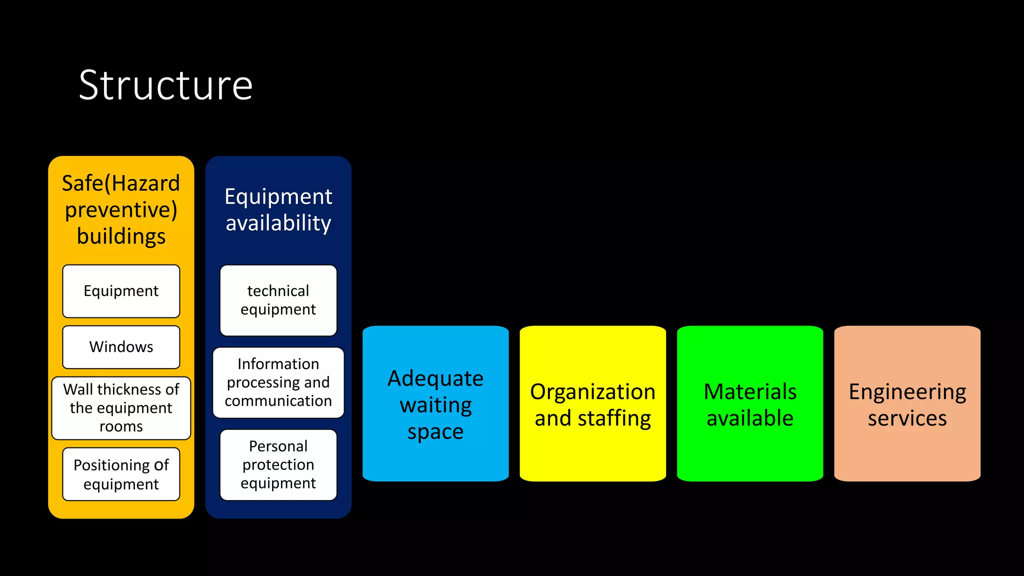
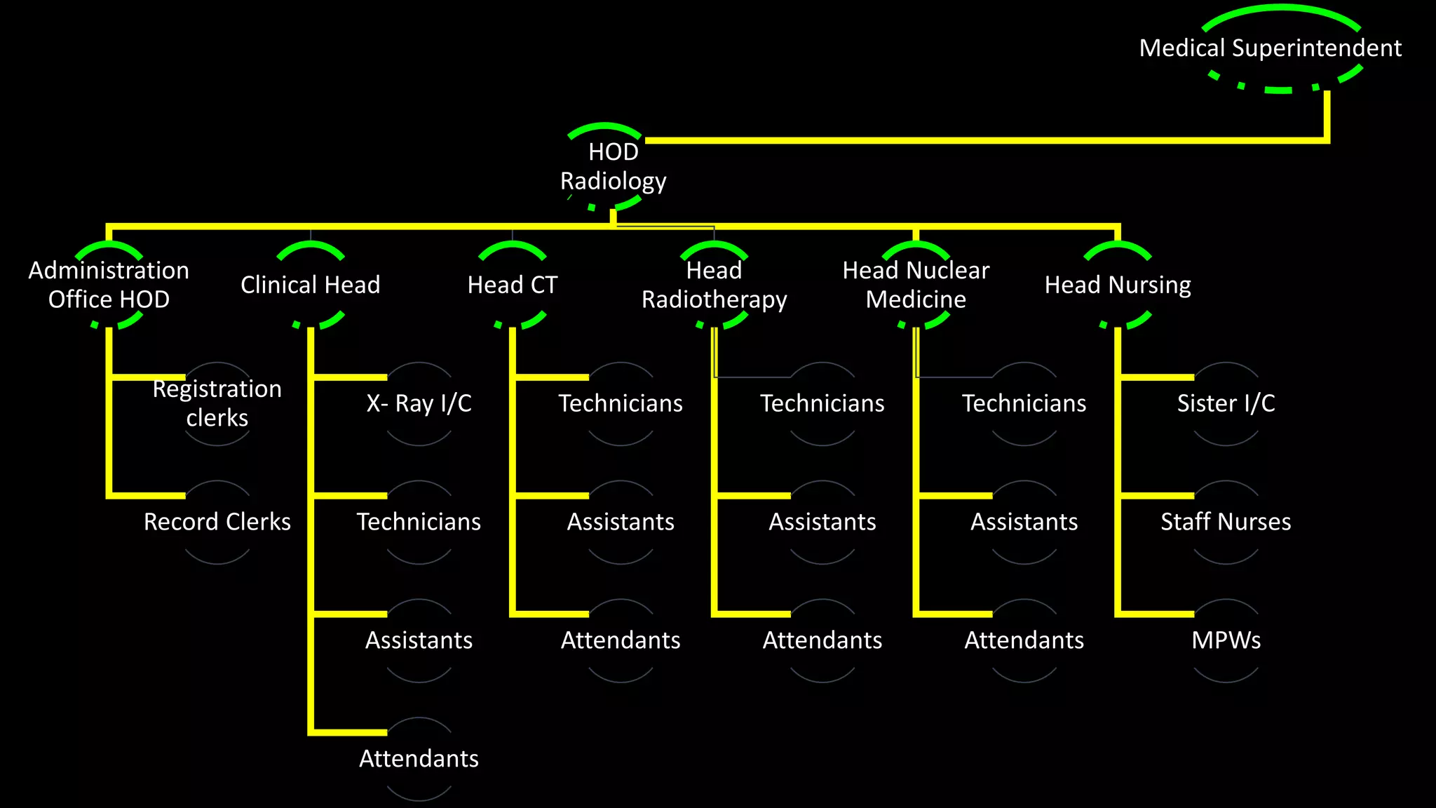
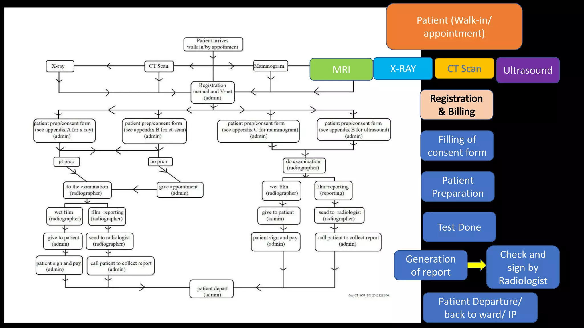
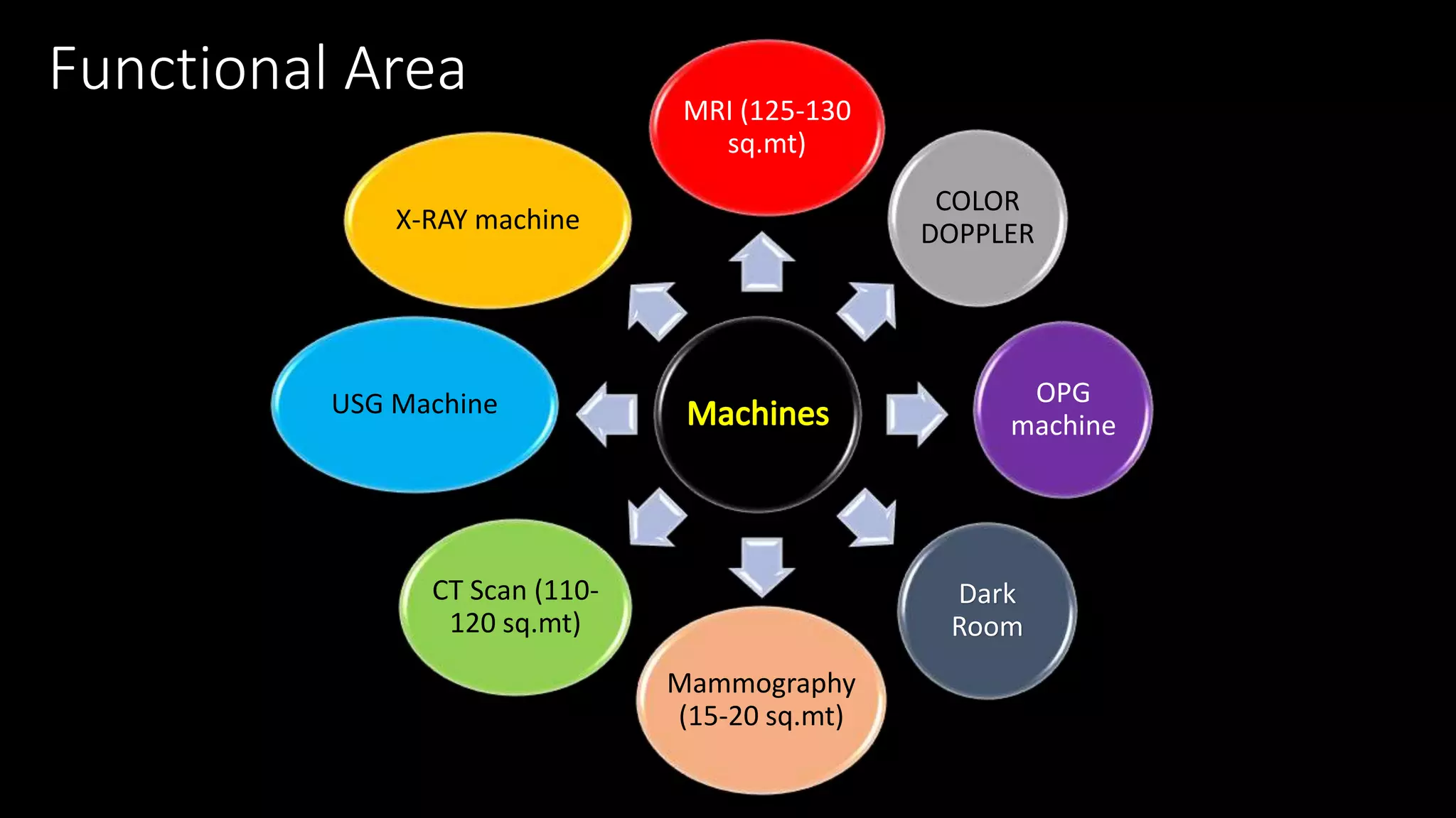
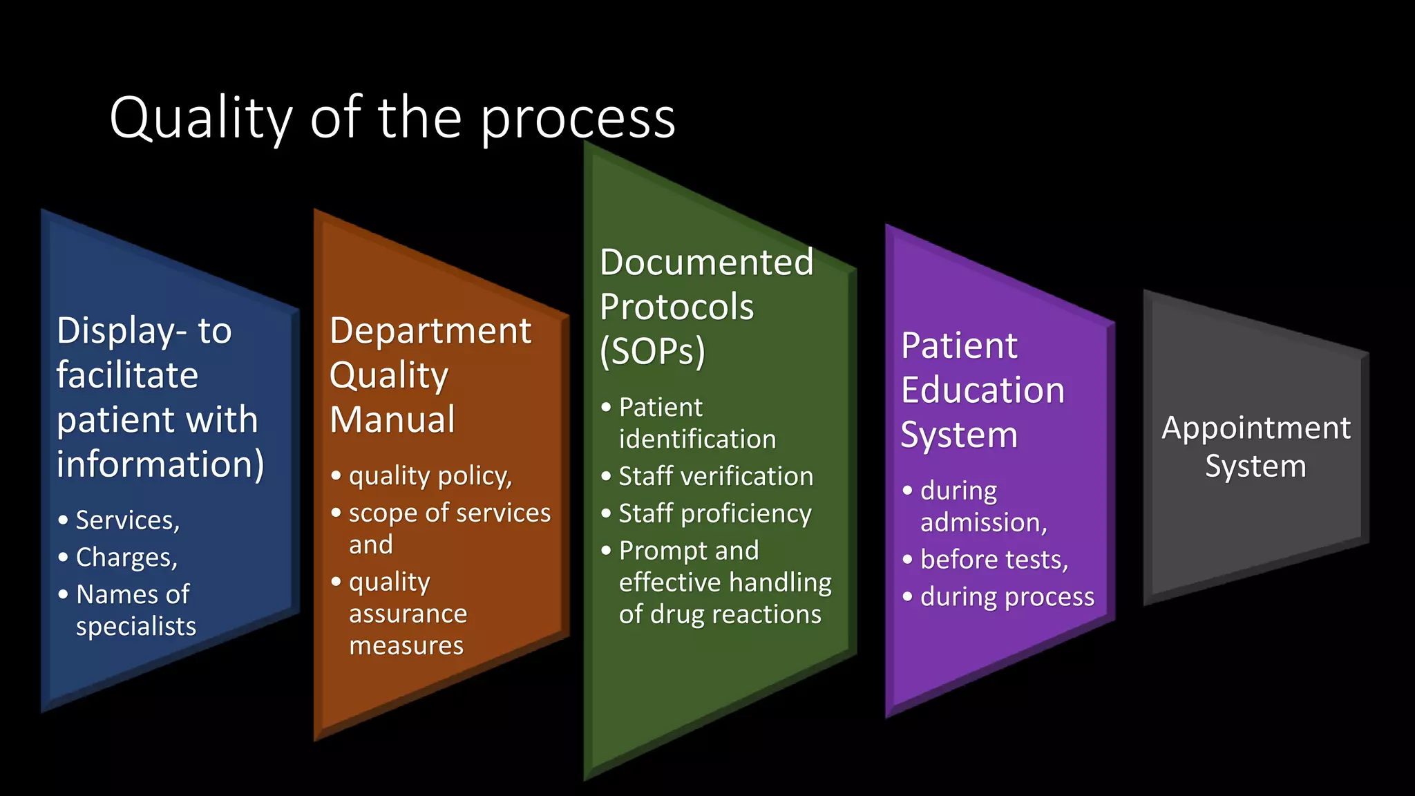
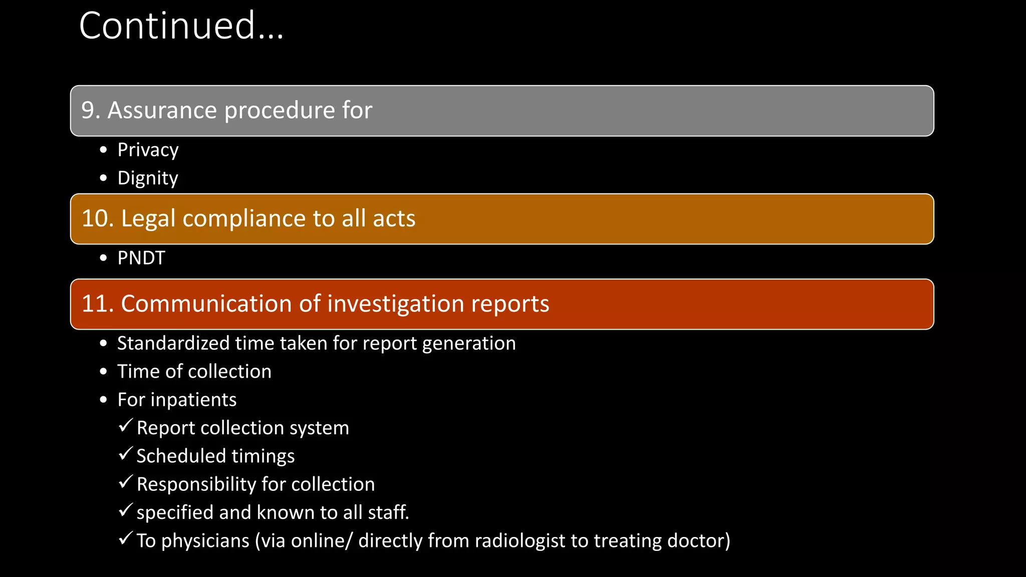
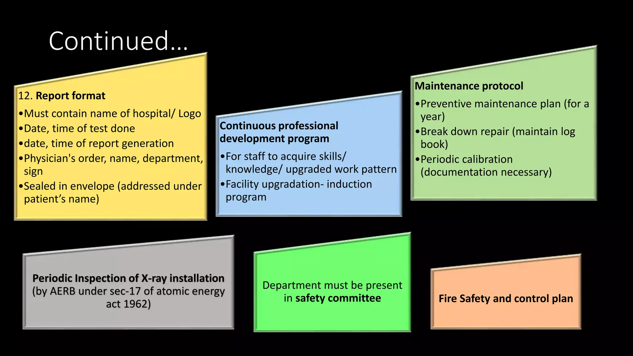
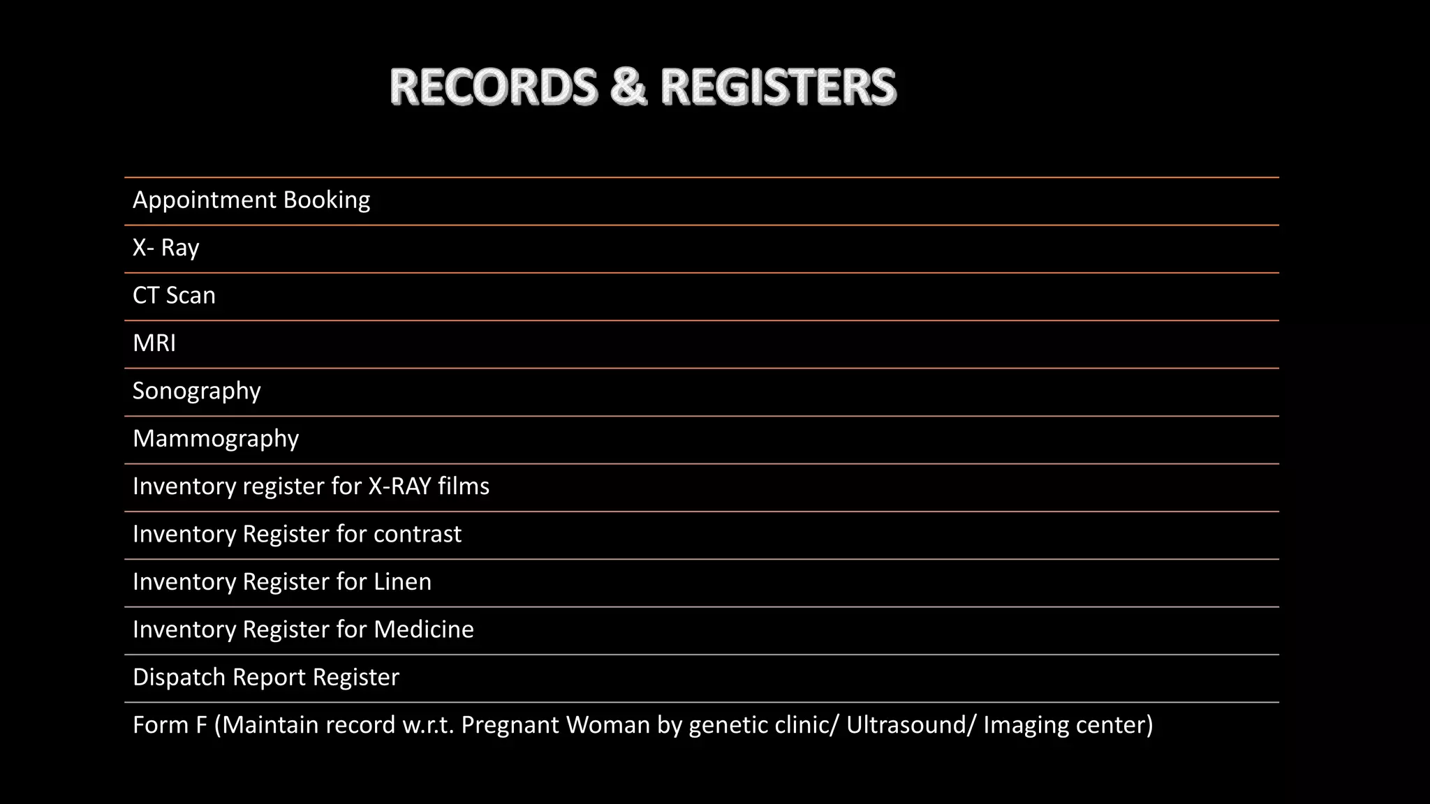
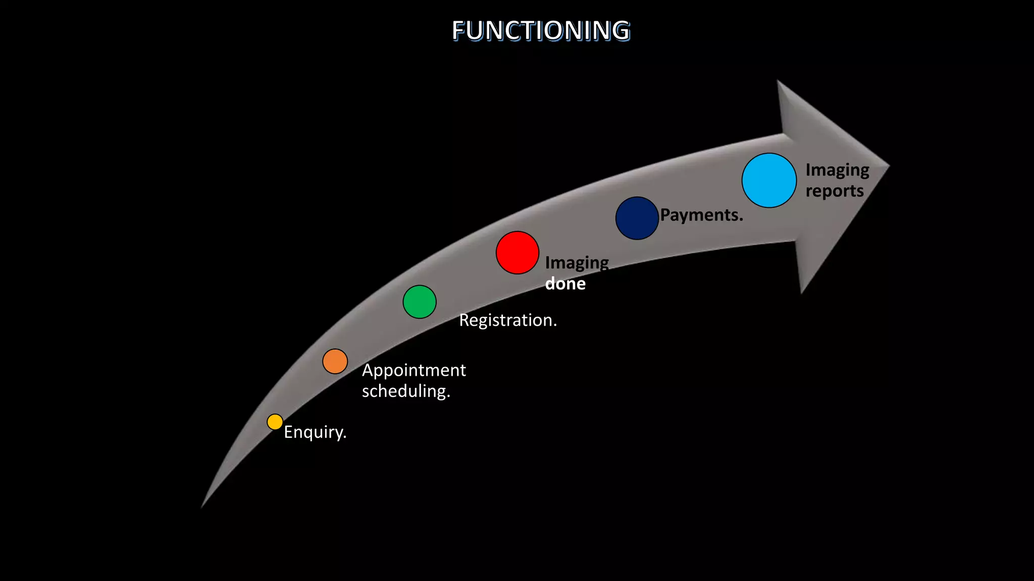
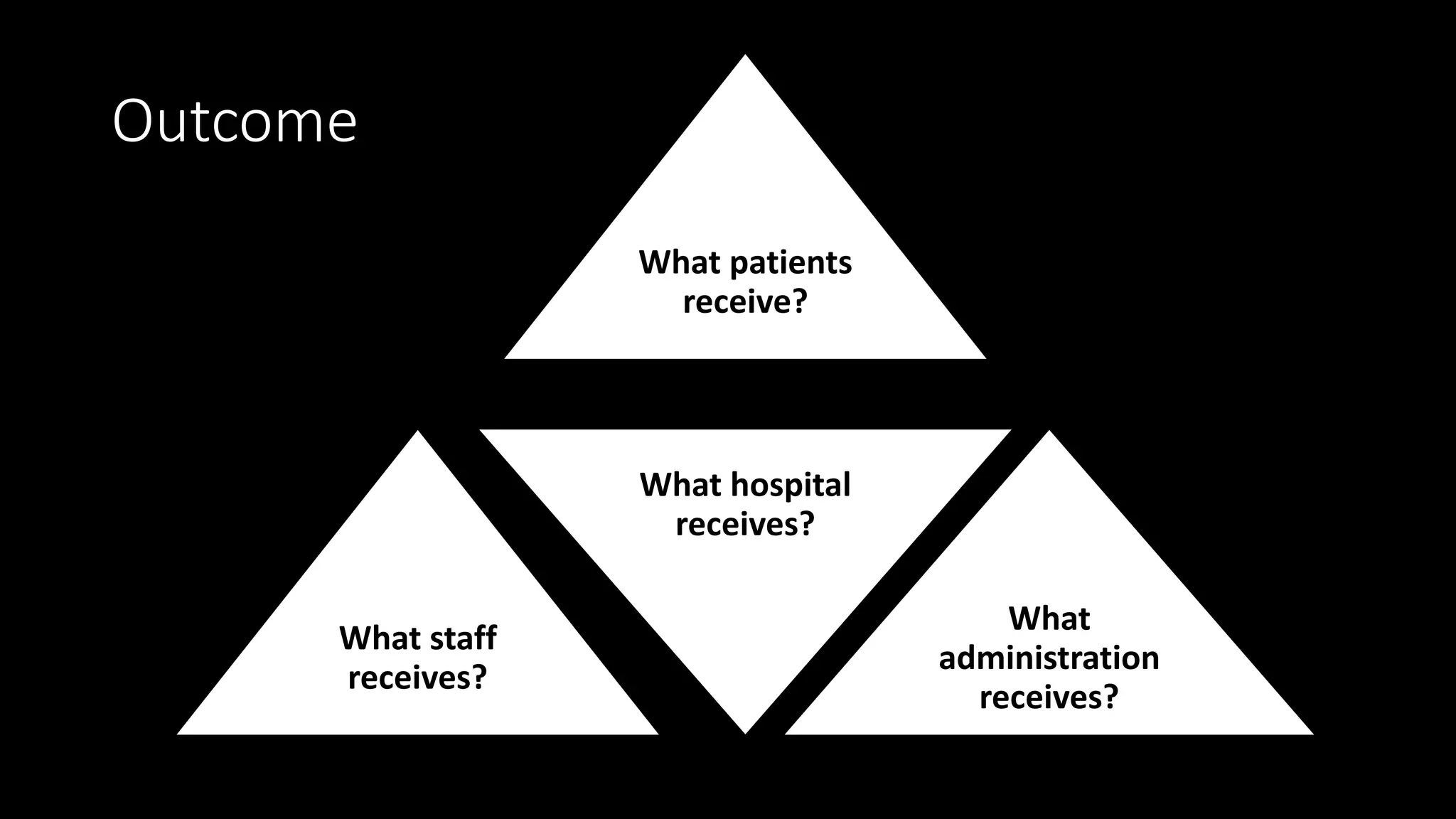
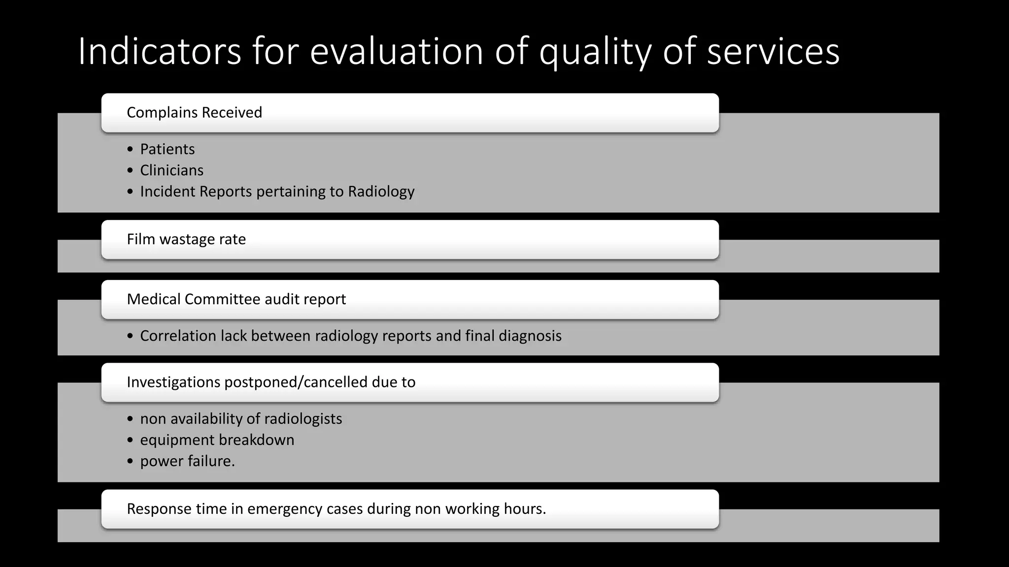
Srishti
hikha
Bhardwaj
hadwal
DONABEDIAN MODEL
Structure
Outcome Process
Structure
Safe(Hazard
preventive)
buildings
Equipment
Windows
Wall thickness of
the equipment
rooms
Positioning of
equipment
Equipment
availability
technical
equipment
Information
processing and
communication
Personal
protection
equipment
Adequate
waiting
space
Organization
and staffing
Materials
available
Engineering
services
Medical Superintendent
HOD
Radiology
Administration
Office HOD
Registration
clerks
Record Clerks
Clinical Head
X- Ray I/C
Technicians
Assistants
Attendants
Head CT
Technicians
Assistants
Attendants
Head
Radiotherapy
Technicians
Assistants
Attendants
Head Nuclear
Medicine
Technicians
Assistants
Attendants
Head Nursing
Sister I/C
Staff Nurses
MPWs
Patient (Walk-in/
appointment)
X-RAY CT Scan UltrasoundMRI
Filling of
consent form
Patient
Preparation
Test Done
Patient Departure/
back to ward/ IP
Generation
of report
Check and
sign by
Radiologist
Functional Area MRI (125-130
sq.mt)
COLOR
DOPPLER
OPG
machine
Dark
Room
Mammography
(15-20 sq.mt)
CT Scan (110-
120 sq.mt)
X-RAY machine
USG Machine
Quality of the process
Display- to
facilitate
patient with
information)
• Services,
• Charges,
• Names of
specialists
Department
Quality
Manual
• quality policy,
• scope of services
and
• quality
assurance
measures
Documented
Protocols
(SOPs)
• Patient
identification
• Staff verification
• Staff proficiency
• Prompt and
effective handling
of drug reactions
Patient
Education
System
• during
admission,
• before tests,
• during process
Appointment
System
Continued…
9. Assurance procedure for
• Privacy
• Dignity
10. Legal compliance to all acts
• PNDT
11. Communication of investigation reports
• Standardized time taken for report generation
• Time of collection
• For inpatients
Report collection system
Scheduled timings
Responsibility for collection
specified and known to all staff.
To physicians (via online/ directly from radiologist to treating doctor)
Continued…
12. Report format
•Must contain name of hospital/ Logo
•Date, time of test done
•date, time of report generation
•Physician's order, name, department,
sign
•Sealed in envelope (addressed under
patient’s name)
Continuous professional
development program
•For staff to acquire skills/
knowledge/ upgraded work pattern
•Facility upgradation- induction
program
Maintenance protocol
•Preventive maintenance plan (for a
year)
•Break down repair (maintain log
book)
•Periodic calibration
(documentation necessary)
Periodic Inspection of X-ray installation
(by AERB under sec-17 of atomic energy
act 1962)
Department must be present
in safety committee Fire Safety and control plan
Appointment Booking
X- Ray
CT Scan
MRI
Sonography
Mammography
Inventory register for X-RAY films
Inventory Register for contrast
Inventory Register for Linen
Inventory Register for Medicine
Dispatch Report Register
Form F (Maintain record w.r.t. Pregnant Woman by genetic clinic/ Ultrasound/ Imaging center)
Enquiry.
Appointment
scheduling.
Registration.
Payments.
Imaging
reports
Imaging
done
Outcome
What patients
receive?
What staff
receives?
What hospital
receives?
What
administration
receives?
Indicators for evaluation of quality of services
• Patients
• Clinicians
• Incident Reports pertaining to Radiology
Complains Received
Film wastage rate
• Correlation lack between radiology reports and final diagnosis
Medical Committee audit report
• non availability of radiologists
• equipment breakdown
• power failure.
Investigations postponed/cancelled due to
Response time in emergency cases during non working hours.
Continued…
6. Turn Around Time of each Report
• radiation safety norms.
• PNDT
• Other similar applicable regulations
7. Instances of breach/ violation of
8. Documented Interaction (between the radiologist and clinician
regarding case diagnosis)
9. Utilization pattern of the equipment
10. Instances of breakdown repairs and downtime thereof. (Log
book)
REGULATORYBODY
Radiation protection rule, 1971, under
Atomic Energy Act 1962.
Safety and protection body of the hospital
and atomic energy regulatory board
Bhaba Atomic Research Center.
Disposal of radio waste as per norm.
Radio monitoring equipment.
Film/TLD badges and monthly monitoring
REFERENCES
1. Quality in hospital
Name of the author SK Joshi.
2. www.slideshare.com/radiology.pptxs/rahulmehta.3208
3. Quality Management powerpoint by NC Das
4. Hospital Planning and Infrastructure
Name of Author- G D Kunders

Radiology Department Quality (SPO- Structure, Process, Outcome)

  • 1.
  • 2.
  • 3.
    Structure Safe(Hazard preventive) buildings Equipment Windows Wall thickness of theequipment rooms Positioning of equipment Equipment availability technical equipment Information processing and communication Personal protection equipment Adequate waiting space Organization and staffing Materials available Engineering services
  • 4.
    Medical Superintendent HOD Radiology Administration Office HOD Registration clerks RecordClerks Clinical Head X- Ray I/C Technicians Assistants Attendants Head CT Technicians Assistants Attendants Head Radiotherapy Technicians Assistants Attendants Head Nuclear Medicine Technicians Assistants Attendants Head Nursing Sister I/C Staff Nurses MPWs
  • 5.
    Patient (Walk-in/ appointment) X-RAY CTScan UltrasoundMRI Filling of consent form Patient Preparation Test Done Patient Departure/ back to ward/ IP Generation of report Check and sign by Radiologist
  • 6.
    Functional Area MRI(125-130 sq.mt) COLOR DOPPLER OPG machine Dark Room Mammography (15-20 sq.mt) CT Scan (110- 120 sq.mt) X-RAY machine USG Machine
  • 7.
    Quality of theprocess Display- to facilitate patient with information) • Services, • Charges, • Names of specialists Department Quality Manual • quality policy, • scope of services and • quality assurance measures Documented Protocols (SOPs) • Patient identification • Staff verification • Staff proficiency • Prompt and effective handling of drug reactions Patient Education System • during admission, • before tests, • during process Appointment System
  • 8.
    Continued… 9. Assurance procedurefor • Privacy • Dignity 10. Legal compliance to all acts • PNDT 11. Communication of investigation reports • Standardized time taken for report generation • Time of collection • For inpatients Report collection system Scheduled timings Responsibility for collection specified and known to all staff. To physicians (via online/ directly from radiologist to treating doctor)
  • 9.
    Continued… 12. Report format •Mustcontain name of hospital/ Logo •Date, time of test done •date, time of report generation •Physician's order, name, department, sign •Sealed in envelope (addressed under patient’s name) Continuous professional development program •For staff to acquire skills/ knowledge/ upgraded work pattern •Facility upgradation- induction program Maintenance protocol •Preventive maintenance plan (for a year) •Break down repair (maintain log book) •Periodic calibration (documentation necessary) Periodic Inspection of X-ray installation (by AERB under sec-17 of atomic energy act 1962) Department must be present in safety committee Fire Safety and control plan
  • 10.
    Appointment Booking X- Ray CTScan MRI Sonography Mammography Inventory register for X-RAY films Inventory Register for contrast Inventory Register for Linen Inventory Register for Medicine Dispatch Report Register Form F (Maintain record w.r.t. Pregnant Woman by genetic clinic/ Ultrasound/ Imaging center)
  • 11.
  • 12.
    Outcome What patients receive? What staff receives? Whathospital receives? What administration receives?
  • 13.
    Indicators for evaluationof quality of services • Patients • Clinicians • Incident Reports pertaining to Radiology Complains Received Film wastage rate • Correlation lack between radiology reports and final diagnosis Medical Committee audit report • non availability of radiologists • equipment breakdown • power failure. Investigations postponed/cancelled due to Response time in emergency cases during non working hours.
  • 14.
    Continued… 6. Turn AroundTime of each Report • radiation safety norms. • PNDT • Other similar applicable regulations 7. Instances of breach/ violation of 8. Documented Interaction (between the radiologist and clinician regarding case diagnosis) 9. Utilization pattern of the equipment 10. Instances of breakdown repairs and downtime thereof. (Log book)
  • 15.
    REGULATORYBODY Radiation protection rule,1971, under Atomic Energy Act 1962. Safety and protection body of the hospital and atomic energy regulatory board Bhaba Atomic Research Center. Disposal of radio waste as per norm. Radio monitoring equipment. Film/TLD badges and monthly monitoring
  • 16.
    REFERENCES 1. Quality inhospital Name of the author SK Joshi. 2. www.slideshare.com/radiology.pptxs/rahulmehta.3208 3. Quality Management powerpoint by NC Das 4. Hospital Planning and Infrastructure Name of Author- G D Kunders